
Karteikarten
micro 2
Karten in diesem Stapel (31)
- 1—VITEK/VITEK2 - antibiotic susceptibility card
- is an automated system for testing the antibiotic susceptibility of bacteria
- system enables identification of Gr+ and Gr- bacteria and other m/o when using particular identification cards
- These cards consist of several micro-channels that contain antibiotics
1) A suspension is made from the previously obtained pure culture
2) Then the plastic tube of the identification card is inserted in the suspension = it serves as a transfer tube for filling up the micro-channels
3) The process following the sample preparation procedure is fully automated and done by the VITEK2 device --> monitor displays information regarding the antibiotic susceptibility of bacteria
4) When testing antibiotic susceptibility the results can be expected after approximately 12-15 hours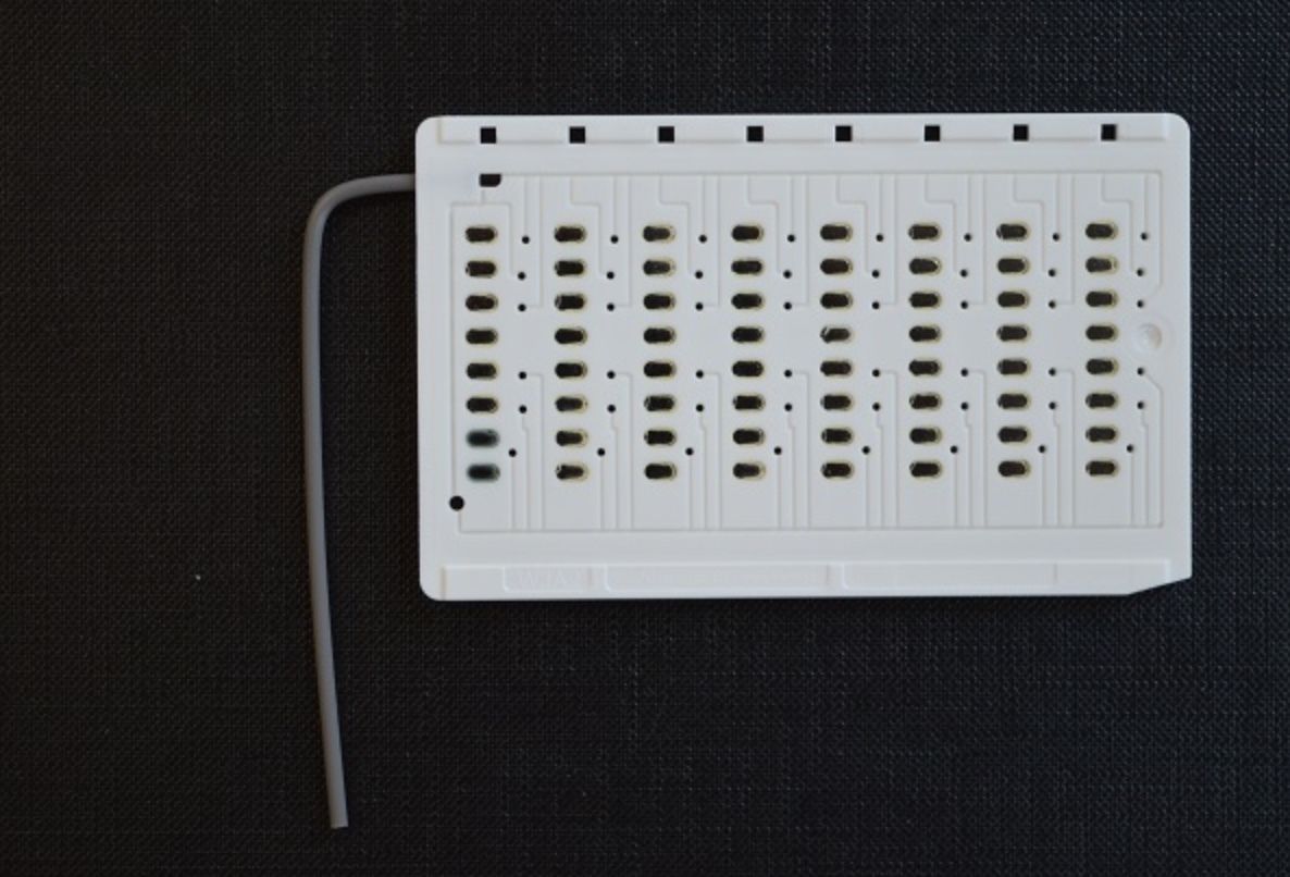
- 2—VITEK/VITEK2 - identification card
- is an automated system for identification of bacteria that detects biochemical properties
- system enables identification of Gr+ and Gr- bacteria and other m/o when using particular identification cards
- These cards consist of several micro-channels that contain dehydrated substrate
1) A suspension is made from the previously obtained pure culture
2) Then the plastic tube of the identification card is inserted in the suspension = it serves as a transfer tube for filling up the micro-channels
3) The process following the sample preparation procedure is fully automated and done by the VITEK2 device
4) After approximately 8 hours the monitor displays information regarding the potential species of bacteria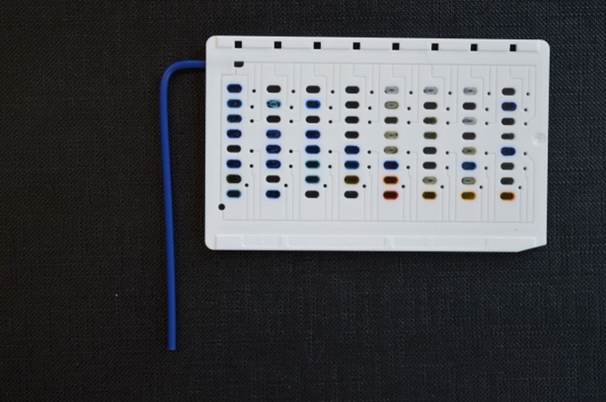
- 3—Simple transport medium - AMIES transport medium (Neisseria gonorrhoeae or Ureaplasma)
Simple transport media are used to transport nonfastidious aerobic or facultative anaerobic microorganisms.
- contains nutrients that are important for bacterial survival (MgCl, KCl, Agar etc.)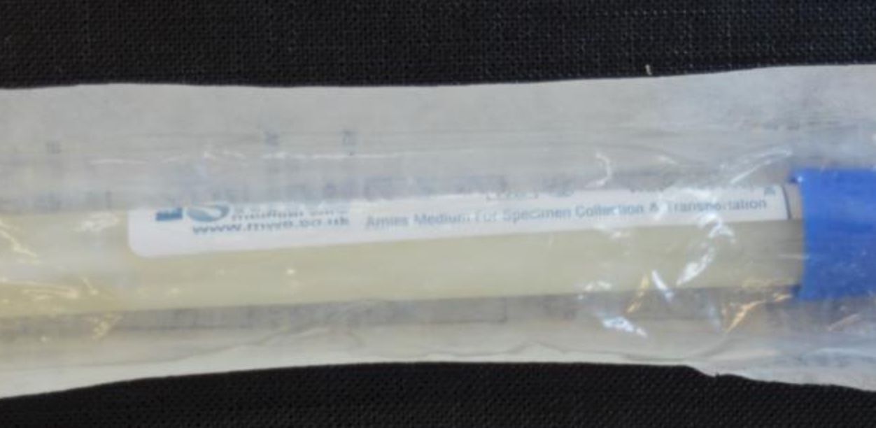
- 4—Tryptic soy broth (Myobacterium tuberculosis)
Tryptic soy broth (TSB) is the most common liquid culture medium and is used for microbial growth, culture and removal of exotoxins of bacteria. Turbidity (cloudiness), formation of sediment and production of a surface membrane (pellicle) can be observed in case of microbial growth.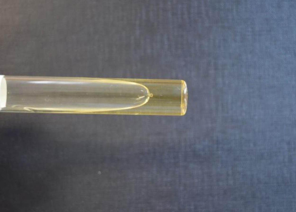
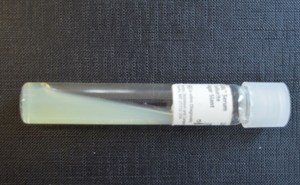
- 5—Tellurite agar
Differential media
- is used to isolate and differentiate biotypes of C.diphtheriae.
- It contains serum agar with added potassium tellurite.
- The "Var gravis" biotype forms gray, rough colonies whereas the other biotypes (var mitis, var intermedius) form smaller, black, sheeny colonies with a smooth surface. - 6—Loeffler Serum Medium
Complex culture media - Enriched
Loeffler’s Serum Medium is prepared by adding 10-12% inactivated (heated for 30 minutes at 56°C) blood serum to broth or TSA that is liquefied and cooled to 50°C. It is used to culture Cornyebacterium species which have proteolytic activity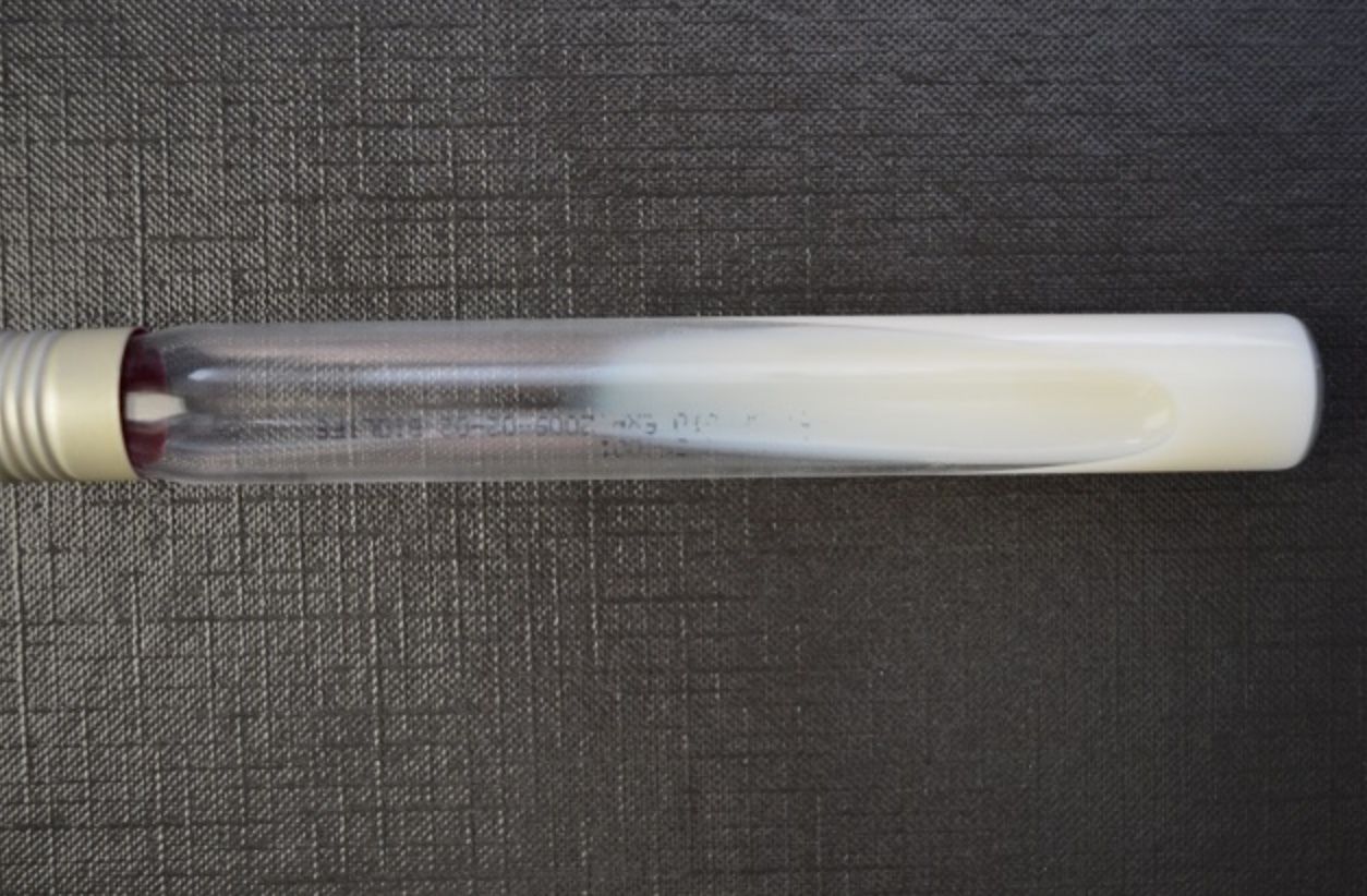
- 7—Chocolate Agar (N. gonorrhoeae, N. meningitidis - respiratory tract)
Complex culture media
- Chocolate agar is prepared by adding sterile 8-10% defibrinated blood to liquefied/molten hot TSA and mixing them thoroughly
- As a result RBC lyse and haemoglobin is released producing growth factors V and X necessary for microorganisms such as H. influenzae
- The medium obtains its chocolate colour as a result of release of haemoglobin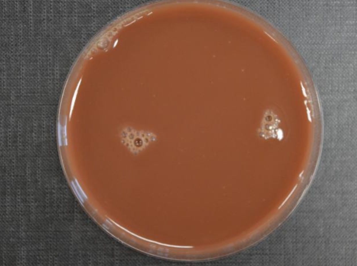
- 8—Slant agar
Solid medium
- broth + agar-agar (2-3% agar)
- is the basic form of agar
- various substances mixed into the agar promote the growth of very specific types of bacteria
- used for cultivation of m/o of pure culture
- m/o will grow different on their size, colour, surface, pigmentation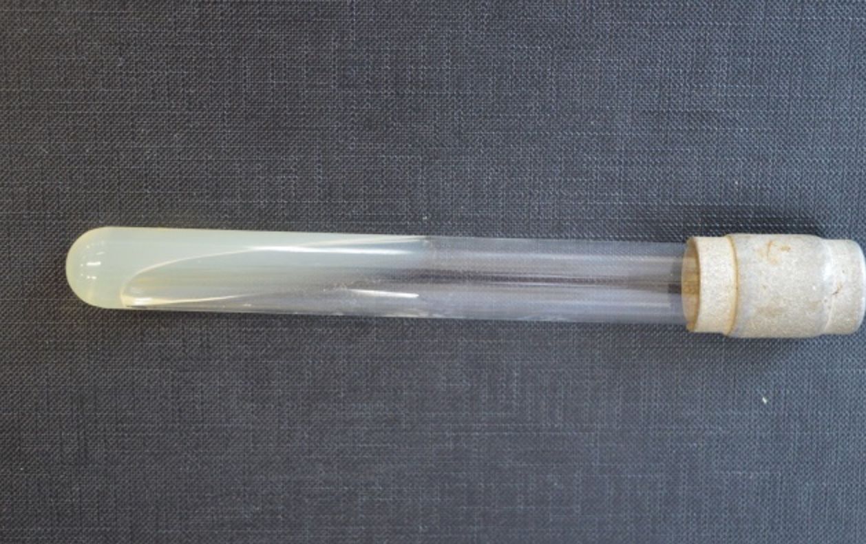
- 9—Sabouraud medium – selective media for cultivating fungi
The medium is prepared by adding 4% dextrose, gentamicin and chloramphenicol (a/b) to TSA
Antibiotics inhibit the growth of Gr- and Gr+ bacteria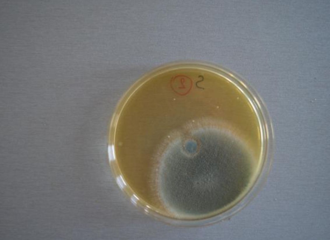
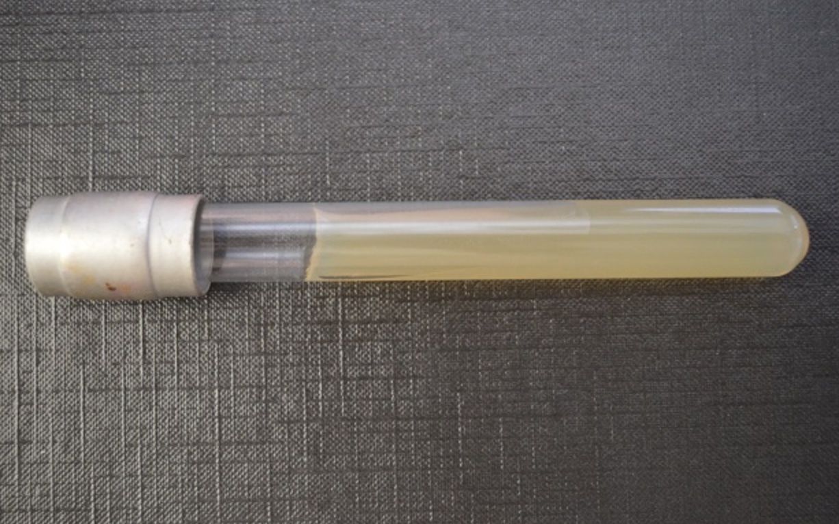
- 10—Stab agar
- 11—Mannitol Salt Agar
Selective media
It is a selective medium for Staphylococcus spp. due to the high concentration of salt and mannitol
- It is also used to identify pathogenic S.aureus which is a mannitol-fermenting bacterium
- The fermentation process increases acidity (pH decreases) and the colonies including the surrounding medium turn yellow
- S.epidermidis, S.saprophyticus that do not ferment mannitol do not alter the colour of the medium and their colonies are pink/red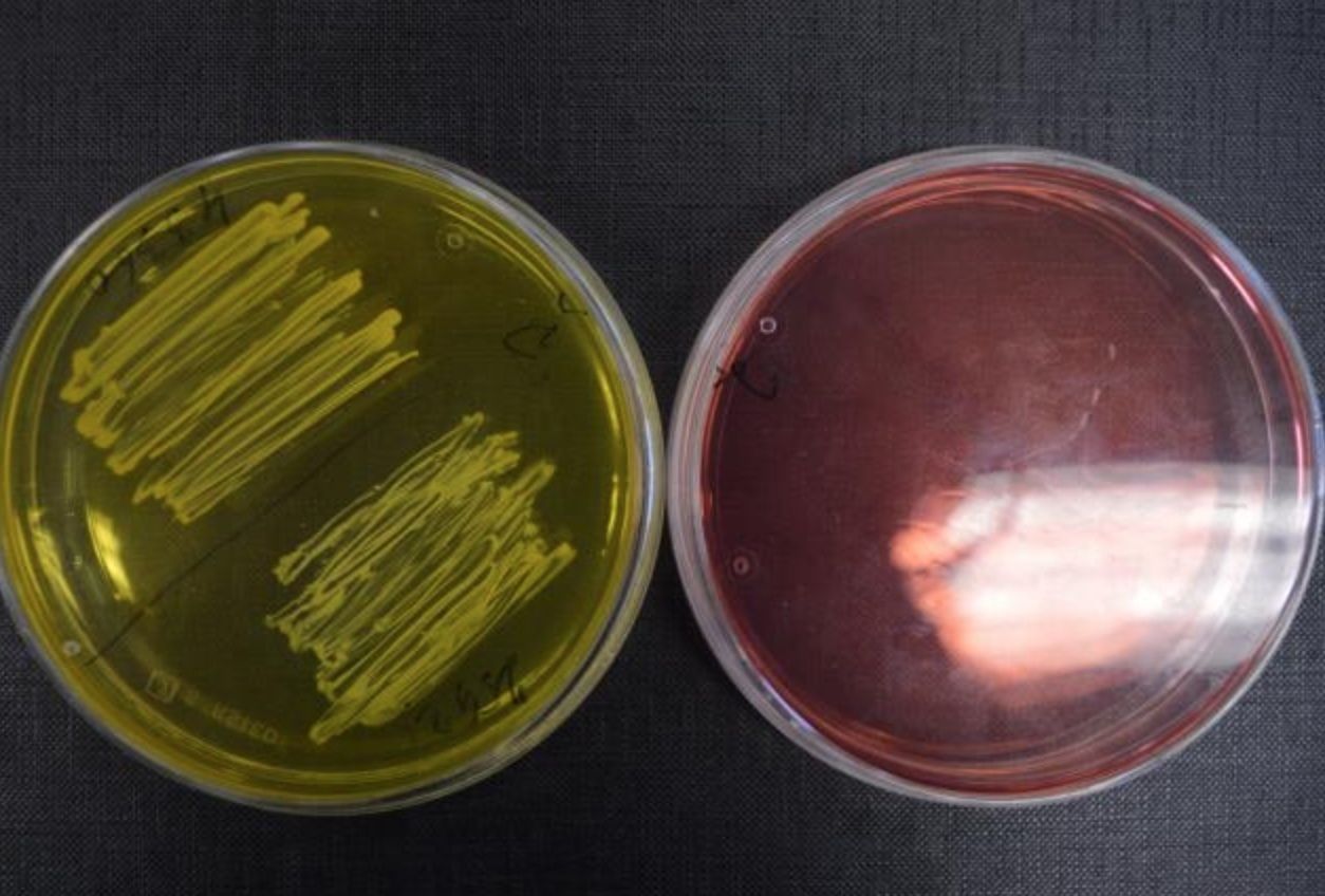
- 12—PLASMA COAGULASE TEST
- is used to differentiate between the pathogenic Staphylococcus aureus (coagulasepositive) and coagulase negative Staphylococcus epidermidis and Staphylococcus saprophyticus
- The test uses the plasma of a rabbit
- A sample of the pure culture to be examined is inoculated into the test-tube
containing the rabbit plasma
- Then the test-tubes are incubated at 37°C for 2 hours
- If the sample of the pure culture contains S.aureus, then the plasma coagulates
(forms clumps) and the reaction is positive
- If the sample of the pure culture contains the coagulase-negative S.epidermidis or
S.saprophyticus, then the rabbit plasma remains liquid and the reaction is negative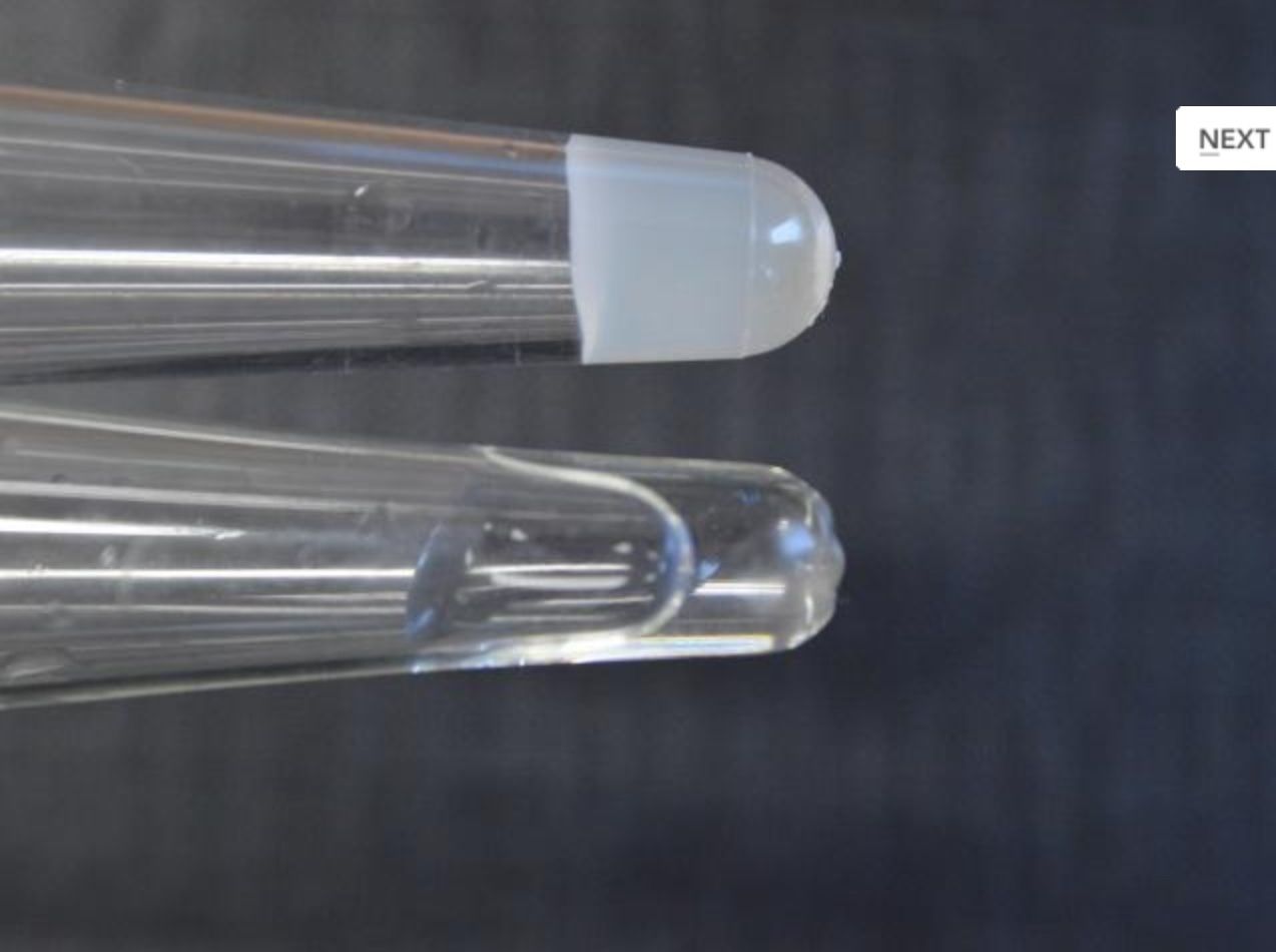
- 13—Port-a-Cul - media
Universal transport media
(Clostridium spp.)
- used to transport aerobic or anaerobic bacteria
- These media are used if the pathogenic agent is suspected to be anaerobic or the oxygen requirements of bacteria are unknown
- Universal transport media contain reducing agents that can bind to molecular oxygen, thus, creating an anaerobic environment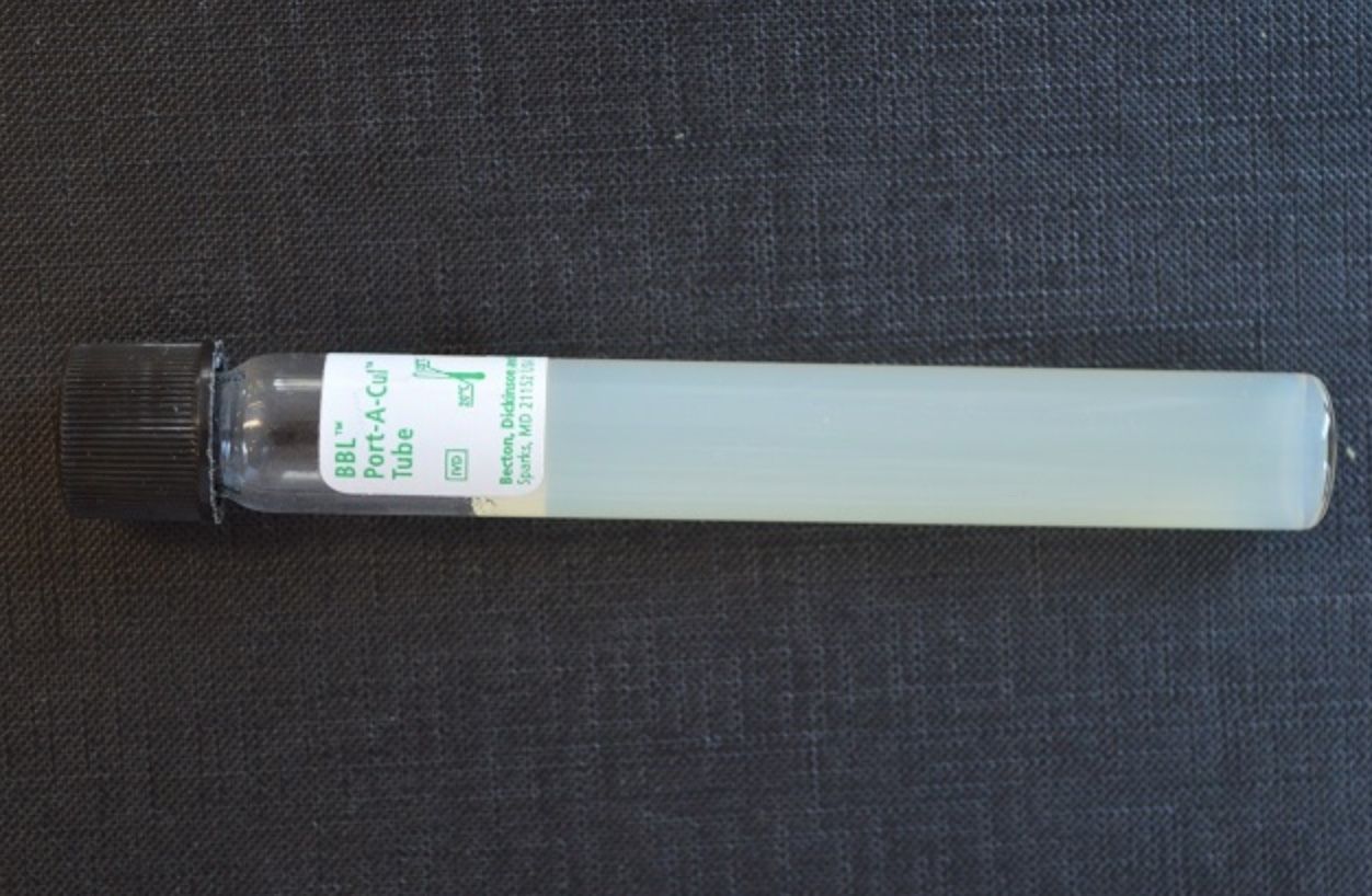
- 14—Egg-Yolk Salt Agar
Selective medium
- to culture Staphylococcus spp.
- It contains an increased level of salt - NaCl (7.5%) that inhibits the growth of other bacteria. - - Only Staphylococcus can survive in such high concentrations of salt
- This medium also enables the detection of lecithinase activity in case of S. aureus. Lecithinase splits lecithin in the egg yolk that results in the formation of a cloudy zone around the S. aureus colonies
- Other species of Staphylococcus produce lecithinase less frequently.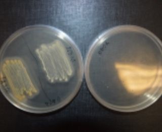
- 15—Mycoline medium
Specialized transport medium
- Sabourraud agar + antibiotics
- is used both to transport and culture samples of fungal infections of the
hair, skin or nails
- if fungal infection is suspectet, a/b inhibits m/o growth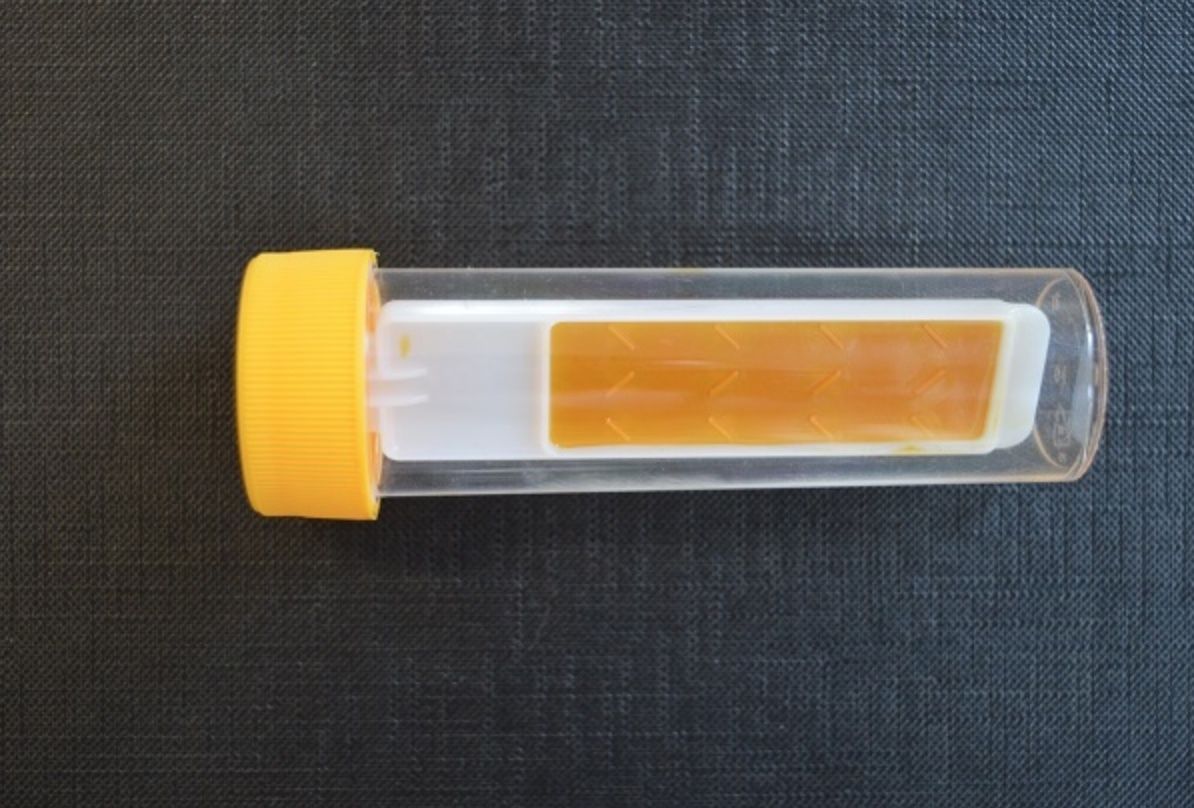
- 16—Levine Agar – eosin and methylene blue agar
Differential media
- media used to differentiate coliform bacteria
- media contain TSA and lactose and indicator: eosin and methylene blue --> they change colour in response to changes in pH
- If coliform bacteria ferment lactose (E.coli), then differences in pH cause the colonies to stain in dark blue
- Colonies of bacteria that do not ferment lactose (Salmonella, Shigella, etc.) remain colourless because the pH does not change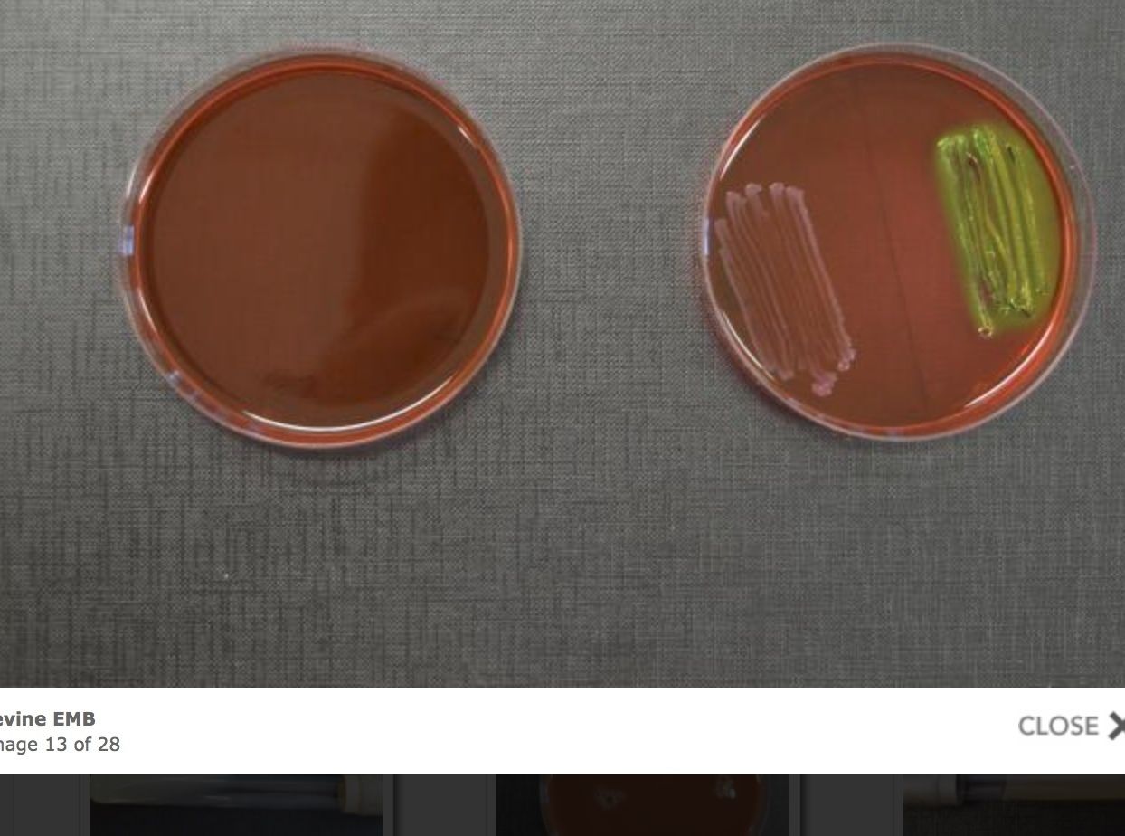
- 17—Gas Pak system or GenerBag - for anaerobic bacteria cultivation
- These Anaerobe systems are widely used to culture anaerobic bacteria
- Culture media with anaerobic bacteria are placed in polyethylene sachets or plastic anaerostats and a gas generator is opened
- The system is hermetically sealed
- As a result of series of chemical reactions that occur in the gas generator, oxygen is absorbed producing anaerobic conditions
- Filter papers with indicators are used to ensure that the conditions are anaerobic
- They change colour when particular anaerobic conditions are achieved.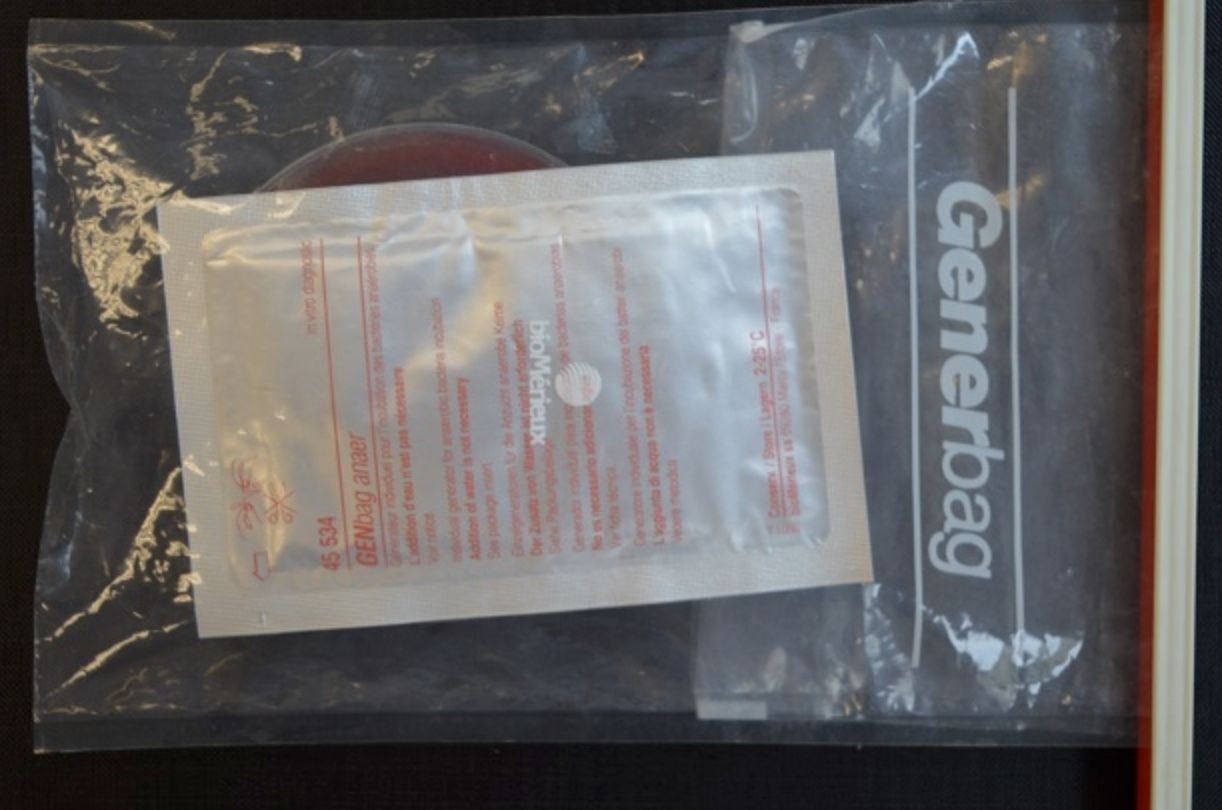
- 18—Gonoline special transport
Specialized transport medium
- contain a generator that maintains the level of CO2 at 5%
- Chocolate agar + a/b
- are used when transporting samples obtained from the urogenital system in cases of sexually transmitted diseases
- The optimal temperature during the transfer is 35±2°C
- The Gonoline special transport medium is used to transport and culture N. gonorrhoeae in the laboratory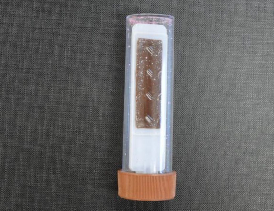
- 19—Hemoline medium
Specialized transport media
- is used to transport blood in cases of sepsis and bacteraemia
- The medium is used to transport and subsequently culture the sample in the laboratory
- It is biphasic as it contains a layer of solid agar and a liquid broth medium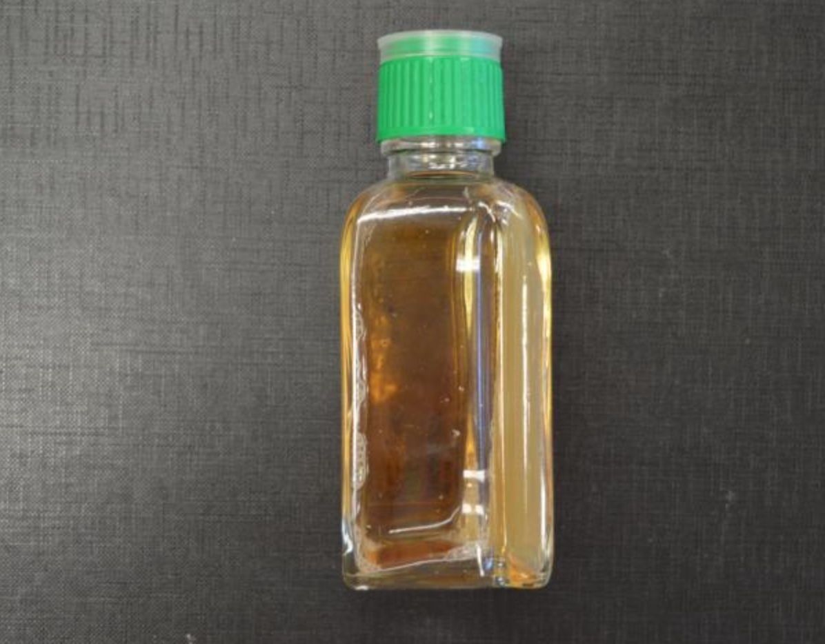
- 20—TSA - tryptic soy agar
Solid culture media
Adding agar (polysaccharide obtained from seaweed) to liquid culture media produces solid culture media. Tryptic soy agar (TSA) is the most commonly used solid culture media in laboratories. Bacteria grow on the surface of TSA forming colonies. TSA can be poured into Petri dishes and used for microbial culture or isolation of pure cultures.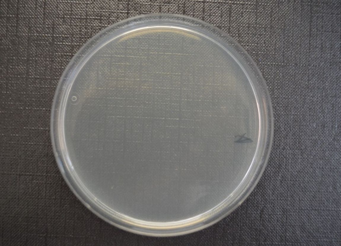
- 21—E-TEST (Epsilometer test, quantitative method)
- E-test is used to detect the minimum inhibitory concentration (MIC)
- A pure culture is inoculated in an even layer onto a sterile culture media
- Afterwards an antibiotic-impregnated plastic strip with an exponential gradient of antibiotic concentrations is placed on the culture media (concentrations are indicated on a scale)
- The culture media is incubated at 37°C for 16-18 hours
- After incubation a symmetrical inhibition ellipse is produced
- The intersection of the sterile and non-sterile area on the scale indicates the MIC value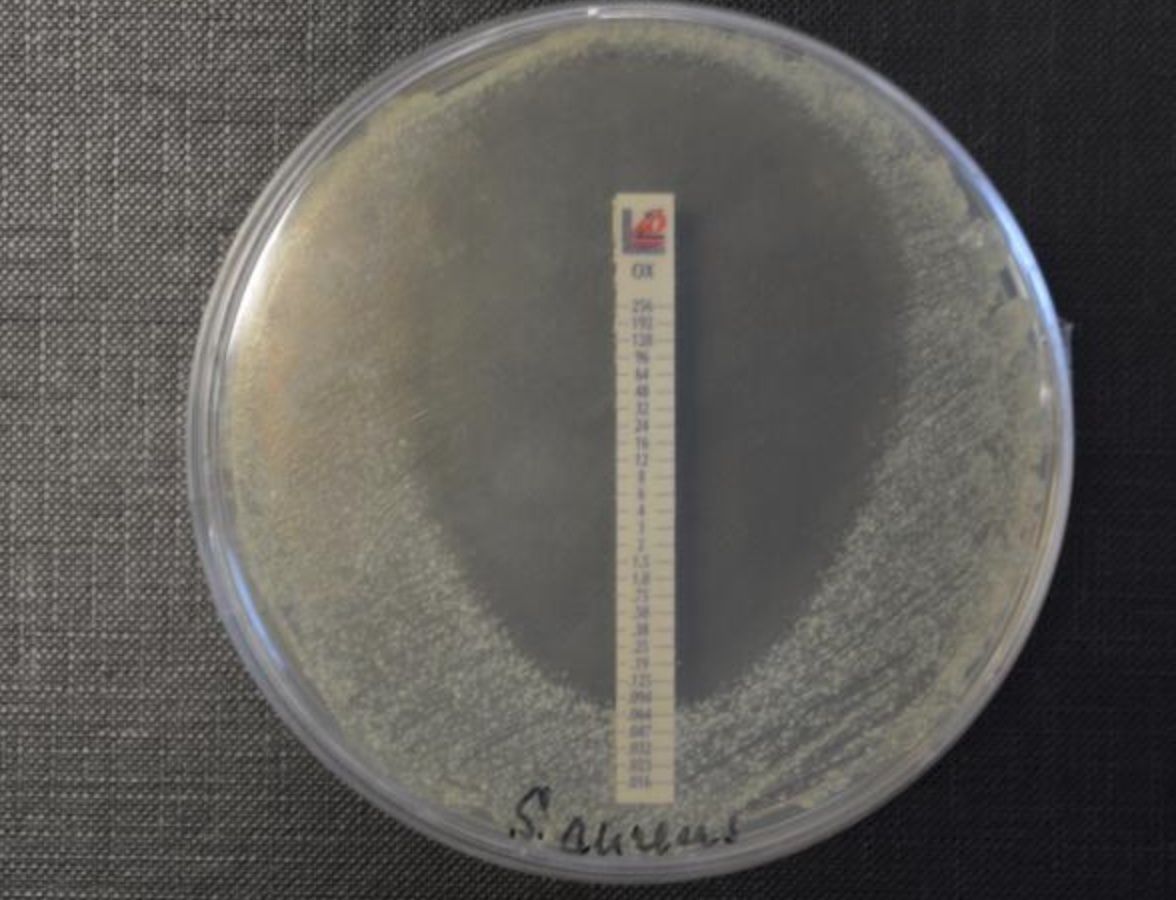
- 22—Endo agar
Differential media
- used to to differentiate coliform bacteria. These media contain TSA and lactose, however the differing ingredient is the indicator (Endo Agar contains basic fuchsin) that changes colour in response to changes in pH
- If coliform bacteria ferment lactose (E.coli), then differences in pH cause the colonies to stain in the particular colour of the indicator (Endo Agar – dark red colour) - Colonies of bacteria that do not ferment lactose (Salmonella, Shigella, etc.) remain colourless because the pH does not change.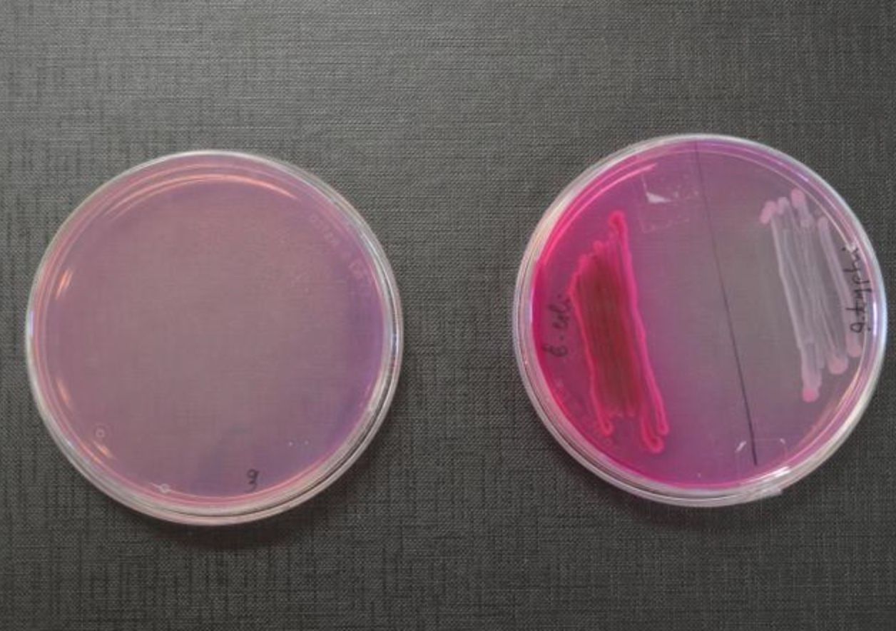
- 23—Candle jar
Cultivation of anaerobes
The culture of anaerobic bacteria is a technically complicated process because microbial growth requires an environment with no oxygen. Such conditions can be produced by applying physical and chemical techniques.
- Candle jars are containers made of glass, metal or plastic
- Culture media inoculated with bacteria are placed in the candle jar
- Afterwards the candle jar is hermetically sealed
- The burning candle uses up all oxygen left in the container
- As a result anaerobic conditions with an increased level of CO2 are produced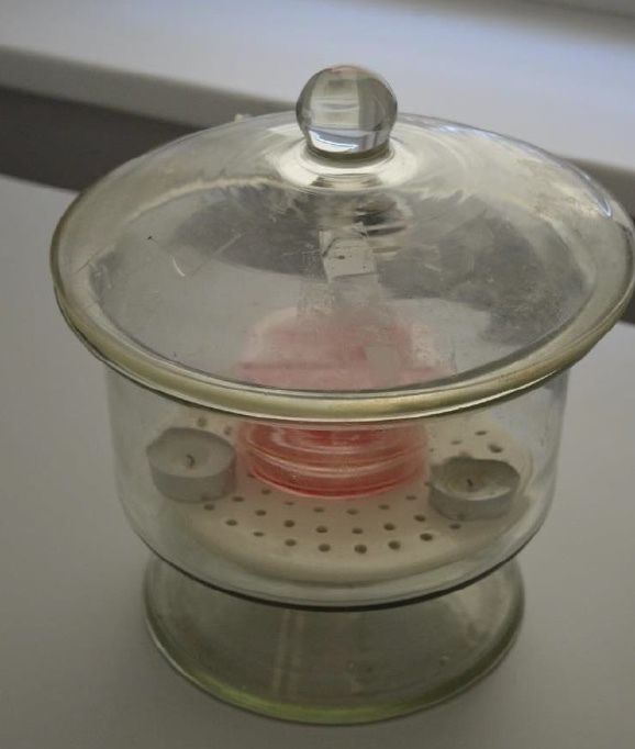
- 24—Disk Diffusion Test (qualitative test)
- A pure culture is inoculated in an even layer onto a sterile culture media
- After inoculation antibiotic-impregnated paper disks are placed on the culture media
- Usually 5-6 different antibiotic-impregnated disks are placed on the media within 2 cm from each other
- The culture media is then incubated at 37°C for 16-18 hours
- Antibiotics diffuse from the paper disk into the gel of the culture media and inhibit the growth of bacteria
- After incubation the diameter of the sterile area (including the diameter of the disk) is measured
- the results are evaluated in accordance with the guidelines.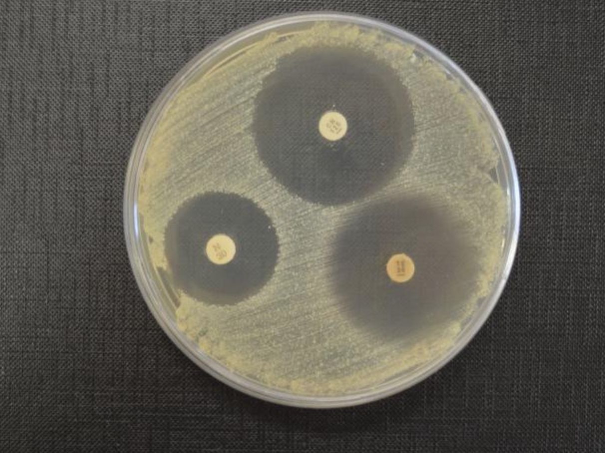
- 25—Bismuth Sulphite Agar
Differential media
- This medium contains bismuth sulphite, ferrous sulphate and brilliant green
- Ferrous sulphate serves as an indicator of H2S production
- Bismuth is a heavy metal that in combination with brilliant green inhibits the growth of many Gr+ and Gr- bacteria except Salmonella that grows and forms black coloured colonies with a metallic sheen
- Therefore the medium is used to identify species of Salmonella.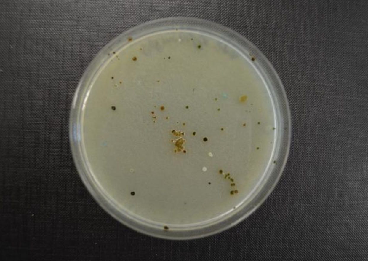
- 26—Blood Agar
Complex culture media - Enriched
- medium is obtained by adding 5-10% sterile defibrinated blood to TSA (liquefied) and cooled to 50°C
- Blood is mixed with agar and under aseptic conditions poured into sterile Petri dishes with no extra sterilization
- used to culture Streptococcus and Staphylococcus species
- enables detection of haemolytic activity of microorganisms (enzymes break down red blood cells)
- α, β or γ-haemolysis can be observed
1) α-haemolysis is partial and appears as a partially transparent area surrounding colonies
2) β-haemolysis is complete and appears as an entirely transparent area around colonies
3) The term γ-hemolysis is used when hemolysis is not observed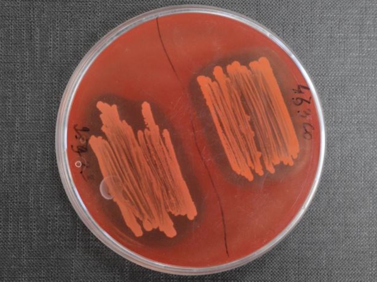
- 27—The API Test System
- is a panel test system where standardized micro tests are used to detect enzymatic
activity in order to identify species of bacteria
- is a panel that consists of 20 mini-test chambers (compartments) that contain
dehydrated substrate (biochemical reagent)
- A suspension is made from a previously obtained pure culture that is poured into
each chamber
- The panel is then incubated at 37°C for 15-18 hours.
- After incubation specific reagents are added to each chamber and change of
colour is observed
- results are compared to the results available in a standardized database and
interpreted by using special software or by using API analytical profile
- Laboratories use different API systems, for example, “API Coryne” to identify
bacteria that cause diphtheria, “API Strep”, “API Staph”, “API E” for identifying
bacteria of the Streptococcaceae, Staphylococcaceae and Enterobacteriacae
family as well as anaerobic bacteria and other microorganisms, including fungi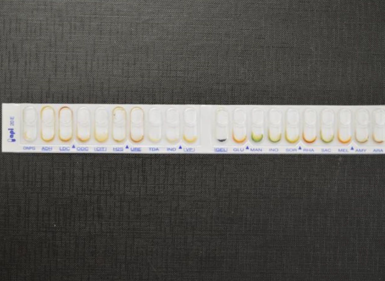
- 28—Löwenstein-Jensen media
Selective media
- This medium is used to culture M.tuberculosis (appear as brown colonies)
- It contains malachite green that inhibits the growth of other microorganisms present in sputum
- The average time of M.tuberculosis culture to grow in this medium is 6 weeks. Colonies are pale yellow, dry and with a rough surface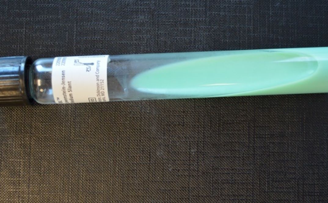
- 29—Streak plate technique
(know procedure)
- After inoculation the culture media is incubated in a thermostat at 37°C for 24 hours
- Following incubation, higher concentration of bacteria can be observed in sections A and B, whereas sections C show discrete colonies that contain microorganisms of the same species and are used to obtain a pure culture
- In order to do this, an agar slant tube is inoculated with a sample of the discrete
colony by using an inoculation loop
- The culture is then incubated in a thermostat at 37°C for 24 hours
- The agar slant is used to identify the species of the bacteria the following day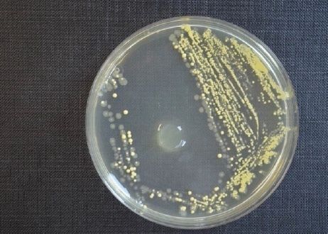
- 30—BACTEC
- are designed for the rapid detection of microorganisms in clinical specimens
1) Inoculate sample into culture medium containing 14C Substrate
2) Bacteria produce 14Co2 from 14C substrate
3) BACTEC system measures amount of CO2 - HIGH if bacteria present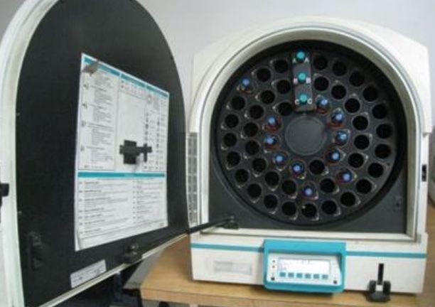
- 31—Mac Conkey agar (MAC)
- Indicator - natural red
- These media contain TSA and lactose and an indicator that changes colour in response to changes in pH. If coliform bacteria ferment lactose (E.coli), then differences in pH cause the colonies to stain in the particular colour of the indicator --> MacConkey Agar – pink colour). Colonies of bacteria that do not ferment lactose (Salmonella, Shigella, etc.) remain colourless because the pH does not change.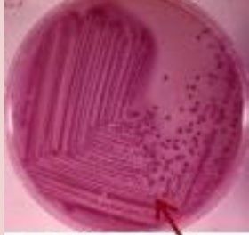
Informationen
- Beschreibung
- Medicine Karteikarten am micro 2, erstellt von Kerim Yildirim am 23/03/2018.
- Erstellt von
- Kerim Yildirim
- Aufrufe
- 0
- Bewertung