GoConqr hat ein neues Design! Sag uns, wie es dir gefällt.

Karteikarten

GIT EM

أطباء 2020 Englisch 29 Karten 1.146 Aufrufe

GIT EM

Karten in diesem Stapel (29)

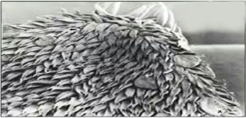
  1. 1—Lingual papilleaLingual papillea
  2. 2—A: Filliform papillea
    Very numerous , elongated
    B: Fungiform papillea
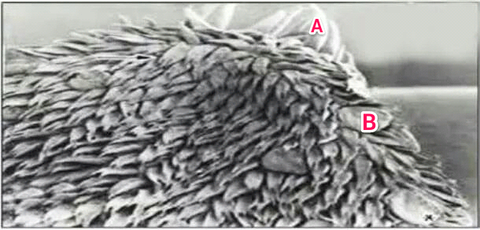
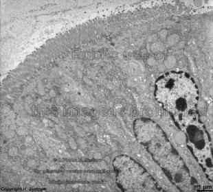
    A: Filliform papillea Very numerous , elongated B: Fungiform papillea
  3. 3—Fungiform papilla :
    Less numerous , mushroom-shaped
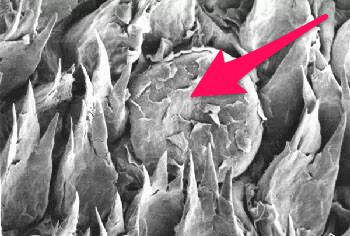
    Fungiform papilla : Less numerous , mushroom-shaped
  4. 4—Parietal cell TEM
    "NOTICE apical occluding junction"
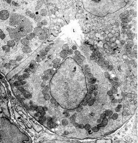
    Parietal cell TEM "NOTICE apical occluding junction"
  5. 5—A : Lumen
    B : Microvilli
    C : Intracellular canaliculas
    D : Mitochondria
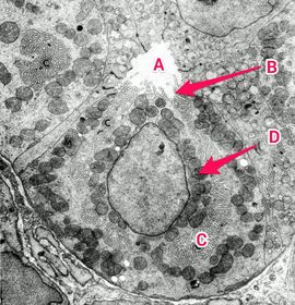
    A : Lumen B : Microvilli C : Intracellular canaliculas D : Mitochondria
  6. 6—Enteroendocrine cell
    (Open type)
    A: Basal electron dense granules
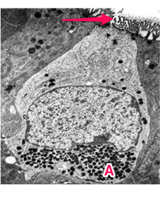
    Enteroendocrine cell (Open type) A: Basal electron dense granules
  7. 7—Enteroendocrine cell
    (Closed type)
    A: basal electron dense granules
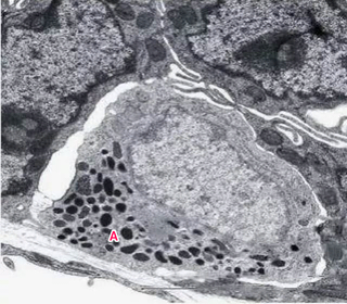
    Enteroendocrine cell (Closed type) A: basal electron dense granules
  8. 8—SEM of small intestine
    A : Villus
    B : Crypt
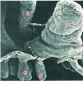
    SEM of small intestine A : Villus B : Crypt
  9. 9—TEM of enterocytes covering the villiTEM of enterocytes covering the villi
  10. 10—Apical surface of enterocytes TEM
    A : Microvilli
    B : Zonula occludens
    C : Zonula adherens
    D : Desmosome
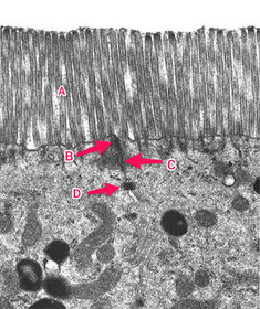
    Apical surface of enterocytes TEM A : Microvilli B : Zonula occludens C : Zonula adherens D : Desmosome
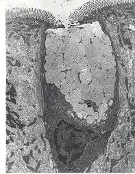
  11. 11—Goblet cellGoblet cell
  12. 12—A : secretory granules containig mucous
    B : Nucleus
    C : rER
    D : Enterocyte
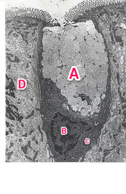
    A : secretory granules containig mucous B : Nucleus C : rER D : Enterocyte
  13. 13—TEM of the basal part
    Of the crypts
    Of small intestine
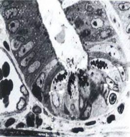
    TEM of the basal part Of the crypts Of small intestine
  14. 14—A : Paneth cell
    B : Apical granules
    C: Entero-endocrine cell
    B : basal granules
    D : blood capillary
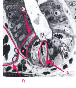
    A : Paneth cell B : Apical granules C: Entero-endocrine cell B : basal granules D : blood capillary
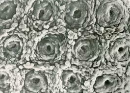
  15. 15—SEM of the ColonSEM of the Colon
  16. 16—A : Goblet cell
    B : opening of a crypt
    *notice absence of villi
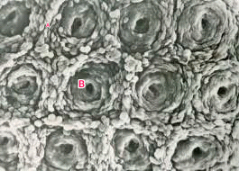
    A : Goblet cell B : opening of a crypt *notice absence of villi
  17. 17—SEM of the Colon (side view)
    A: smooth surface
    B: Crypts
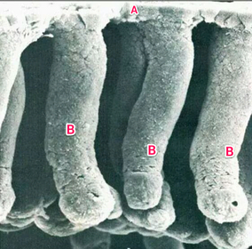
    SEM of the Colon (side view) A: smooth surface B: Crypts
  18. 18—TEM of coloumnar absorbtive cells
    Lining the colon
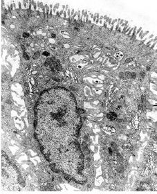
    TEM of coloumnar absorbtive cells Lining the colon
  19. 19—A : short microvilli
    B : lateral membrane infoldings
    ( for water absorbtion)
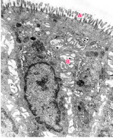
    A : short microvilli B : lateral membrane infoldings ( for water absorbtion)
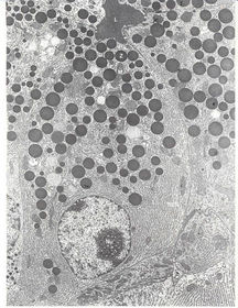
  20. 20—TEM OF Pancreatic acinar cellTEM OF Pancreatic acinar cell
  21. 21—A : Lumen
    B : Zymogen granules
    C : Nucleus
    With evident nucleolus
    D : rER
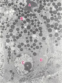
    A : Lumen B : Zymogen granules C : Nucleus With evident nucleolus D : rER
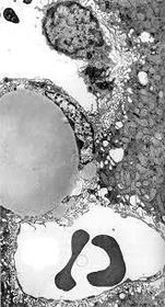
  22. 22—TEM of a part of the liverTEM of a part of the liver
  23. 23—A : large fat droplet inside Ito cell
    B : Kupffer cell
    C : Blood sinusoid
    D : Space of Disse
    E : Microvilli
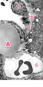
    A : large fat droplet inside Ito cell B : Kupffer cell C : Blood sinusoid D : Space of Disse E : Microvilli
  24. 24—TEM of part of Liver
    A: Nucleus of endothelial cell
    B : Microvilli
    C: Blood sinusoid
    D : Hepatocyte
    E: Space of Disse
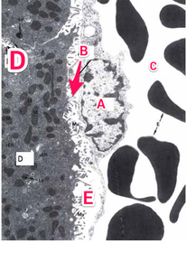
    TEM of part of Liver A: Nucleus of endothelial cell B : Microvilli C: Blood sinusoid D : Hepatocyte E: Space of Disse
  25. 25—A : Hepatocyte
    B : Kupffer Cell
    C : fat droplets inside
    Ito cell
    E : Space of Disse
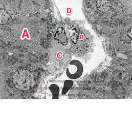
    A : Hepatocyte B : Kupffer Cell C : fat droplets inside Ito cell E : Space of Disse
  26. 26—TEM of a bile canaliculus
    A: Hepatocytes
    B: Microvilli
    C: Lumen of bile canaliculus
    D: tight junction
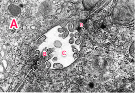
    TEM of a bile canaliculus A: Hepatocytes B: Microvilli C: Lumen of bile canaliculus D: tight junction
  27. 27—TEM OF BILE CANALICULUS
    A: Hepatocyte
    B : Lumen of bile canaliculus
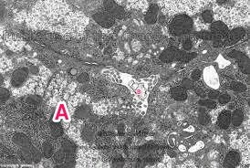
    TEM OF BILE CANALICULUS A: Hepatocyte B : Lumen of bile canaliculus
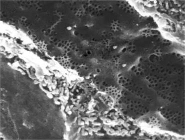
  28. 28—SEM of Fenestrated sinusoid of LiverSEM of Fenestrated sinusoid of Liver
  29. 29—A : Space of Disse
    B : Hepatocyte
    C : fenestrea of epithelial cells
    ( Sieve plate)
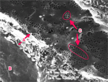
    A : Space of Disse B : Hepatocyte C : fenestrea of epithelial cells ( Sieve plate)

Informationen

Beschreibung
Karteikarten am GIT EM, erstellt von أطباء 2020 am 28/11/2014.
Erstellt von
أطباء 2020 · Nov 2014
Aktualisiert
23 Okt 2017
Aufrufe
1.146
Bewertung

Feedback

Worum geht es?

Wie gefällt dir das neue Design?

Dein Eindruck