
Karteikarten
Basic tissues- slideshow 3
Karten in diesem Stapel (20)
- 1TendonsDense regular connective tissue
• Features bundles of parallel type I collagen
• Provides great strength (but little stretch) in binding together components of the musculoskeletal system. - 2Tendons facts• Attach skeletal muscle to
bone
• Densely packed collagen bundles, sparse ground substance
• Fibroblasts (tendinocytes) oriented, elongated nuclei
• Poor vascularization
• Heal slowly when damaged
• Thin external sheet of dense CT: Epitendineum - 3Ligaments• Connect bone to bone or cartilage
• Robust but flexible bands of
collagenous tissue
• Limit joint motion to appropriate
ranges and directions
• Similar structure to tendons, but
• Less dense and more elastic fibers - 4Ligament typesWhite ligaments:
Rigid, Type I collagen fibers
Example: anterior cruciate ligament of the knee
Yellow ligaments
Flexible, Elastic fibers
Example: ligamentum flavum of the vertebral column - 5Ligament clinical condition- sprains• Torn, overstretched ligament
• Usually mild (not complete
rupture) - 6Ligament clinical condition- Enthesitis• Inflammation in the entheses (the
places where the tendons and
ligaments insert into bone)
• Usually affects the heel, knee, hip,
toes, elbow, and fingertips.
• Presents as pain and stiffness
when moving the joint - 7Adipose tissueAdipose tissue: loose connective tissue dominated by adipocytes.
• Locations: Deep to the skin, especially at sides, buttocks, breasts; padding around eyes and kidneys
• Functions: Padding and cushions shocks; insulates; stores energy
• In standard histological views, the cells appear empty as their lipid inclusions dissolve during section preparation. - 8Two types of adipose tissueWhite adipose tissue (unilocular):
Fat storage, structural fill, thermal
insulator, shock absorption
Brown adipose tissue (multilocular):
Generate heat, mainly in fetus,
brown color, numerous mitochondria - 9Adipose tissue - clinical conditionAdipocytes become hypertrophic
with a gigantic lipid droplet
• Many dead adipocytes (30 times
more) and debris from dead cells
• Large macrophages (remove
damaged cells and cellular debris,
alter secretion of adipokines)
• Chronic low-grade inflammation
• Number and size of macrophages
correlates with insulin resistance - 10CartilageTough, durable supporting connective tissue
• High concentration of GAGs and
proteoglycans interacting with collagen and elastic fibers
• Bears mechanical stresses without
permanent distortion.
• Provides low-friction surfaces, structural
support and cushioning
• Guides development and growth of long bones
• No vascularization, no nerves.
• Chondrocytes receive nutrients by diffusion from surrounding connective tissue = perichondrium
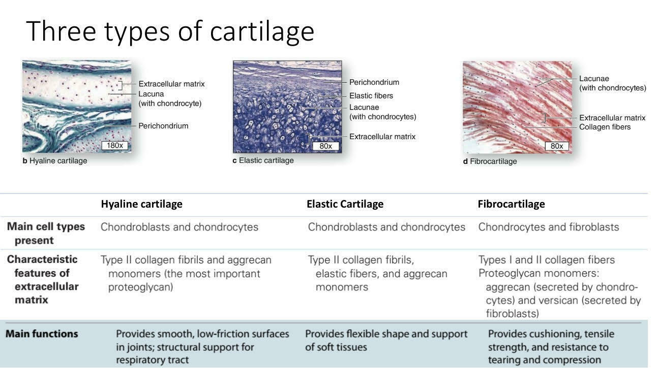
• Slow healing - 11Three types of cartilage
- 12Hyaline cartilage- LocationsLocations: Between tips of ribs and bones of sternum, covering bone surfaces at synovial joints, supporting larynx (voice box), trachea, bronchi, forming part of nasal septum
- 13Hyaline cartilage- FunctionsProvides stiff but somewhat flexible support; reduces friction between bony surfaces
- 14Hyaline cartilage – articular cartilage• Depth dependent properties
Collagen alignment
• Superficial: Parallel to bearing surface
• Deep: Perpendicular to subchondral bone
Chondrocytes
• Aligned with collagen fibres - 15Hyaline Cartilage - Clinical conditionOsteoarthritis
• Chronic disease
• Major cause of pain and
disability in elderly
• Gradual loss of hyaline
cartilage in articular joints
• Chondrocyte signaling and
metabolism altered
• Bone-to-bone contact
• Pain and inflammation
• Deterioration of movement
and function
Chondrosarcoma
• Slow-growing malignant
tumors
• Originates from chondrocytes
• Secretion of cartilage matrix - 16Elastic Cartilage similar to hyaline cartilage except that it contains an abundant network of elastic fibersLOCATIONS: Auricle of external ear; epiglottis;
auditory canal; cuneiform cartilages of larynx
FUNCTIONS: Provides support, but tolerates distortion without damage andhsturns to original shape - 17Fibrous Cartilage Fibroblast a mixture of hyaline cartilage and dense connective tissueLOCATIONS: Pads within knee joint;
between pubic bones of pelvis; intervertebral discs
FUNCTIONS: Resists compression;
prevents bone-to-bone contact; limits relative movement - 18Growth of cartilage – Type 1: AppositionalAppositional growth: The cartilage grows at its external surface through the differentiation of fibroblasts into
chondrocytes within the cellular layer of the perichondrium - 19Growth of cartilage – Type 2: InterstitialInterstitial Growth: The cartilage expands from within as chondrocytes in the matrix divide, grow, and
produce new matrix - 20Growth of cartilage – SummaryAll forms of cartilage form from embryonic
mesenchyme.
• Cartilaginous structures grow by mitosis of
existing chondroblasts in lacunae (interstitial
growth) or formation of new chondroblasts
peripherally from progenitor cells in the
perichondrium (appositional growth).
• Repair or replacement of injured cartilage is
very slow and ineffective, because of the
tissue’s lack of blood vessels and
low metabolic rate
Informationen
- Beschreibung
- Medicine Karteikarten am Basic tissues- slideshow 3, erstellt von i i am 14/12/2022.
- Erstellt von
- i i
- Aufrufe
- 0
- Bewertung