
Karteikarten
Histology Nervous Tissue
Karten in diesem Stapel (39)
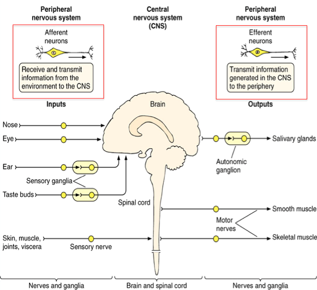
- 1Nervous system: Overview Anatomical Divisions CNS? PNS?CNS:
Brain & Spinal cord
PNS:
Peripheral nerves & Ganglia - 2Functional divisions: (4 of them)-Afferent neurons (Arriving): info. from periphery to the CNS
-Efferent neurons (Exit): info. from CNS out to the periphery
Somatic
(voluntary)
&
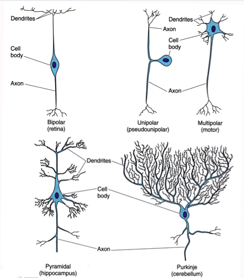
Autonomic (involuntary) - 3Two types of cells in the nervous system Neuronsconduct electrical impulses
- 4Two types of cells in the nervous system Glial cellssupport, nurture, and protect neurons.
NEED SUPPORT CELLS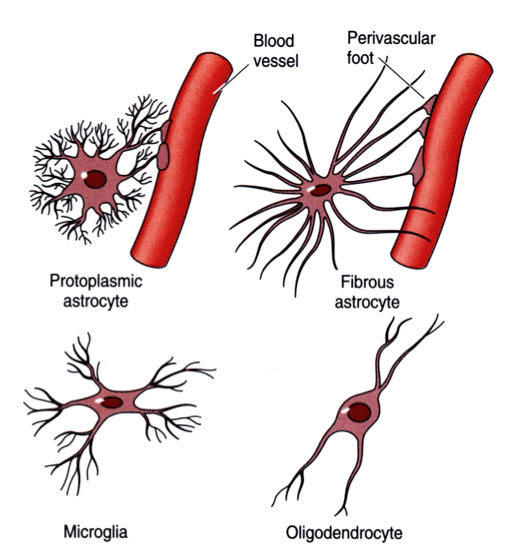
- 5Types of neurons: Bipolar neuronsSingle axon and single dendrite
Located in sense organs
*isolated to the eye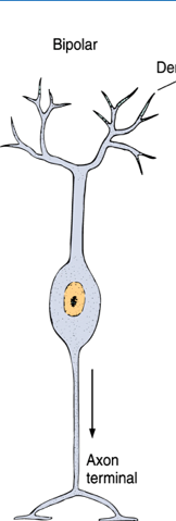
- 6Types of neurons: Multipolar neuronsSingle axon and multiple dendrites
Most common type
"work horse"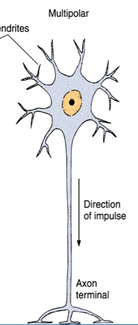
- 7Types of neurons: Pseudounipolar neuronsSingle process extends from cell body and branches into an axon and dendrite
(cell body is b/w two stalks of an axon)
First developed in the embryo as bipolar cells whose axon and dendrite later fuse into a single process
Located in spinal and cranial ganglia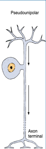
- 8DendritesReceive stimuli from sensory cells, axons, or other neurons
Convert these signals into small electrical impulses which are transmitted to soma (cell body)
Often have arborized terminals which permit a neuron to receive very LARGE number of STIMULI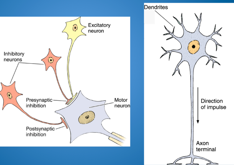
- 9Neuronal Cell Body: (a.k.a. soma or perikaryon) Nucleus Nissl bodies Neurofilaments Microtubules Axon HillockNucleus:
Large, with dispersed chromatin and large nucleolus (centrally placed, "owl eye")
Nissl bodies:
Polyribosomes and rER (LOTS of rER & Golgi-protein being made)
Abundant in larger neurons
Neurofilaments:
Intermediate filaments
Part of cytoskeleton
Microtubules:
Move organelles within cell
Direct axonal flow
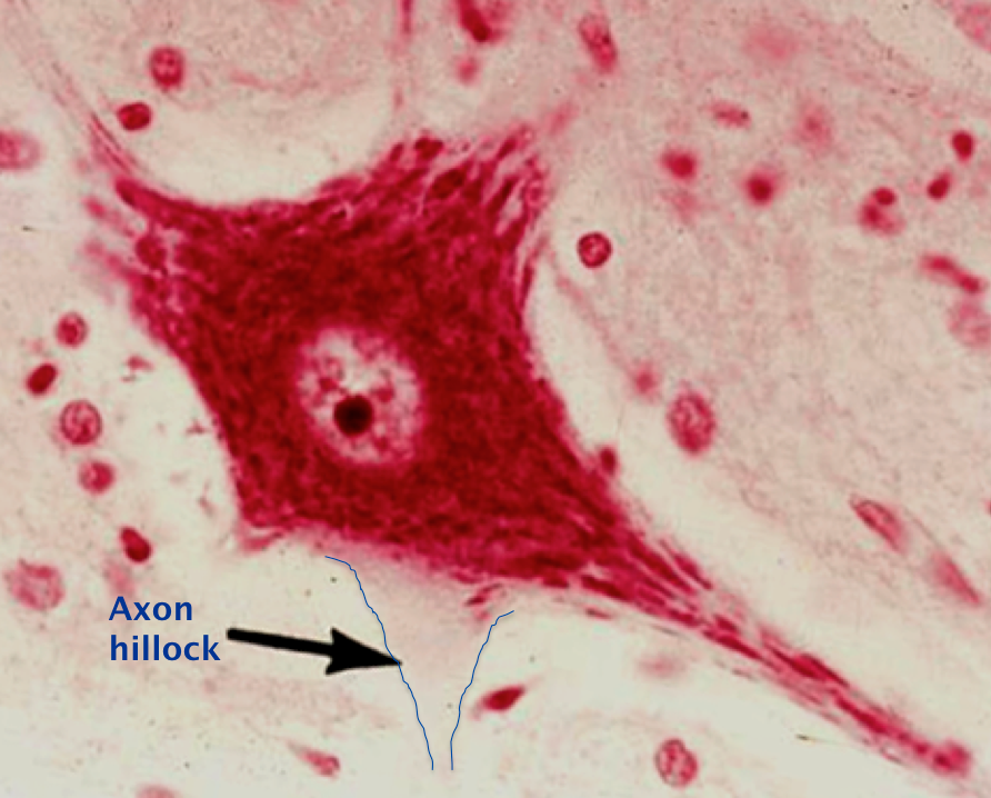
Axon hillock:
LACKS rER and ribosomes (lacks staining: like a shadow)
Contains many microtubules & neurofilaments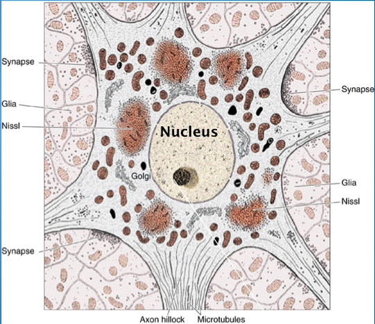
- 10What type of neurons are shown in the image? What are the arrows pointing at?**Note the BLUE Nissl substance in cell body, many have nice nucleolus
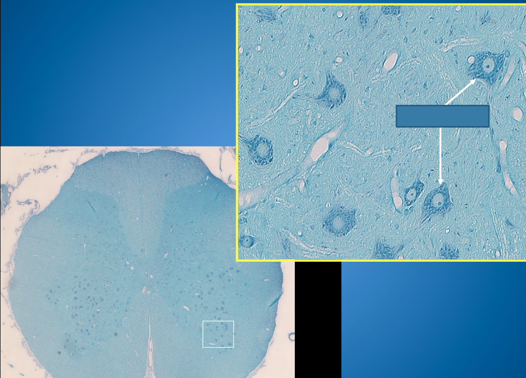
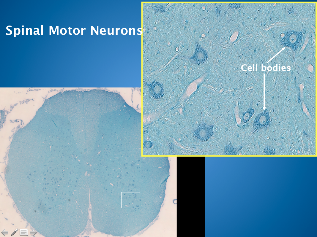
- 11Axons Called what in cytoplasm? in plasma membrane?Conduct impulses AWAY from the soma to the axon terminals (ONLY ONE DIRECTION)
While dendrites taper, axons retain a CONSTANT diameter along their course. Diameter is PROPORTIONAL with SIZE of cell BODY.
Length is highly variable.
Axons may branch!
Cytoplasm = axoplasm (devoid of rER)
Plasma membrane = axolemma
Terminate in axon terminals from which impulses are passed to another neuron or other type of cell (muscle or gland)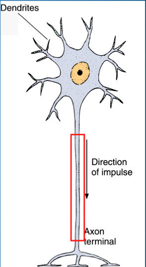
- 12—Lacks stain b/c lacks rER & ribosomes
*like a SHADOW
contains many microtubules & microfilaments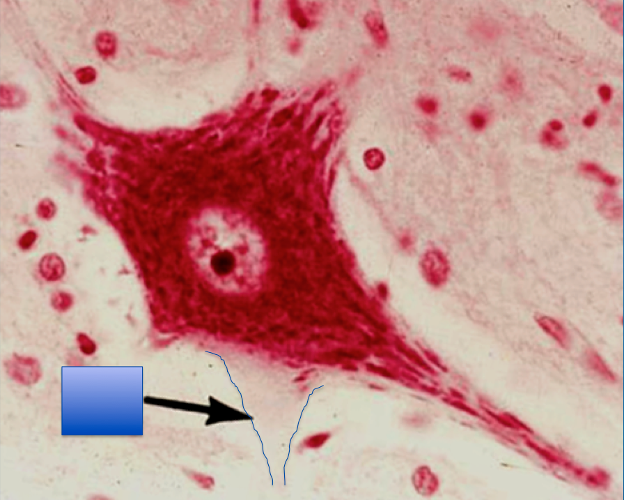
- 13From axon to nerve fiber Axon enveloped by?If an Individual axon is enveloped by a
MYELIN SHEATH,
a nerve fiber
*myelinated = FASTER impulse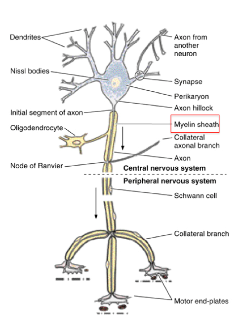
- 14Myelin sheath is produced by: what in the CNS? what in the PNS?Oligodendrocytes in CNS
Schwann cells in PNS
**KEY POINT: ALL neurons have SUPPORT cells that protect it or serve some other function (none are their own entity)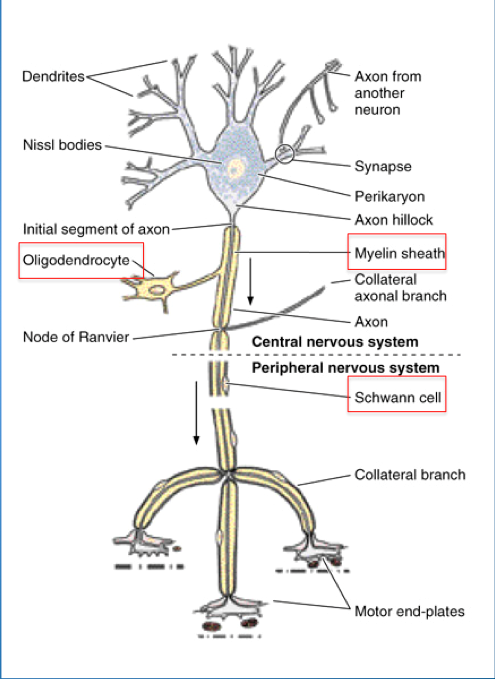
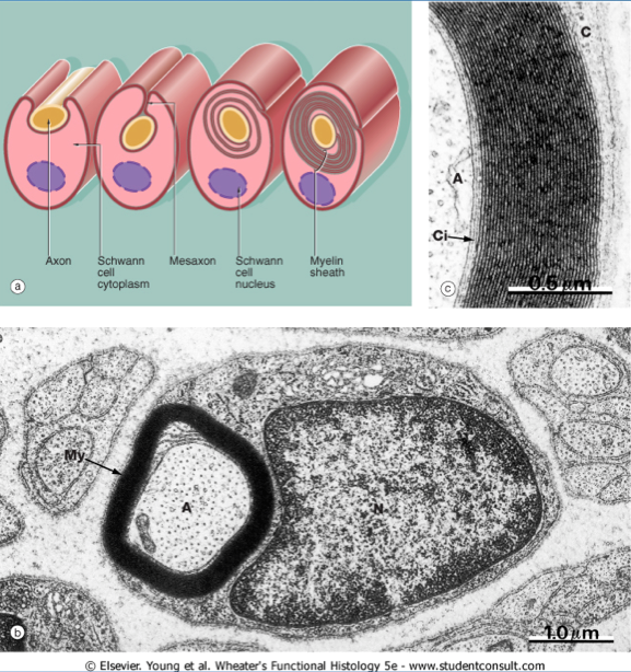
- 15Myelin sheath in the PNSLipoprotein sheath formed by several layers of plasma membrane of a SCHWANN cell wrapping around an axon
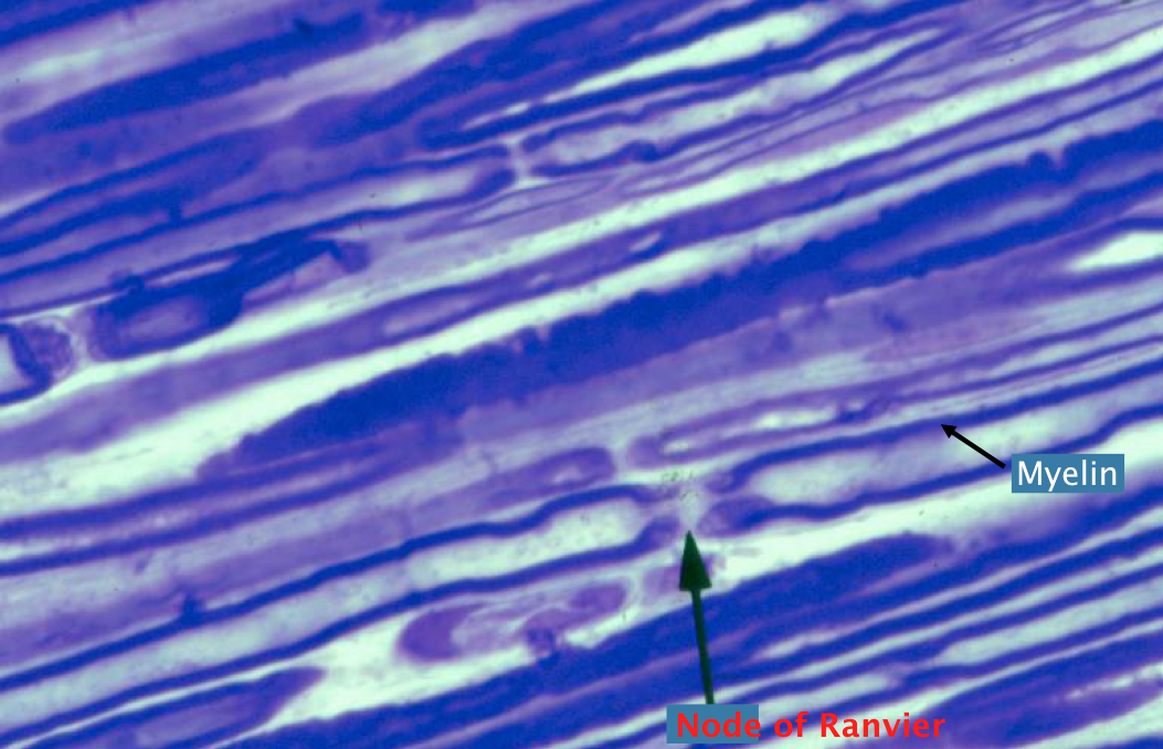
Segments covered by ONE Schwann cell are separated by NODES OF RANVIER
It is of constant thickness along the length of an axon (thicker axons have thicker myelin sheaths.)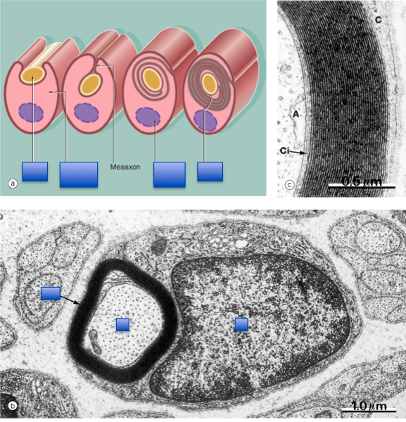
- 16Myelin sheath in the PNSNodes of Ranvier:
represent a discontinuity between adjacent Schwann cells (NOT very long); lack myelin
Schmidt-Lantermann clefts:
cone-shaped, oblique discontinuities in the myelin sheath;
formed by Schwann cell cytoplasm that is not displaced to the periphery during myelin formation
S1 = Schmidt-Lantermann clefts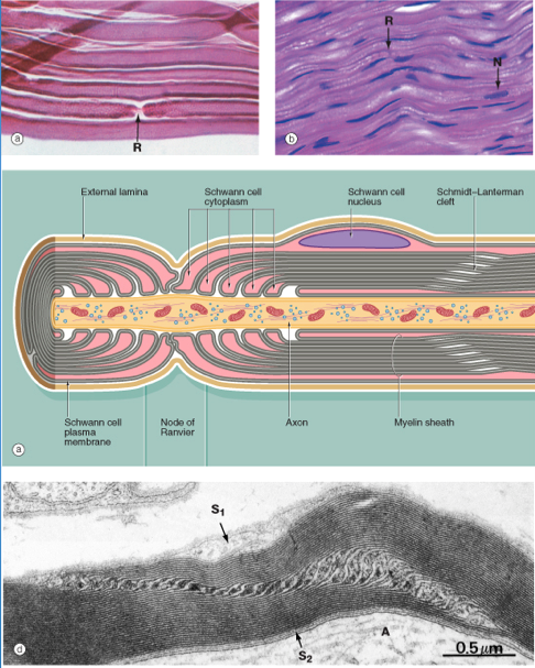
- 17Myelin sheath in the CNS:
- 18Unmyelinated Nerve Fibers in the PNS:In unmyelinated nerve fibers, the Schwann cells engulf ~8-10 axons (protects it)
**EMBEDDED in Schwann-cell cytoplasm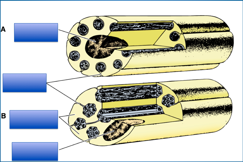
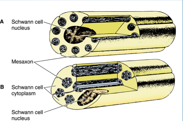
- 19The coverings of axons - SUMMARY CNS versus PNS Myelinated versus UnmyelinatedAxons are never completely “naked”
They may be myelinated or unmyelinated
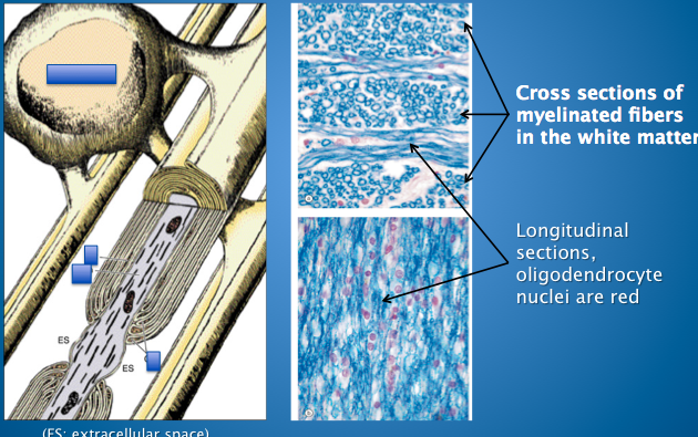
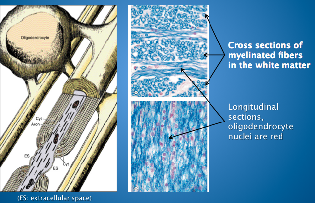
In the CNS, myelin is formed by oligodendrocytes;
In CNS, unmyelinated axons are insulated by astrocyte processes
In the PNS, myelin is formed by Schwann cells,
In PNS, unmyelinated axons (singly or in groups) are
EMBEDDED in Schwann-cell cytoplasm - 20Disorders of Myelin: Multiple SclerosisMOST COMMON disease of myelin in CNS
-damaged oligodendrocytes
Most common demyelinating disease
Unknown etiology; immune/viral factors suspected
Multiple areas of demyelination in brain and spinal cord; progressive
Weakness of lower extremities, visual problems, sensory disturbances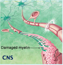
- 21Disorders of Myelin: Guillain-Barre syndrome-Schwann Cell degeneration
-Usually viral infection
-Pain in extremities
Acute inflammatory demyelinating disease involving peripheral nerves
Often preceded by viral infection, immunization, or allergic reactions
Considered to be of autoimmune etiology
Ascending muscle weakness and PARALYSIS
(moving from lower extremity upward)
Most patients recover but respiratory failure may lead to death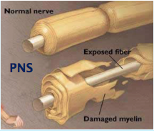
- 22Peripheral nerves
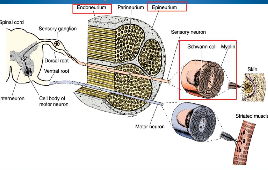
- 23Connective Tissue Investments (in periphery): Epineurium Perineurium EndoneuriumEpineurium:
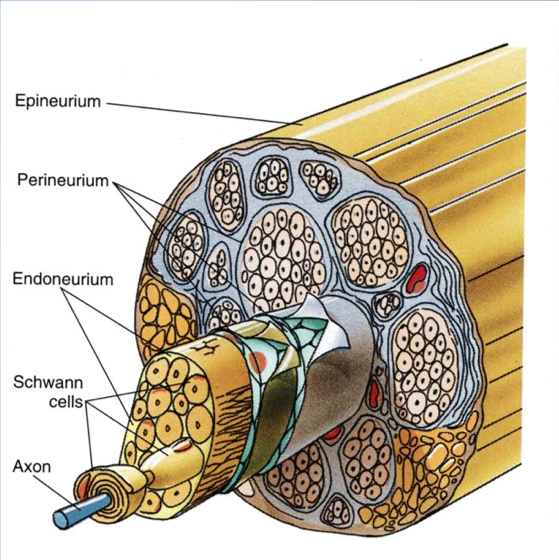
Fibrous dense connective tissue that forms the external coat of nerves
Perineurium:
Layer of modified dense connective tissue that surrounds each bundle (fascicle) of nerve fibers
**Inner surface consists of flattened EPITHELIOID cells joined by tight junctions (barrier function)
Endoneurium:
Thin layer of loose connective tissue surrounding individual nerve fibers (in b/w axons)
-fibroblasts make endoneurium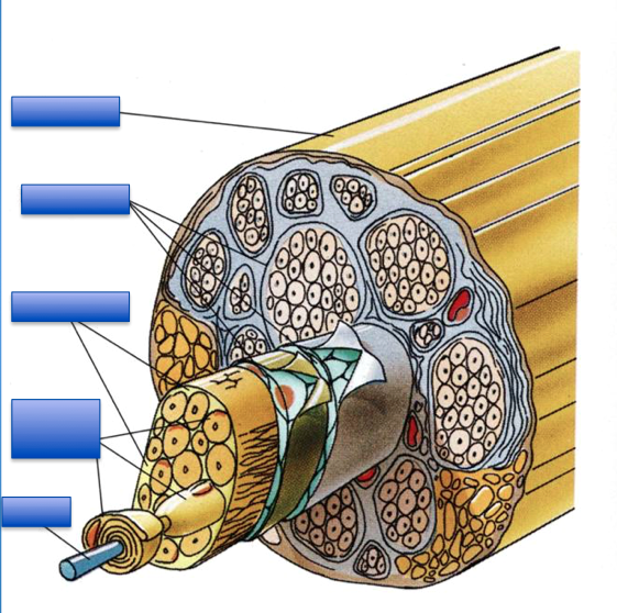
- 24Peripheral Nerve Fascicle/Bundle (toluidine blue stain)Perineurium surrounds nerve bundles/fascicles
Unmyelinated axons tend t be clustered into "donut shape"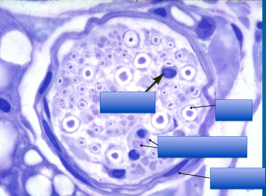
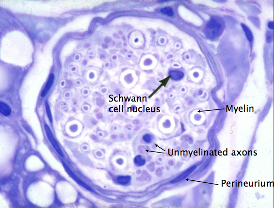
- 25Peripheral nerve (cross section, osmium stain) What is myelin made of?Myelin = Lipids
*can see the myelin because of stain
(osmium + something else because osmium alone appears black)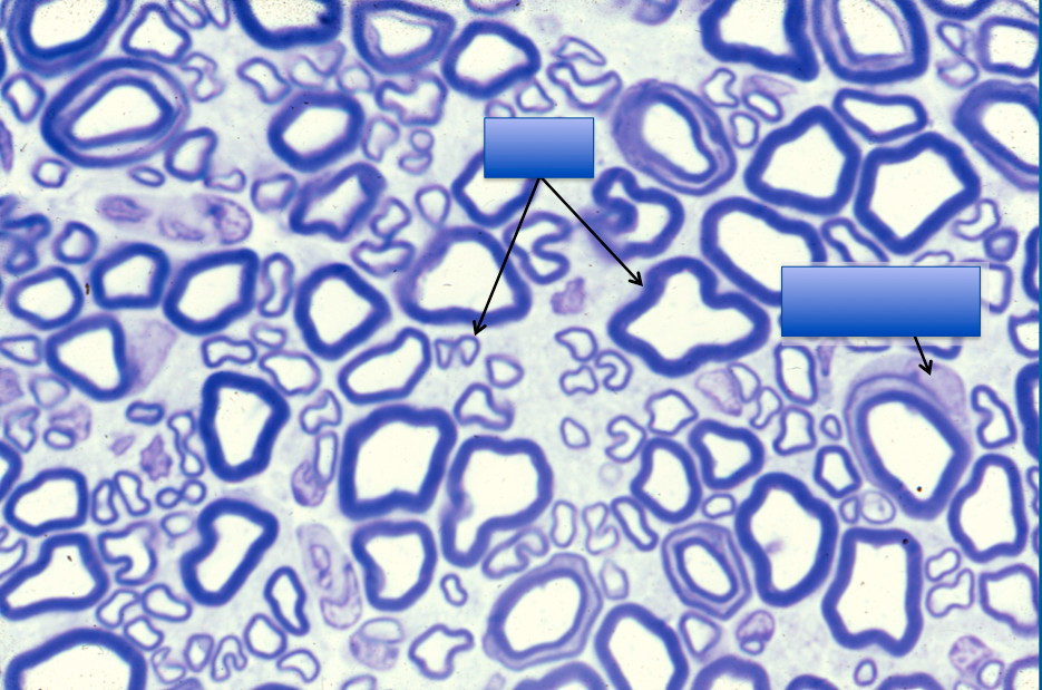
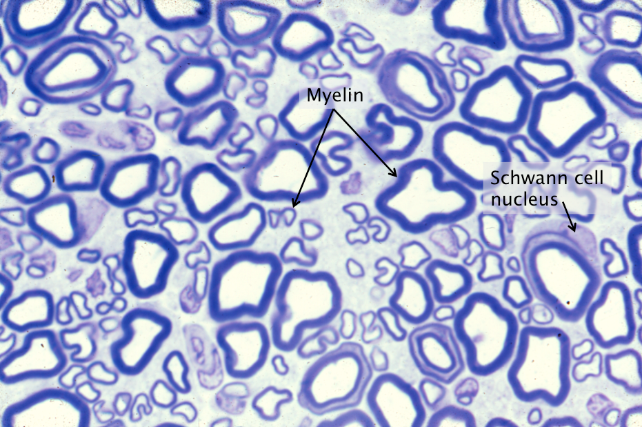
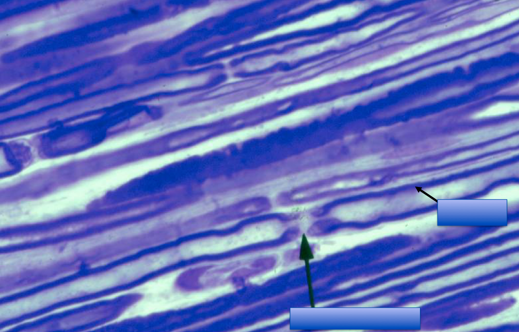
- 26Peripheral nerve fibers stained with osmium
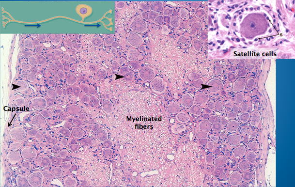
- 27Dorsal root or sensory ganglion: pseudounipolar neuronsBlack arrows (w/o stems) are pointing at
Dorsal root or sensory ganglion
(ROUND)
-have a lot of myelin (fairly unique to DRG)
*Support cells = Satellite cells
*Pseudounipolar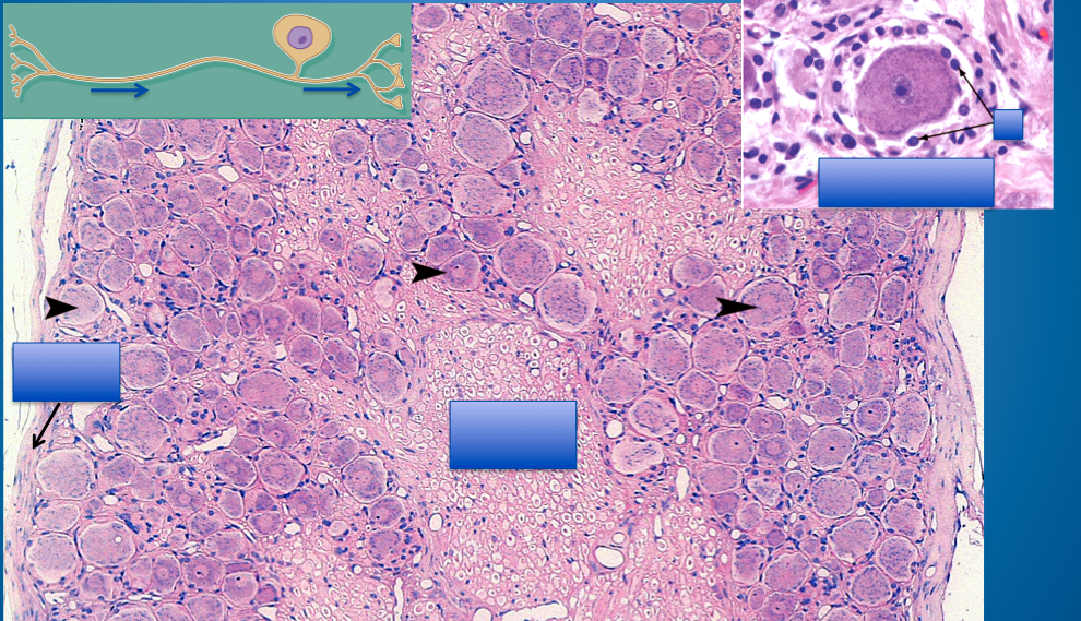
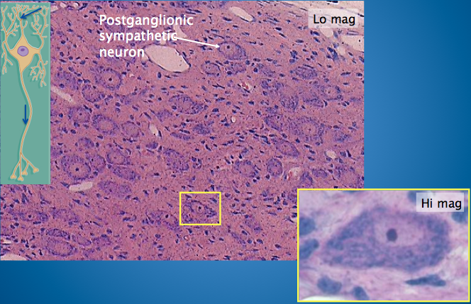
- 28Sympathetic GanglionPreganglionic fibers from spinal cord enter ganglion and terminate on dendrites (upper blue arrow) and cell bodies of postganglionic neurons
*NOT as ROUND
-More multipolar
-less myelin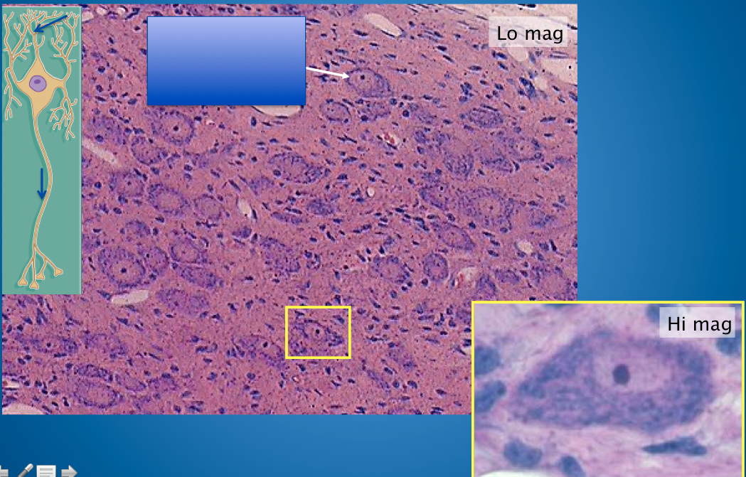
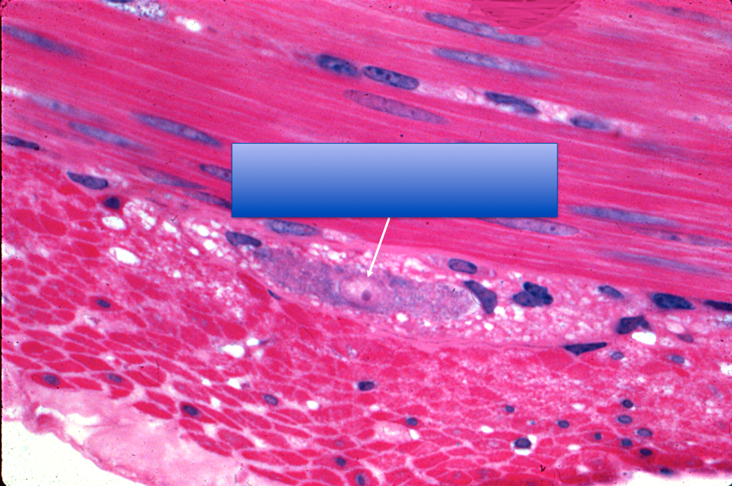
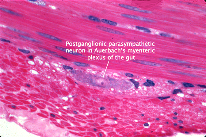
- 29Parasympathetic GanglionNote: smooth muscle (peristalsis in intestine) involuntary!
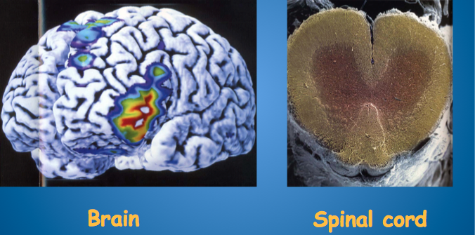
- 30CNS 2 components??
- 31Spinal Cord
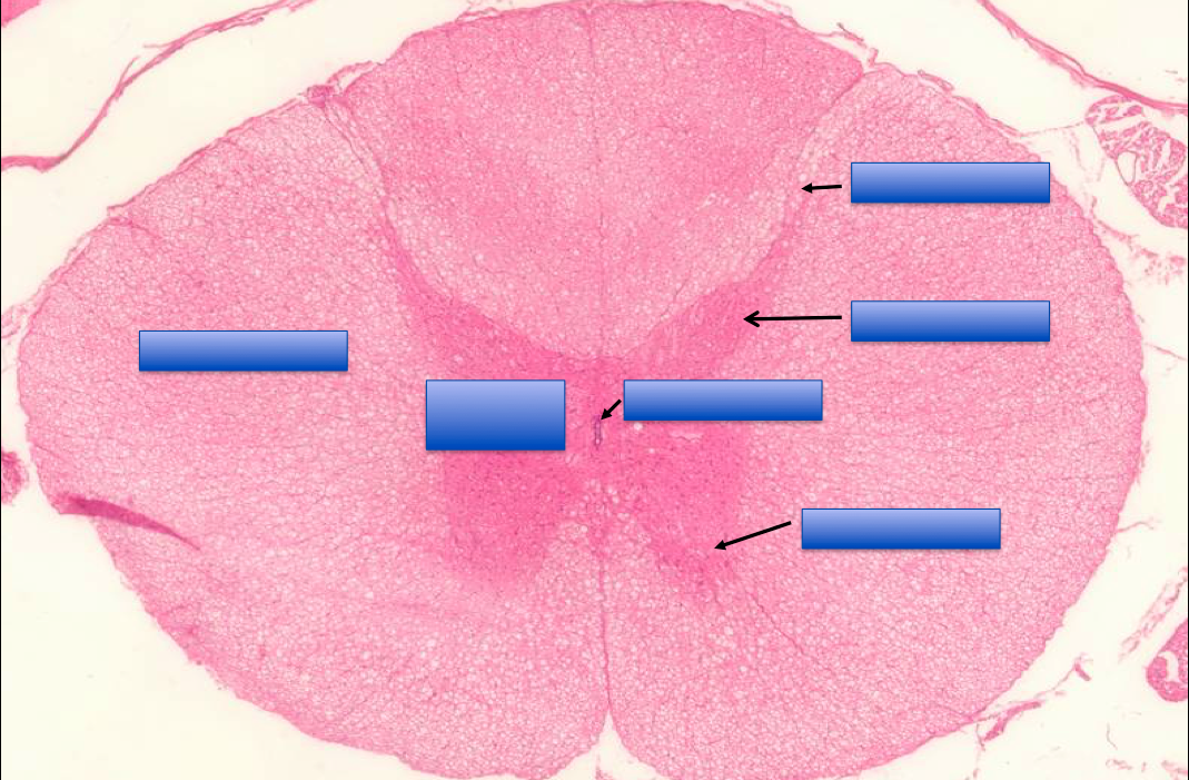
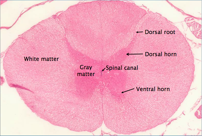
- 32White matter of Spinal Cord: Mostly myelinated or unmyelinated?White Matter:
Contains MOSTLY MYELINATED but also some unmyelinated nerve fibers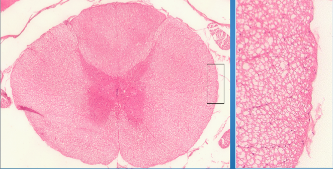
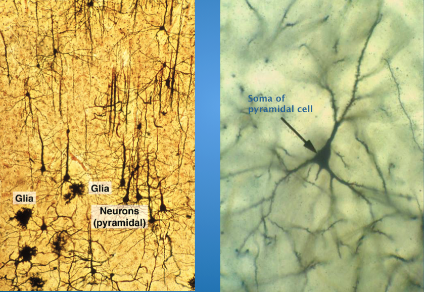
- 33PYRAMIDAL cells from CEREBRAL cortex Golgi silver impregnation method"no spikes"
Function:
Spatial recognition
Long axons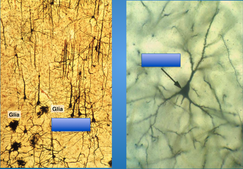
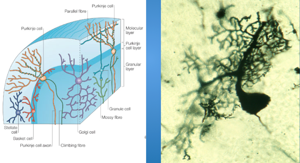
- 34PURKINJE cells in CEREBELLAR cortex Golgi silver impregnation methodVERY EXTENSIVE dendrites/dendritic tree
Functions:
Proper movement & Pain receptors in cerebellar cortex
*Also found in the HEART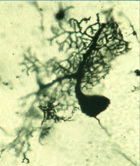
- 35Specialized Receptors 2 Types:1. Meissener corpuscles
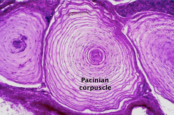
2. Pacinian corpuscles - 36—Encapsulated receptor in dermis, joints, and mesenteries
Multilayered capsule of fibroblasts, collagen, and fluid, surrounding a nerve terminal
Pressure, touch, vibration
DEEP CORE touch
(Palm of your hands)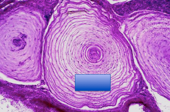
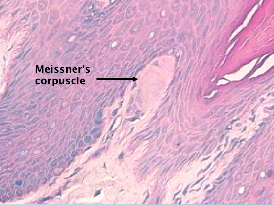
- 37—Encapsulated receptors in dermis (skin)
CT capsule enveloping nerve terminal and its Schwann cell
Fine/LIGHT touch
-how you get your finger prints
-axon leading up to the nerve ending (bulge)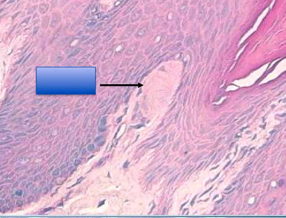
- 38Degeneration and Regeneration of Nerve TissueA. Normal nerve fiber
B. Injured nerve fiber
C. Muscle fiber atrophies; Scwann cells proliferate; axon penetrates into “track” laid down by Schwann cells;
grows an average of 2.5
mm (1/10 of an inch)/day.
D. Regeneration is complete. (regernation doesn't happen often)
KEY POINT:
Death to the CELL BODY is IRREVERSIBLE,
but DAMAGED AXONS can REGENERATE.
*Also, doesn't regenerate, just reattaches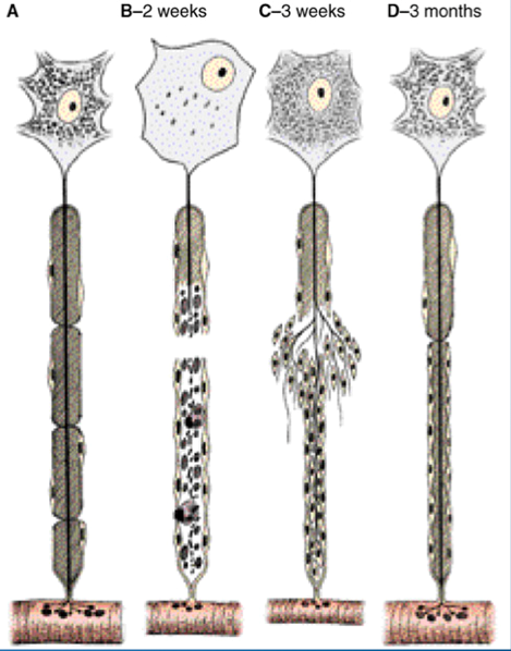
- 39Exam Question: The Axon Hillock of a Spinal Motor Neuron: A. Contains an abundance of rER B. Stains intensely histologicaly C. Is where the nucleus resides D. Contains microtubules and neurofilamentsD. Contains microtubules and neurofilaments