
Mindmap
NEPHROLOGY 2018
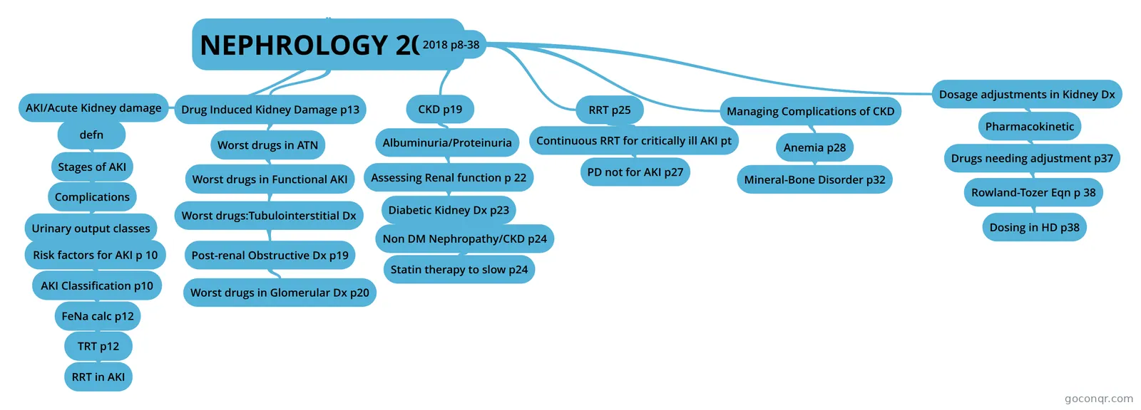
Mind map: nephrology 2018
Inhalt der Mindmap
NEPHROLOGY 2018
2018 p8-38
- AKI/Acute Kidney damage
- defn
- acute decrease in GFR over days/weeks see increased SCr of 0.3 mg/dl or more within 48 hrs or and increase of 1.5 x in SCr from baseline taken within 7 days or urine volume less than 1/2 your wt per hour ie 0.5ml/kg/hr for at least 6 hrsa)KDIGO improving global outcomes grp) and AKIN(acute drug inj netwrk) use RIFLE criteria to define
- Stages of AKI
- AKIN (acute kidney injury network) will define/diagnose AKI when: a) absolute increase in SCr >0.3mg/dl same as KDIGO b) an increase in baseline SCr by 50% or more (same as 1.5 x increase? like KDIGO) c)urinary output <0.5ml/kg/h for 6 hours or more(same as KDIGO)
- AKIN and KDIGO have same criteria for staging ito SCr but KDIGO also looks at urine output PTO p3 (verify this info?) see stages table 1 p9by heartito of urine outputstage 1 crcl reduced by half over 6-12 hrsstage 2 if this happens over 12 hrs or longerstage 3 if even less in crcl happens 1/3 of wt/hr over 24 hrs or moreor if no urine being produced for 1/2 a day
- ito SCr stage 1: SCr 0.3 increase or more or 1.5- under 2 x baseline SCr stage 2: 2 to under 3 times baseline SCr stage 3 = 3x baseline SCr or SCr rise of 0.4 or had to initiate RRT (renal replacement therapy) or child <18 and crcl <35
- RIFLE developed by Acute Dialysis Quality Initiative to also know when someone is ESRD looks at SCr and urine output this is different from above and defines stages ito kidney function RIFLE stands for a) R risk of renal dysfunction can exist if SCr increases to 1.5 x baseline or GFR decrease by 25% (like KDIGO and AKIN stage 1) b)I Injury to Kidney : SCr increases to 2x baseline or GFR decrease by >50% (like stage 2) c)F Failure of kidney function SCr increase to 3x baseline or GFR decrease by >75% or SCr>4mg/dl with acute increase in SCr of 0.5mg/dl (like stage 3) d) L loss of kidney function have complete loss of kidney function >4 weeks e) END STAGE KIDNEY Dx: Complete loss beyond a month now and is >3 months
- So....... ESRD is complete loss of kidney function for >3 months ito of GFR R Risk of RF is decrease by 25% GFR I Injury is decrease by 50% GFR F Failure is decrease by 75% GFR or SCr>4mg/dl ito of urine output it overlaps with satges 1-3 of AKIN
- Complications
- Urinary output classes
- p 10 Urinary output classificationi. Anuric: Less than 50 mL/24 hours; associated with worse outcomesii. Oliguric: 50–500 mL/24 hoursiii. Nonoliguric: More than 500 mL/24 hours; associated with better patient outcomes and easierto manage because of fewer problems with volume overload
- also look at community acquired vs hospital acquired vs ICU pt
- Risk factors for AKI p 10
- p 12 prevention of AKI similar to just preventing risks
- AKI Classification p10
- AKI can be classified as 1. pre-renal Characterized by hypoperfusion to the kidney i. Systemic hypoperfusion: Hemorrhage, volume depletion, drugs, CHF ii. Isolated kidney hypoperfusion: Renal artery stenosis, emboli also on ACEI, NSAIDS, Cyclosporine2. Functional (same info as for pre-renal) 3.Intrinsic (Long-standing renal hypoperfusion Nephrotoxins (e.g., contrast or antibiotics) Vasculitis Glomerulonephritis ** see muddy brown casts and sediments and casts4. Post renal (CA, BPH, kidney stones)Good to know table 3
- FeNa calc p12
- Sodium ratio / creatinine ratio x 100 this fractiona; excretion of sodium defines what RF id due to prerenal or intrinsic or post renal FeNa = (urine Na/plasma Na) divided by (urine Creatinine/palsma creatinine) FeNa<1% = prerenal ARF (azotemia) FeNa> 1%= ATN acute tubular necrosis caution Lasix and loop diuretics cause natriuresis (urinary Na increases) FeNa can be high with diuretic use FeNa can be low due to intrinsic renal problems ( acute glomerular nephritis), ATN, rhabdomyolysis, contrast, sepsis) in CKD FeNa can be high constantly this is % of sodium filtered by kidney which is excreted in urine It is measured ito plasma and urine sodium concentration because urinary sodium concentration alone
- TRT p12
- Prerenal azotemia: Correct primary hemodynamics. a. Normal saline if volume depletedb. Pressure management if neededc. Blood products if neededd. Hold or discontinue medications that affect renal hemodynamics (i.e., ACEIs, ARBs, NSAIDs
- Intrinsic: No specific therapy universally effective a. Eliminate the causative hemodynamic abnormality or toxin. b. Avoid additional insults. c. Manage fluid and electrolytes to prevent volume depletion or overload and electrolyte imbalances. d. Nutrition support is important
- should not use ffl in intrinsic AKI Loop diuretics except to manage hypovolemia for intrinsic AKI Fenoldopam Dopamine Atrial natriuretic peptide Recombinant human insulin like growth factor 1 all not to be used
- 3. Postrenal AKI: Relieve obstruction.
- RRT in AKI
- a. Blood urea nitrogen (BUN) greater than 100 mg/dL b. Volume overload unresponsive to diuretics c. Uremia or encephalopathy d. Life-threatening electrolyte imbalance: Hyperkalemia, hypermagnesemia e. Refractory metabolic acidosis
- Urinary output classes
- defn
- CKD p19
- CKD defn: Kidney Dx outcomes quality initiative KDOQI defines CKD as i)kidney damage for more than 3 months ii)may see GFR change (not always) iii)structural or functional abnormality of kidneys iv) see markers like abnormal composition of blood/urine or abnormal imaging tests v) or see GFR changes <60ml/min for 3 months with or without kidney damage
- Stages of CKD table 4 p 20 ito of kidney damage and GFR stage 5 (worst ) RF is level of damage and GFR<15 stage 4 severe decr GFR < 30 (15-29 next increment) stage 3 moderate decr GFR<60 (30-59 next increment) stage 2 Kidney damage with mild decr in GFR <90 (60-89 next increment) stage 1 Kidney damage but with normal GFR >90 Increased risk group with GFR>90 (normal) eg DM, HPT, family hx
- P20 table 5 KDIGO also categorizes pts but as G G5 is same as stage 5 <15 G4 same as stage 4 <30 G3*** is where its different KDIGO speaks of mild 3a and moderate 3b ,based on GFR GFR 3a 30-44 is moderate to severe GFR 3b 45-59 is mild to moderate GFR decrease G2 is like stage 2 GFR 60-89 G1 is like stage 1 GFR>90 no talk of risk group GFR>90
- C. Risk factors discussed p20
- Albuminuria/Proteinuria
- p 22 Albuminuria/Proteinuria basically protein and albumin will be a marker for kidney damage, heart dx and indicates the progression of the dx ACR = albumin/creatinine ratio. ACR is used to assess proteinuria. Can do untmed test but in more severe cases do a 24 hr urine collection
- table 6 p 22 KDIGO has categorized CKD based on ACR and daily excretion in 24 hrs Albuminuria categories A1 to A3 pretty much same for ACR(mg/g albumin/Cr ratio) and daily excretion(mg/24h) Stage A1 :both ACR and daily excretion are <30 (normal to mild increases) Stage A2 :30-300 moderately increase in protein in urine Stage A3: >300 severely increased albumin in urine Nephrotic range when 24 hour urine sample shows >3000 mg/24h
- Assessing Renal function p 22
- don't use just one method i)use Scr (affected by age, sex, muscle mass) ii)use CrCl eqn Cockgroft-gault Eqn affected by muscle mass and can overestimate bc of creatinine secretion in tubulescan use cockgroft gault for low muscle mass, amputations, HDiii)can use GFR- uses inulin , iothalmate...not used muchiv)eGFR only accurate if GFR<60 eGFR (mL/minute/1.73 m2) = 175 × SCr−1.154 × age−0.203 (× 0.742 if female) (× 1.212 if African American) v)CKD-EPI eqn is good for using, race etc , good for GFR>60 vi) for kids use Scwartz bedside formula
- Diabetic Kidney Dx p23
- how to diagnose: Diagnosis a. Long history of diabetes b. Proteinuria c. Retinopathy (suggests microvascular disease how to monitor Type 1 vs II for Type 1 DM begin to monitor 5 years after diagnosis for Type II begin right away as may have had it for a while
- Pathogenesis of Diabetic KD: a) pt has hypertension thus monitor and manage it aggressively see next section pto b) protein in urine are glycosylated (carb becomes attached to it) in glomerulus So want to keep glycosylated hemoglobin (A1C) <7% Also some evidence that low protein diet will slow progression of stages 4-5 and decrease risk of ESRD eg 0.8g/kd/day like 60g protein(>1.3g/kg/day bad eg 100g)c) genetic predisposition
- Aggressively manage HPT: a) ADA and JNC say 140/90 ok for BP for any level of albuminuria KDIGO subdivides albuminuria into moderate to severe : BP goal 130/80 mild to moderate albuminuria get same BP goal as ADA , JNC ie 140/90
- drugs for HPT i) even if not hypertensive- use ACEI or ARB for any level of albuminuria and need to be aggressive - use moderate to hih doses of ACEI/ARB but exception is with hyoerkalemia...hold ACEI/ARB if K>5.6 or if SCr increases > 30% ii)Diuretics added for most pts but choice depends on stage stage 1-3 choose thiazide stage 4-5 choose loop diuretic BP>160/90 needs 2 drugs to begin with (start both ACEI + diuretic) iii)D&V (non DHP) are 2nd line to ACEI/ARB and combining them is an option Non Drug methods to control BP: decrease salt 2.6g/day Na and modify DASH diet to restrict K also
- Non DM Nephropathy/CKD p24
- Even if no DM ir pt has albuminuria need to treat HPT trt: ACEI or ARB still doc in albuminuria and HPT. Need 2 drugs often so add on diuretic (just as for CKD in DM pt)
- HPT goals same as before JNC says >18 yo with CKD all need to have BP goal <140/90 KDIGO again subdivides into mild and moderate mild /normal albuminura: goal140/90 moderate to severe albuminuria goal 130/80
- Statin therapy to slow p24
- basically everyone with CKD can get a statin. even transplant pt gets statin (only exception is HD pt not on statin- dont start statin)and G3-G5 can get statin or statin + ezetimibeonly exception is HD pt:dont start statinsbut is they were on statin or Zetia before then ok to continue it
- another intervention is to stop smoking
- RRT p25
- HD and PD AEIOU indications Acidosis, Electrolytes (incr K, Phos) , Intoxication (barbiturates, Li, salicylates, methanol, theophylline ,chloral hydrate), Overload of fluid(pulm symptoms) Uremia (pericarditis and wt loss)
- Can measure if HD is adequate i) Kt/V 1.2-1.4 okay (KDOQI goal 1.4) K clearance x time of dialysis over Vd of urea ii) URR is a ratio looking at diff bet BUN before and after over pre BUN as a % URR= BUN pre- post/preBUN x100 >65% is goal target= 70%
- Complications of HD: a)Hypotension to treat Trendelburg , give fluids thus to prevent: give midodrine before HD: 2.5-10mg before and limit fluids bet sessions b)Vascular access infection Staph treat aggressively
- Continuous RRT for critically ill AKI pt
- p27 Hemofiltration type uses convection rather than diffusion and needs high fluid replacement bc operates at high rateSC = sieving coefficient and it allows you to det' how much drug is removedSC is a ratio of concentrations and is conc of drug in ultrafiltrate divided by con of drug in blood continuous hemodialysis uses diffusion instead of convection like above to remove solutes
- Continuous hemodiafiltration type is combo of previous 2 uses dialysis and filtration and needs both replacement fluids and dialysate to be replaced
- PD not for AKI p27
- PD is not usually for AKI Catheter fills fluid into peritoneum where it sits for a while then is drained out. Solutes and fluid cross the peritoneal membrane
- complications of PD a) infection in peritoneum The most common gram-positive organisms include Staphylococcus epidermis, S. aureus, and streptococci. The most common gram-negative organisms include Escherichia coli and Pseudomonas aeruginosa. b) WBC>100 or neutrophils>50% in peritoneal dialysate TRT: a) Empiric treatment should cover gram-positive and gram-negative bacteria. (b) Intraperitoneal administration of i)vancomycin or ii)first-generation cephalosporin and intraperitoneal third-generation cephalosporin or aminoglycoside (c) Adjust as needed depending on culture and sensitivity.
- Other complications: Hyperglycemia Ca levels can be affected high or low Ca low Albumin levels
- Managing Complications of CKD
- Anemia p28
- initiate evaluation when crcl<60 and Hgb <13 in men and Hgb<12 in females How often depends on stages stage 5 worse if on HD: so check q 3 mo stage 4-5 and non HD 2x pa anything else annual check ok monitoring labs: Mean corpuscular volume iii. Reticulocyte count iv. Iron studies (a) TSAT (serum iron/total iron-binding capacity × 100): Assesses available iron (b) Ferritin: Measures stored iron v. Serum vitamin B12 and folate concentrations
- so if Hgb < 13 or Hgb < 12: first check folate, iron ferritin and TIBC, Vit B12(all for RBC) if iron low is low give po/iv iron to correct anemia. If corrected just give iron MD dose If anemia is not corrected: p29 Next tackle Hgb a)Hgb< 10 start erythropoetin stimulation agent + iron Monitor Hgb every 2-4 weeks aim for Hgb<11 (if you aim for 13 see greater risk of Cv events) So hold dose in nonHD CKD pt if Hgb >10and hold dose in HD pt if Hgb>11 or max 11.5not to dose if during monitoring:in 2 weeks Hgb>1 increase then reduce dose by 25%if in 4 weeks Hgb drops by 1 then increase dose by 25%Recommend to start ESA trt if Hgb <10 in HD and nonHD pt
- ESA Maintenance therapy 1)Epoetin (Epogen, Procrit) 50-100 iu weekly subq dose 30% less than iv dose 2)Darbe-poetin Aranesp duration thus dosing once weekly, or once every 2-3 weeks b) dose is 0.45mcg/kg for non HD CKD pt: once every 4 weeks for HD pt: it s more frequent ie once per week or 0.75 mcg/kg every 2 weeks c)Mircera (Methoxy-PEG Erythropeitin Beta) Onset of Hgb increase occurs 7–15 days after initial dose. (c) Less-frequent dosing; initial dose is 0.6 mcg/kg of body weight once every 2 weeks (d) Once the Hgb stabilizes, can double the dose and administer once monthly Monitoring: i)Monitoring Hgb with ESA with CKD pt: a)during initiation - monitor every 2-4 weeks in beginning b) MD: monitor monthly for HD pt c) MD for non HD pt monitor q3 mo ii)Monitor BP with ESA trt bc it can go up iii)Monitor Iron stores oral iron is not recommended in CKD, but can be used pre-dialysis as a trialKDIGO and KDOQI have guidelines re storagecheck TSAT (serum iron/total iron-binding capacity × 100):KDOQI says it should be >20% and KDIGO says should be >30%Also monitor Ferritin:KDOQI says Ferritin should be> 100 ng/mL (non-dialysis CKD; peritoneal dialysis)and >200 for HDand KDIGO says Ferritin should be >500
- Parenteral Iron: a)Iron Dextran (Dexferrum): test dose 25mg one time due to possible anaphylaxis Dose same like Venofer also can do IVPB 500-1000mg in 250ml NS over 1 h or longer for non HD even MD same as venofer i)sodium ferric gluconate Ferrlecit: for HD need 8x 125mg (=1g TD) given 3x weekly during HD then MD is 31.25125mg /week for 10 weeks(no test dose) ii) Iron sucrose(venofer) 100mg iv 3 times /week during HD x 10 doses (1 g TD) for non-HD CKD pt : give 200mg x 5 doses MD 25-100mg/week x 10 weeks (no test dose needed)
- ron products for replacement continued... d)Ferric carboxymaltose (injectafer) 15mg/kg iv upto 750mg may repeat after at least 7 days (max 1500mg elemental iron=2x 750mg) no MD, no test dose e)Ferumoxytol (Ferraheme) 510mg then 2nd 510mg iv dose 3-8 days later for CKD no MD, no test dose
- Mineral-Bone Disorder p32
- Consequences of CKD-MBD: renal osteodystrophy + vascular calcification What factors contribute? a) incr phos b)decreased 1.25-dihydroxyvit D3 production c)decr Ca absorption in gut d)decr ionized free Ca once e) direct stimulation of PTH secretion f)high PTH causes decr reabsorption of phosphorus and increased reabsorption of Ca in proximal tubule and this adpation is lost when GFR<30 and since gut Ca absorption is low at this point, bone sesorption of Ca is the way the body maintains Ca concentrations (thru elevated PTH)...and eventually can lead to bone osteodystrophy.
- Laboratory abnormalities (Table 13) i. Phosphorus ii. Corrected calcium (measured calcium + 0.8[4 − serum albumin]) iii. Intact PTH iv. Alkaline phosphatase v. 25-Hydroxyvitamin D How to monitor Ca, Phos etc Table 13 p 32
- Therapy goals p 32 differ in different stages stage 3-5 : aim to avoif hypercalcemia and want to bring Phos levels back to normal In stage 5 want to keep PTH 2-9 times the normal Non Diet goal: limit Phos intake 800-1000mg per day
- Phos BInders taken with meals to bind Phos in gut a)Calcium carbonate (40% elemental Ca) typical dose 1250mg b)Calcium actetate (PhosLo, Phoslyra) 25% elemental Ca 667mg capsule/tabs or 667mg/5ml soln Typical dose 2001 mg (3 caps/tabs) c)Sevelamer Hcl (Renagel) 400 or 800mg tab typical dose 800mg, 2400mg Sevelamer: a)non absorbable phos binder, binds phos in gut from food (another caveat: decreases LDL and increases HDL) b) as with Ca , primary therapy in stage 5 CKD, but look at Ca levels c) but Metabolic Acidosis may worsen with Sevelamer^^^ d) Sevelamer carbonate (Renvela) 800mg, 0.8 or 2.4 g powder typical dose 800-2400mg e) Lanthanum carbonate (Fosrenol) 500, 750, 1000mg chewable tablet typical dose 250-500mg f) Sucroferric oxyhydroxide (Velphorol) 500mg chewable tab typical dose 500mg g)Aluminium Hydroxide 320mg/5ml susp Typical dose 300-600mgAluminium phos binders can be an issue thus only short term can cause bone dx, encephalopathy, erythropeitin resistanc h)Ferric citrate (Auryxia) 1g tab dose 2g = 420mg ferric ion
- Besides Phos binders, can use Vit D analogues: d. Vit D analogs: a was trt goals c was Phos binders now d is Vit D analogues Suppress PTH synthesis and reduce PTH concentrations... but can result in high Ca thus this limits it's use i) in CKD G3a-G5: they don't routinely use calcitriol or Vit D analogues per KDIGO so keep itfor more severe G4-G5 with progressing hyperparathyroidism ii)ergocalciferol Vit D2 is inactive Vit D for low 25-hydroxyvit D levels. Dose weekly or monthly. Check every 6 mo iii)Cholecalciferol in active Vit D. need daily dose. iv)Calcifediol (25 hydroxyvit D3) becomes calcitriol after hydrolysis (alternative t0 ergocalciferol in stage 3-4
- Vit D replacement is weekly doses or monthly but depends on levels is very low <5 then weekly for 12 weeks( 3 mo) then monthly or x1 imi dose if 5-15 then weekly for 1 month then go to monthly 16-30 monthly po
- Calcitriol (Caljex/Roacltrol) oral and parental formulation for hypocalcemia and prevention/trt of secondary hyperparathyroidism no hepatic/renal activation needed can incr Ca to hypercalcemic levels thus adjust dose every 4 weeks
- emplar Pari-calcitol for treatment/prevention of hyperparathyroidism Parental and po formulations again no activation like Calcitriol Advantage over Calcitriol - less hypercalcemia ( which means less Ca mobilization from bone, less Ca absorption from gut)
- Hectorol{ Doxercalciferol}: Vit D analogue approved to treat /prevent secondary hyperparathyroidism. also prodrug Advantage over calcitriol: Lower incidence of hypercalcemia than calcitriol than with calcitriol bc decreased Ca loss from bone and decr absorptn in gut...same as with Zemplar
- Besides Phos Binders w/food and Vit D analogues, Can use Calcimimetics Calcimimetics: also for secondary hyperparathyroidism advantage is can be used with high Ca and phosphate levels when you can't use Vit D analogues or can't increase their doses i)Sensipar: Cinacalcet atthaches to Ca receptor on parathyroid and increases sensitivity to Ca conc and thus decreases PTH 1. Initially, 30mg qd irrespective of of PTH concentration/Phos binder used/Vit D analogue used 2. 5% Risk of hypocalcemia (vs hypercalcemia with Vit D analogues) so don't start with Ca < 8.4 and check Ca every 1-2 weeks. Hypocalcemia can exacerbate seizures so caution in seizure pt 3.Dose reductions may be needed with flecainide, tricyclic antidepressants, thioridazine bc Cincalcet inhibits 2D6 4. Dose reduction of Cinacalcet concentrations upto 2 fold may be needed with ketoconazole bc ketoconazole inhibits CYP3A Parsa-biv Etel-calcetide also works at receptor for Ca sensing on parathyroid gland thus reducing PTH 1. initially 5mg ivi tiw AFTER HD 2.How is dose increased? 2.5 -5 mg increments every 4 weeks 3. monitoring: serum Ca baseline, 1 week after start and then every 4 weeks 4. If switching from cina-calcet need to dc cinacalcet for 7 days then start etel-calcetide
- Osteoprosis Trt: 1. for osteoporosis in CKD pt G1-G3 and high risk of fractur ..treat as with general nonCKD pt population 2. For mpore severe CKD G3-5 with Ca and Phos abnormalities and fracture risk see how reversible the Ca and Phos abnormalities are and consider progression of CKD. Consider a bone biopsy.
- Anemia p28
- Dosage adjustments in Kidney Dx
- Pharmacokinetic
- Changes that occur on absorption, distribution, elimination in CKD: 1. Absorption a orally decreased in CKD b. ph more alkaline in stomach (uremia) c. edema d. Phos binders bind drugs too
- 2. Distribution issues in CKD a. water soluble drugs change in concentration b. As elimination worsens and toxins accumulate- acidic and neutral drugs get displaced of the protein binding sites or alternatively these sites change and the drugs bound to them are not as much bound so their free plasma levels increase eg Phenytoin free fraction increases from 10 to 30% if CKD pt has ESRD and hypoalbuminemia ith CKD: pt can be more sensitive to benzo's
- Distribution continues.... Free fraction calculations for adjusted concentration: both for hypoalbuminemia and renal failure look at measured concentration and divide by [measured albumin +0.1] for hypoalbuminemia: measured albumin x0.2 for renal failure: measured albumin x 0.1 important to note that even tho have more free concentration the total concentration is lower and for phenytoin dose adjustment is not needed excretion is affected- it's decreased
- Drugs needing adjustment p37
- table 16 Byheart this table: Dose adjustments Anitbiotics: CC, L , MMN A/C: all E, F + A, R, E, D Cardiac: SAN, ACEI , K sparing diuretics( triamterene, amiloride, eplerenone, spironolactone, Lipid lowering drigs: statins esp Crestor, fibrates: clofibrate, fenofibrate Narcotics: codeine and avoid meperidine in poor renal function Antipsychotics/Epilepsy meds: chloral hydrate,gabapentin, Keppra, Li, Lacosamide, topiramate, paroxetine, primidone,trazodone, vigabatrin 3 L's PPTT Keppra, gaba, are on our list Diabetic meds: common ones: insulins, metformin, chlorpropamide glyburide, glipizide,acarbose,exenatide gliptins: alogliptin, saxagliptin,sitagliptin gliflozins: canagliflozin, dapagliflozin Antivirals: individualize - monitor CD4, viral load, SE vir: tenofovir and adefovir dine: stavudine , lamivudine and zidovidine sine- didanosine emtricitabine
- Rowland-Tozer Eqn p 38
- Rowland Tozer estimation tells you if you should give 50% or 30% etc of full dose if crcl is poor Basicially take fraction of pt crcl KF and say 1-that fraction say crcl is 60 normal crcl is 120ml/min 60/120=0.5 1-0.5=1 this is multiplied to unchanged drug excreted in urine eg if 98% is excreted unchanged its about 1 Q the adjustment factor is 1- (unchanged % x (1-crcl fraction)) Q= 1-[(Fe(1-KF)] eg Calc Lyrica dose in pt with crcl 60ml/min 1 approx 98% of administered dose is unchanged in urine so approx =1 2. KF = 60/120 = 0.5 3. Q= 1-[0.98 x(1-60/120)] Q= 1-[1x(1-0.5)] Q=1-(0.5)=0.5 so give 50% of dose so give 1/2 of dose with normal dosing interval or give normal dose but make dosing interval longer like double
- Dosing in HD p38
- n HD : how would you adjust dosing 1. Why adjust? a) accumulation due to RF b)may need to reduce BP meds bc dialysis causes hypotension c)dialysis removed drugs i)vanco etc are larger molecules but with use of high flux membranes these can be removed too along with smaller molecules ii)these drugs insoluble in water are unlikely to be removed (fatty drugs) iii)albumin cannot pass thru membranes thus any highly bound protein bound drugs will not likely will not pass thru the membranes and will not be removed iv) Vd affects removal via dilaysis too: small Vd ie staying in central circulation- these will be easily removed. Those with large Vd will not be removed easily eg digoxin and TAD even if not highly protein bound so reduce digoxin and TAD doses.
- Pharmacokinetic
- AKI/Acute Kidney damage
Drug Induced Kidney Damage p13
- Worst drugs in ATN
- Worst Drugs common drug-induced kidney disease in the inpatient setting Most implicated medications: Aminoglycosides, NSAIDs, ACEIs, intravenous contrast dye,amphotericin
- Pseudo-Nephrotoxicity a. Drugs that inhibit the tubular secretion of Cr: Trimethoprim, cimetidine b. Drugs that increase BUN: Corticosteroids, tetracycline c. Drugs that interfere with SCr assay: Cefoxitin and other cephalosporins Nephrology ACCP Updates in Therapeutics® 2018: Pharm
- ATN acute tubular necrosis a)worst one aminoglycosides Caused by proximal tubular damage leading to obstruction of the lumen ii. Cationic charge of drug leads to binding to tubular epithelial cells and uptake into those cells. iii. Accumulation of phospholipids and toxicity see gradual increase on SCr after 6-10 days See nonoliguric RF (>500ml urine output/24 h) see low K, low Mg sometimes
- Patient related: Preexisting kidney disease or damage, advanced age, poor nutrition, shock, gram-negative bacteremia, liver disease, hypoalbuminemia, obstructive jaundice, dehydration, and K and magnesium deficiencies
- Prevention i. Avoid use in high-risk patients. ii. Maintain adequate hydration. iii. Limit the total cumulative aminoglycoside dose. iv. Avoid the use of other nephrotoxins. v. Use extended-interval (once daily) dosing; monitor these and other high-risk patients closely.
- Next drug causing damage is Contrast occurs in 2-50% depending on pt How does it damage? a)Renal ischemia caused by alteration in intrarenal hemodynamics (a) Osmotic diuresis and dehydration. Contrast media based on osmolality: High-osmolar contrast media about 2000 mOsm/kg, low-osmolar contrast media 600–800 mOsm/kg, iso-osmolar contrast media 290 mOsm/kg b)Some contrast agents also cause systemic hypotension on injection and renal vasoconstriction caused by the release of adenosine, endothelin, and other vasoconstrictors
- how does it present with dye? Presentation i. Initial transient osmotic diuresis, followed by tubular proteinuria ii. SCr increases within 24 hours and peaks 2–5 days after the procedure. iii. 50% of patients develop oliguria, and some will need dialysis Risk for Dye issues: DM, older >75 yo, crcl <60 or SCr >1.5, dehydrated, larger doses >140ml or very osmotic dyes, CHF , hypotension etc or any condition decreasing blood flo to kidney, anemia, other drugs which are bad for kidney
- Prevention of damage with dyes: p 15 use low osmolar types or use alternative imaging agents, hydrate pt with saline/ bicarb 6-12 h before procedure to keep urinary output>150ml/h Acetylcysteine 3-4 doses before) po ***Antioxidant and vasodilatory mechanism. Accumulation of glutathione takes time; thus, it may not be as effective in emergencies. Various dosing recommendations. Widely used Vit C *** evidence good 3g before then 4 doses of 2g after do not use HD, Theophylline, Fenoldopam with Dye
- Gadolinium dyes(iodine free) see nephrogenic sustemic fibrosis/dermatitiis Occurs in patients with moderate CKD to end-stage renal disease (ESRD) given intravenous contrast, and systemic acidosis seems to be a risk factor (Magnevist, Omniscan, and OptiMARK considered inappropriate for use in patients with AKI or CKD) see Onset 2–18 days after exposure iv. Presents as burning; itching; swelling, hardening, or tightening of skin; skin patches; spots on eyes; joint stiffness; and muscle weakness v. Can cause organ damage, and deaths
- Cisplat and Carboplat are direct tubular toxins, may be irreversible see SCr peak in 10 - 12 days after and continues to rise with subsequent cycles Prevention Avoid concurrent use of nephrotoxins. ii. Use smallest dose possible and decrease frequency of administration. iii. Aggressive intravenous hydration: 1–4 L within 24 hours of high-dose cisplatin or carboplatin iv. ********Amifostine: Cisplatin-chelating agent that should be considered in patients at risk of nephrotoxicity
- Amphotericin B a)direct toxicity and also causes vasoconstriction b) i)Increases as cumulative dose increases ii. Approaches 80% with cumulative doses of 4 g or more c)Presentation i. Manifests after administration of 2–3 gii. Loss of tubular function leads to electrolyte wasting (especially K+, Na+, and magnesium) anddistal tubular acidosis.iii. Patients may need substantial K+ and magnesium replacement.iv. SCr increases and GFR decreases because of a decrease in kidney blood flow from vasoconstrictioncaused by amphotericin ii. Approaches 80% with cumulative doses of 4 g or more Prevention i. Avoid other nephrotoxins (especially cyclosporine) and limit the total cumulative dose. ii. Intravenous hydration with 0.9% sodium chloride at least 1 L/day before each dose iii. Use a liposomal product in high-risk patients.***********
- Worst drugs in Functional AKI
- Functional or Hemodynamically mediated: Caused by a decrease in intraglomerular pressure through the vasoconstriction of afferent arterioles orthe vasodilation of efferent arteriolesACEI
- affects GFR bc of vasodilation caused by ACE which causes drop in hydrostatic pressure in glomerulus and then decr in GFR or rise in Scr (usually rises by 30%) usually seen in 2-5 days after starting and stabilizes by 3 weeks. >30% increase in Scr maybe harmful. Who is at risk? Risk factors for toxicity: Patients with bilateral (or unilateral with a solitary kidney) renal artery stenosis, decreased effective kidney blood flow (CHF, cirrhosis), preexisting kidney disease, and volume depletion Prevention: start at lower doses and titrate and move to longer acting products eventually initially: monitor SCr daily in hospital or weekly( outpt) Avoid Diuretics and NSAIDS when starting
- NSAIDS mechanism of damage: Vasodilatory prostaglandins help maintain glomerular hydrostatic pressure by afferent arteriolar dilation, especially in times of decreased kidney blood flow. ii. Administration of an NSAID in the setting of decreased kidney perfusion reduces this compensatory mechanism by decreasing the production of prostaglandins, resulting in afferent vasoconstriction and reduced glomerular blood flow. Avoid use to prevent recovery is rapid COX2 not any better, may be worse
- Cyclosporin and tacrolimus also causes vasoconstriction of afferent arterioles thus increases Scr. usually need biopsy to make sure it's not organ rejection Prevention : Monitor serum cyclosporine and tacrolimus concentrations closely. ii. Use lower doses in combination with other nonnephrotoxic immunosuppressants (e.g., steroids, mycophenolate mofetil). iii. Calcium channel blockers may help antagonize the vasoconstrictor effects of cyclosporine by dilating afferent arterioles
- Worst drugs:Tubulointerstitial Dx
- affects tubules & interstitium Acute onset generally involves interstitial inflammatory cell infiltrates, rapid loss of kidney function, and systemic symptoms (i.e., fever and rash). b. Chronic onset shows interstitial fibrosis, slow decline in kidney function, and no systemic symptoms
- a)acute type: caused by allergic response in interstitium. May need biopsy to confirm. DC offending drug and start steroids for allergic response.i) B lactams show classic type of response.occur about 1–2 weeks after therapy initiation and include fever, maculopapular rash, eosinophilia, pyuria, hematuria, and proteinuria. Eosinophiluria also.b)NSAIDs more seen in chronic older user after 6 mo and don't see the classic systemic responses
- b)chronic interstitial type often progressive and irreversible so more damaging i) Li over 10 years may see it- SCr and BUN may increase slowly so monitor these and stress hydration and use lower doses ii)Cyclosporine/Tacrolimus drugs prwsent later (more like 6-12 mo) cf to in hemodynamic AKI
- c) Papillary Necrosis affects papillae and see necrosis of collecting ducts- difficult to diagnose. more see in females i)NSAIDS eg Phenacetin may cause this. Esp seen with combinations of NSAIDs ii) Caffeine products can also put pt at risk
- Post-renal Obstructive Dx p19
- post renal : after filtration occurs have obstruction of urine flowing Look at renal, extra-renal then nephrolitiasis
- a) Renal obstruction: cause by drugs ppt or tissue degradation byproducts accumulating in tubules Prevention : Prevention includes pretreatment hydration, maintenance of high urinary volume, and alkalinization of the urine.i) Tissue degradation products may cause a rapid decline in kidneys causing oliguric or anuric renal failureeg1. uric acid build up after tumor is lysed by chemo drugseg2. Myoglobin may ppt after drugs may induce rhabdomyolysis eg statinsii) Drugs can induce it too:Drug precipitation: Sulfonamides, methotrexate, acyclovir, ascorbic acid; needle-like crystals observed in leukocytes found on urinalysis can prompt diagnosis
- b)Extrarenal urinary tract obstruction eg with anticholinergics and cyclophos use a. Benign prostatic hypertrophy can be worsened by anticholinergics. b. Bladder outlet or ureteral obstruction from fibrosis after cyclophosphamide for hemorrhagic cystitis
- c)Nephrolisthiasis usually kidney stones will not cause classic symptoms eg more common with certain drugs i)triamterene ii)Sulfadiazine iii) Ephedrine products iii)indinavir
- Worst drugs in Glomerular Dx p20
- Glomerular Dx: hallmark is proteinuria sometimes see GFR decreases A few drugs can cause it pto
- Drugs causing Gomerular Dx: a) NSAIDS associated with allergic interstitial nephritis b) Heroin : from toxicity effects of heroin...leads to ESRD often c)Gold ivi: see immune complex formation along glomerular capillary loops
- Worst drugs in ATN
Informationen
- Beschreibung
- Mindmap am NEPHROLOGY 2018, erstellt von Salosh Mood am 27/09/2017.
- Erstellt von
- Salosh Mood
- Aufrufe
- 0
- Bewertung