
Mindmap
NERVOUS SYSTEM 3
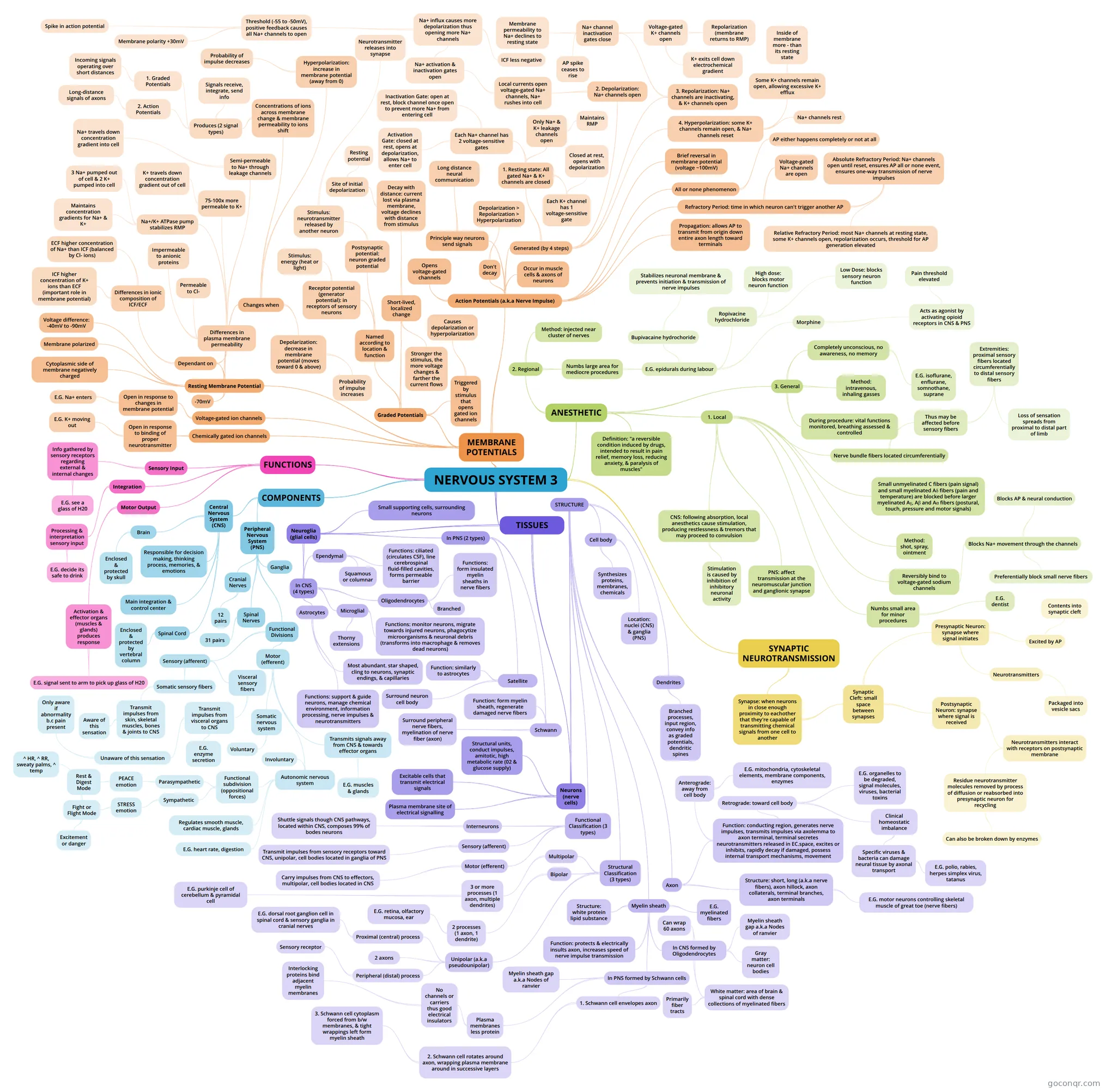
Mind map: Nervous system
Inhalt der Mindmap
NERVOUS SYSTEM 3
FUNCTIONS
- Sensory Input
- Info gathered by sensory receptors regarding external & internal changes
- E.G. see a glass of H20
- Info gathered by sensory receptors regarding external & internal changes
- Integration
- Processing & interpretation sensory input
- E.G. decide its safe to drink
- Processing & interpretation sensory input
- Motor Output
- Activation & effector organs (muscles & glands) produces response
- E.G. signal sent to arm to pick up glass of H20
- Activation & effector organs (muscles & glands) produces response
- Sensory Input
COMPONENTS
- Central Nervous System (CNS)
- Brain
- Enclosed & protected by skull
- Spinal Cord
- Enclosed & protected by vertebral column
- Main integration & control center
- Responsible for decision making, thinking process, memories, & emotions
- Brain
- Peripheral Nervous System (PNS)
- Spinal Nerves
- 31 pairs
- Cranial Nerves
- 12 pairs
- Ganglia
- Functional Divisions
- Sensory (afferent)
- Somatic sensory fibers
- Transmit impulses from skin, skeletal muscles, bones & joints to CNS
- Aware of this sensation
- Transmit impulses from skin, skeletal muscles, bones & joints to CNS
- Visceral sensory fibers
- Transmit impulses from visceral organs to CNS
- Unaware of this sensation
- Only aware if abnormality b.c pain present
- E.G. enzyme secretion
- Unaware of this sensation
- Transmit impulses from visceral organs to CNS
- Somatic sensory fibers
- Motor (efferent)
- Somatic nervous system
- Voluntary
- Autonomic nervous system
- Involuntary
- Regulates smooth muscle, cardiac muscle, glands
- Functional subdivision (oppositional forces)
- Sympathetic
- STRESS emotion
- Fight or Flight Mode
- ^ HR, ^ RR, sweaty palms, ^ temp
- Excitement or danger
- Fight or Flight Mode
- STRESS emotion
- Parasympathetic
- PEACE emotion
- Rest & Digest Mode
- PEACE emotion
- Sympathetic
- E.G. heart rate, digestion
- Transmits signals away from CNS & towards effector organs
- E.G. muscles & glands
- Somatic nervous system
- Sensory (afferent)
- Spinal Nerves
- Central Nervous System (CNS)
TISSUES
- Neuroglia (glial cells)
- Small supporting cells, surrounding neurons
- In CNS (4 types)
- Astrocytes
- Most abundant. star shaped, cling to neurons, synaptic endings, & capillaries
- Functions: support & guide neurons, manage chemical environment, information processing, nerve impulses & neurotransmitters
- Microglial
- Thorny extensions
- Functions: monitor neurons, migrate towards injured neurons, phagocytize microorganisms & neuronal debris (transforms into macrophage & removes dead neurons)
- Ependymal
- Squamous or columnar
- Functions: ciliated (circulates CSF), line cerebrospinal fluid-filled cavities, forms permeable barrier
- Oligodendrocytes
- Branched
- Functions: form insulated myelin sheaths in nerve fibers
- Astrocytes
- In PNS (2 types)
- Satellite
- Surround neuron cell body
- Function: similarly to astrocytes
- Schwann
- Surround peripheral nerve fibers, myelination of nerve fiber (axon)
- Function: form myelin sheath, regenerate damaged nerve fibers
- Satellite
- Neurons (nerve cells)
- Excitable cells that transmit electrical signals
- Structural units, conduct impulses, amitotic, high metabolic rate (02 & glucose supply)
- Plasma membrane site of electrical signalling
- STRUCTURE
- Cell body
- Synthesizes proteins, membranes, chemicals
- Location: nuclei (CNS) & ganglia (PNS)
- Dendrites
- Branched processes, input region, convey info as graded potentials, dendritic spines
- Axon
- Structure: short, long (a.k.a nerve fibers), axon hillock, axon collaterals, terminal branches, axon terminals
- E.G. motor neurons controlling skeletal muscle of great toe (nerve fibers)
- Function: conducting region, generates nerve impulses, transmits impulses via axolemma to axon terminal, terminal secretes neurotransmitters released in EC.space, excites or inhibits, rapidly decay if damaged, possess internal transport mechanisms, movement
- Anterograde: away from cell body
- E.G. mitochondria, cytoskeletal elements, membrane components, enzymes
- Retrograde: toward cell body
- E.G. organelles to be degraded, signal molecules, viruses, bacterial toxins
- Clinical homeostatic imbalance
- Specific viruses & bacteria can damage neural tissue by axonal transport
- E.G. polio, rabies, herpes simplex virus, tatanus
- Specific viruses & bacteria can damage neural tissue by axonal transport
- Anterograde: away from cell body
- Structure: short, long (a.k.a nerve fibers), axon hillock, axon collaterals, terminal branches, axon terminals
- Myelin sheath
- Structure: white protein lipid substance
- Function: protects & electrically insults axon, increases speed of nerve impulse transmission
- E.G. myelinated fibers
- In PNS formed by Schwann cells
- Plasma membranes less protein
- No channels or carriers thus good electrical insulators
- Interlocking proteins bind adjacent myelin membranes
- No channels or carriers thus good electrical insulators
- 1. Schwann cell envelopes axon
- 2. Schwann cell rotates around axon, wrapping plasma membrane around in successive layers
- 3. Schwann cell cytoplasm forced from b/w membranes, & tight wrappings left form myelin sheath
- 2. Schwann cell rotates around axon, wrapping plasma membrane around in successive layers
- Myelin sheath gap a.k.a Nodes of ranvier
- Plasma membranes less protein
- In CNS formed by Oligodendrocytes
- Can wrap 60 axons
- Myelin sheath gap a.k.a Nodes of ranvier
- White matter: area of brain & spinal cord with dense collections of myelinated fibers
- Primarily fiber tracts
- Gray matter: neuron cell bodies
- Structural Classification (3 types)
- Multipolar
- 3 or more processes (1 axon, multiple dendrites)
- E.G. purkinje cell of cerebellum & pyramidal cell
- 3 or more processes (1 axon, multiple dendrites)
- Bipolar
- 2 processes (1 axon, 1 dendrite)
- E.G. retina, olfactory mucosa, ear
- 2 processes (1 axon, 1 dendrite)
- Unipolar (a.k.a pseudounipolar)
- Peripheral (distal) process
- Sensory receptor
- Proximal (central) process
- E.G. dorsal root ganglion cell in spinal cord & sensory ganglia in cranial nerves
- 2 axons
- Peripheral (distal) process
- Multipolar
- Functional Classification (3 types)
- Sensory (afferent)
- Transmit impulses from sensory receptors toward CNS, unipolar, cell bodies located in ganglia of PNS
- Motor (efferent)
- Carry impulses from CNS to effectors, multipolar, cell bodies located in CNS
- Interneurons
- Shuttle signals though CNS pathways, located within CNS, composes 99% of bodes neurons
- Sensory (afferent)
- Cell body
- Neuroglia (glial cells)
MEMBRANE POTENTIALS
- Resting Membrane Potential
- -70mV
- Cytoplasmic side of membrane negatively charged
- Voltage difference: -40mV to -90mV
- Membrane polarized
- Dependant on
- Differences in plasma membrane permeability
- Impermeable to anionic proteins
- Semi-permeable to Na+ through leakage channels
- Na+ travels down concentration gradient into cell
- 75-100x more permeable to K+
- K+ travels down concentration gradient out of cell
- Permeable to Cl-
- Na+/K+ ATPase pump stabilizes RMP
- Maintains concentration gradients for Na+ & K+
- 3 Na+ pumped out of cell & 2 K+ pumped into cell
- Differences in ionic composition of ICF/ECF
- ECF higher concentration of Na+ than ICF (balanced by Cl- ions)
- ICF higher concentration of K+ ions than ECF (important role in membrane potential)
- Differences in plasma membrane permeability
- Changes when
- Concentrations of ions across membrane change & membrane permeability to ions shift
- Produces (2 signal types)
- 1. Graded Potentials
- Incoming signals operating over short distances
- 2. Action Potentials
- Long-distance signals of axons
- Signals receive, integrate, send info
- 1. Graded Potentials
- Produces (2 signal types)
- Hyperpolarization: increase in membrane potential (away from 0)
- Probability of impulse decreases
- Depolarization: decrease in membrane potential (moves toward 0 & above)
- Probability of impulse increases
- Concentrations of ions across membrane change & membrane permeability to ions shift
- Graded Potentials
- Short-lived, localized change
- Stronger the stimulus, the more voltage changes & farther the current flows
- Decay with distance: current lost via plasma membrane, voltage declines with distance from stimulus
- Site of initial depolarization
- Resting potential
- Site of initial depolarization
- Triggered by stimulus that opens gated ion channels
- Causes depolarization or hyperpolarization
- Named according to location & function
- Receptor potential (generator potential): in receptors of sensory neurons
- Stimulus: energy (heat or light)
- Postsynaptic potential: neuron graded potential
- Stimulus: neurotransmitter released by another neuron
- Neurotransmitter releases into synapse
- Stimulus: neurotransmitter released by another neuron
- Receptor potential (generator potential): in receptors of sensory neurons
- Short-lived, localized change
- Action Potentials (a.k.a Nerve Impulse)
- Principle way neurons send signals
- Long distance neural communication
- Occur in muscle cells & axons of neurons
- Brief reversal in membrane potential (voltage ~100mV)
- Don't decay
- Depolarization > Repolarization > Hyperpolarization
- Opens voltage-gated channels
- Generated (by 4 steps)
- 1. Resting state: All gated Na+ & K+ channels are closed
- Only Na+ & K+ leakage channels open
- Maintains RMP
- Each Na+ channel has 2 voltage-sensitive gates
- Activation Gate: closed at rest, opens at depolarization, allows Na+ to enter cell
- Inactivation Gate: open at rest, block channel once open to prevent more Na+ from entering cell
- Each K+ channel has 1 voltage-sensitive gate
- Closed at rest, opens with depolarization
- Only Na+ & K+ leakage channels open
- 2. Depolarization: Na+ channels open
- Local currents open voltage-gated Na+ channels, Na+ rushes into cell
- Na+ activation & inactivation gates open
- Na+ influx causes more depolarization thus opening more Na+ channels
- Threshold (-55 to -50mV), positive feedback causes all Na+ channels to open
- Spike in action potential
- Membrane polarity +30mV
- ICF less negative
- Threshold (-55 to -50mV), positive feedback causes all Na+ channels to open
- Na+ influx causes more depolarization thus opening more Na+ channels
- Na+ activation & inactivation gates open
- Local currents open voltage-gated Na+ channels, Na+ rushes into cell
- 3. Repolarization: Na+ channels are inactivating, & K+ channels open
- Na+ channel inactivation gates close
- Membrane permeability to Na+ declines to resting state
- AP spike ceases to rise
- Voltage-gated K+ channels open
- K+ exits cell down electrochemical gradient
- Repolarization (membrane returns to RMP)
- Na+ channel inactivation gates close
- 4. Hyperpolarization: some K+ channels remain open, & Na+ channels reset
- Some K+ channels remain open, allowing excessive K+ efflux
- Inside of membrane more - than its resting state
- Na+ channels rest
- Some K+ channels remain open, allowing excessive K+ efflux
- 1. Resting state: All gated Na+ & K+ channels are closed
- All or none phenomenon
- AP either happens completely or not at all
- Propagation: allows AP to transmit from origin down entire axon length toward terminals
- Refractory Period: time in which neuron can't trigger another AP
- Voltage-gated Na+ channels are open
- Absolute Refractory Period: Na+ channels open until reset, ensures AP all or none event, ensures one-way transmission of nerve impulses
- Relative Refractory Period: most Na+ channels at resting state, some K+ channels open, repolarization occurs, threshold for AP generation elevated
- Principle way neurons send signals
- Chemically gated ion channels
- Open in response to binding of proper neurotransmitter
- E.G. K+ moving out
- Open in response to binding of proper neurotransmitter
- Voltage-gated ion channels
- Open in response to changes in membrane potential
- E.G. Na+ enters
- Open in response to changes in membrane potential
- Resting Membrane Potential
SYNAPTIC NEUROTRANSMISSION
- Synapse: when neurons in close enough proximity to eachother that they're capable of transmitting chemical signals from one cell to another
- Synaptic Cleft: small space between synapses
- Presynaptic Neuron: synapse where signal initiates
- Neurotransmitters
- Packaged into vesicle sacs
- Excited by AP
- Contents into synaptic cleft
- Neurotransmitters
- Postsynaptic Neuron: synapse where signal is received
- Neurotransmitters interact with receptors on postsynaptic membrane
- Residue neurotransmitter molecules removed by process of diffusion or reabsorbed into presynaptic neuron for recycling
- Can also be broken down by enzymes
- Residue neurotransmitter molecules removed by process of diffusion or reabsorbed into presynaptic neuron for recycling
- Neurotransmitters interact with receptors on postsynaptic membrane
- Presynaptic Neuron: synapse where signal initiates
- Synaptic Cleft: small space between synapses
- Synapse: when neurons in close enough proximity to eachother that they're capable of transmitting chemical signals from one cell to another
ANESTHETIC
- Definition: "a reversible condition induced by drugs, intended to result in pain relief, memory loss, reducing anxiety, & paralysis of muscles"
- 1. Local
- Method: shot, spray, ointment
- Numbs small area for minor procedures
- E.G. dentist
- Reversibly bind to voltage-gated sodium channels
- Blocks Na+ movement through the channels
- Blocks AP & neural conduction
- Preferentially block small nerve fibers
- Blocks Na+ movement through the channels
- Small unmyelinated C fibers (pain signal) and small myelinated Aδ fibers (pain and temperature) are blocked before larger myelinated Aγ, Aβ and Aα fibers (postural, touch, pressure and motor signals)
- Nerve bundle fibers located circumferentially
- Thus may be affected before sensory fibers
- Extremities: proximal sensory fibers located circumferentially to distal sensory fibers
- Loss of sensation spreads from proximal to distal part of limb
- Extremities: proximal sensory fibers located circumferentially to distal sensory fibers
- Thus may be affected before sensory fibers
- CNS: following absorption, local anesthetics cause stimulation, producing restlessness & tremors that may proceed to convulsion
- Stimulation is caused by inhibition of inhibitory neuronal activity
- PNS: affect transmission at the neuromuscular junction and ganglionic synapse
- 2. Regional
- Method: injected near cluster of nerves
- Numbs large area for mediocre procedures
- E.G. epidurals during labour
- Bupivacaine hydrochoride
- Stabilizes neuronal membrane & prevents initiation & transmission of nerve impulses
- Ropivacine hydrochloride
- High dose: blocks motor neuron function
- Low Dose: blocks sensory neuron function
- Morphine
- Acts as agonist by activating opioid receptors in CNS & PNS
- Pain threshold elevated
- Acts as agonist by activating opioid receptors in CNS & PNS
- Bupivacaine hydrochoride
- E.G. epidurals during labour
- 3. General
- Completely unconscious, no awareness, no memory
- E.G. isoflurane, enflurane, somnothane, suprane
- Method: intravenous, inhaling gasses
- During procedure: vital functions monitored, breathing assessed & controlled
- Completely unconscious, no awareness, no memory
Informationen
- Beschreibung
- Mindmap am NERVOUS SYSTEM 3, erstellt von Marina Acquaroli am 18/11/2015.
- Erstellt von
- Marina Acquaroli
- Aufrufe
- 0
- Bewertung