
Mindmap
Exchange
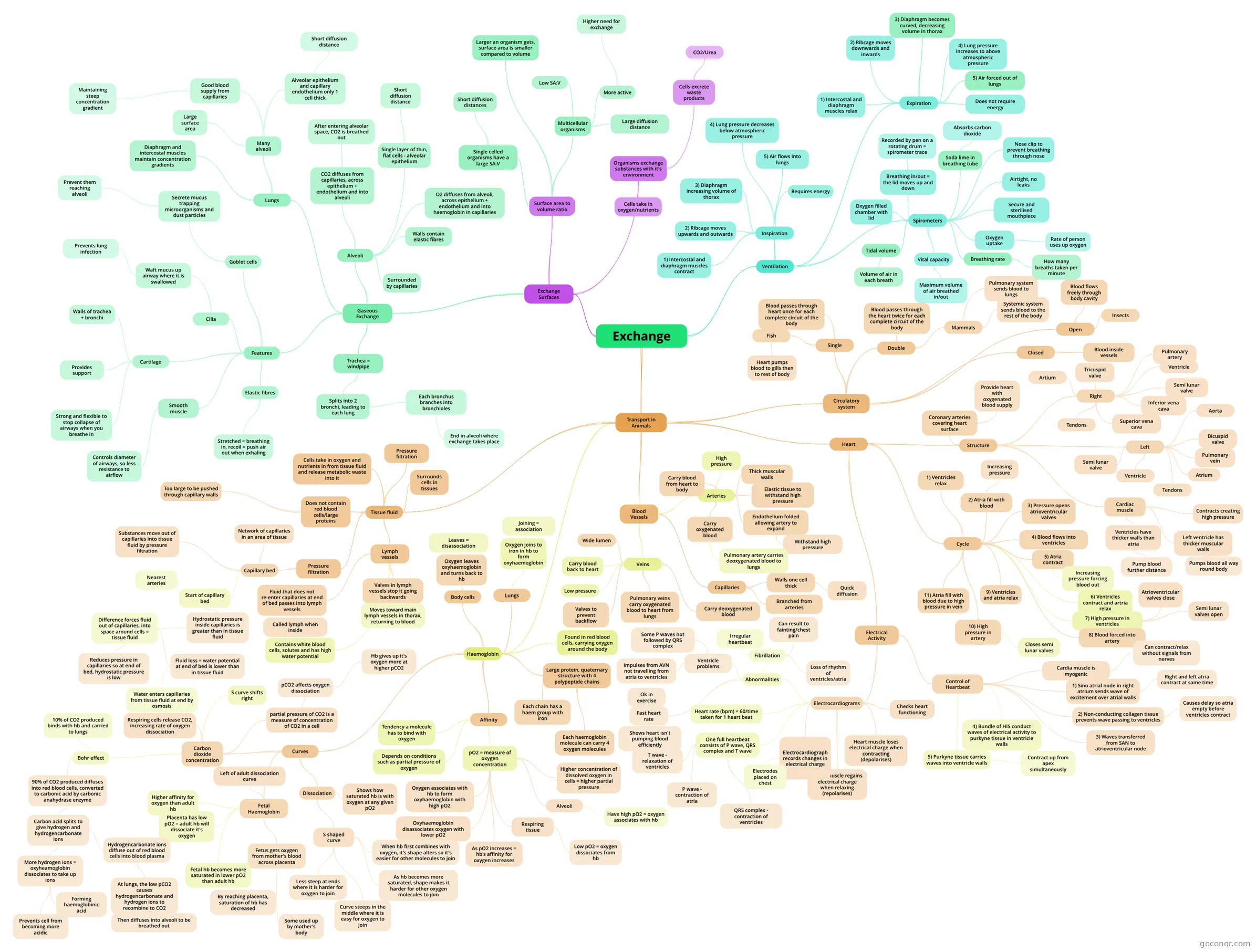
Mind map: Exchange biology
Inhalt der Mindmap
Exchange
Exchange Surfaces
- Organisms exchange substances with it's environment
- Cells take in oxygen/nutrients
- Cells excrete waste products
- CO2/Urea
- Surface area to volume ratio
- Larger an organism gets, surface area is smaller compared to volume
- Single celled organisms have a large SA:V
- Short diffusion distances
- Multicellular organisms
- Large diffusion distance
- Low SA:V
- More active
- Higher need for exchange
- Gaseous Exchange
- Trachea = windpipe
- Splits into 2 bronchi, leading to each lung
- Each bronchus branches into bronchioles
- End in alveoli where exchange takes place
- Each bronchus branches into bronchioles
- Splits into 2 bronchi, leading to each lung
- Alveoli
- Surrounded by capillaries
- Single layer of thin, flat cells - alveolar epithelium
- Short diffusion distance
- Walls contain elastic fibres
- O2 diffuses from alveoli, across epithelium + endothelium and into haemoglobin in capillaries
- CO2 diffuses from capillaries, across epithelium + endothelium and into alveoli
- After entering alveolar space, CO2 is breathed out
- Lungs
- Many alveoli
- Large surface area
- Alveolar epithelium and capillary endothelium only 1 cell thick
- Short diffusion distance
- Good blood supply from capillaries
- Maintaining steep concentration gradient
- Diaphragm and intercostal muscles maintain concentration gradients
- Many alveoli
- Features
- Goblet cells
- Secrete mucus trapping microorganisms and dust particles
- Prevent them reaching alveoli
- Secrete mucus trapping microorganisms and dust particles
- Cilia
- Waft mucus up airway where it is swallowed
- Prevents lung infection
- Waft mucus up airway where it is swallowed
- Elastic fibres
- Stretched = breathing in, recoil = push air out when exhaling
- Smooth muscle
- Controls diameter of airways, so less resistance to airflow
- Cartilage
- Walls of trachea + bronchi
- Strong and flexible to stop collapse of airways when you breathe in
- Provides support
- Goblet cells
- Trachea = windpipe
- Organisms exchange substances with it's environment
Ventilation
- Inspiration
- 1) Intercostal and diaphragm muscles contract
- 2) Ribcage moves upwards and outwards
- 3) Diaphragm increasing volume of thorax
- 4) Lung pressure decreases below atmospheric pressure
- 5) Air flows into lungs
- Requires energy
- Expiration
- 1) Intercostal and diaphragm muscles relax
- 2) Ribcage moves downwards and inwards
- 3) Diaphragm becomes curved, decreasing volume in thorax
- 4) Lung pressure increases to above atmospheric pressure
- 5) Air forced out of lungs
- Does not require energy
- Spirometers
- Oxygen filled chamber with lid
- Breathing in/out = the lid moves up and down
- Recorded by pen on a rotating drum = spirometer trace
- Soda lime in breathing tube
- Absorbs carbon dioxide
- Nose clip to prevent breathing through nose
- Airtight, no leaks
- Secure and sterilised mouthpiece
- Tidal volume
- Volume of air in each breath
- Vital capacity
- Maximum volume of air breathed in/out
- Breathing rate
- How many breaths taken per minute
- Oxygen uptake
- Rate of person uses up oxygen
- Inspiration
Transport in Animals
- Circulatory system
- Single
- Blood passes through heart once for each complete circuit of the body
- Fish
- Heart pumps blood to gills then to rest of body
- Double
- Blood passes through the heart twice for each complete circuit of the body
- Mammals
- Pulmonary system sends blood to lungs
- Systemic system sends blood to the rest of the body
- Closed
- Blood inside vessels
- Open
- Blood flows freely through body cavity
- Insects
- Single
- Heart
- Structure
- Right
- Inferior vena cava
- Superior vena cava
- Pulmonary artery
- Semi lunar valve
- Ventricle
- Artium
- Tricuspid valve
- Tendons
- Left
- Aorta
- Pulmonary vein
- Atrium
- Semi lunar valve
- Bicuspid valve
- Tendons
- Ventricle
- Coronary arteries covering heart surface
- Provide heart with oxygenated blood supply
- Cardiac muscle
- Contracts creating high pressure
- Left ventricle has thicker muscular walls
- Pumps blood all way round body
- Ventricles have thicker walls than atria
- Pump blood further distance
- Right
- Cycle
- 1) Ventricles relax
- 2) Atria fill with blood
- Increasing pressure
- 3) Pressure opens atrioventricular valves
- 4) Blood flows into ventricles
- 5) Atria contract
- Increasing pressure forcing blood out
- 6) Ventricles contract and artria relax
- 7) High pressure in ventricles
- Atrioventricular valves close
- Semi lunar valves open
- 8) Blood forced into artery
- 9) Ventricles and atria relax
- 10) High pressure in artery
- Closes semi lunar valves
- 11) Atria fill with blood due to high pressure in vein
- Electrical Activity
- Control of Heartbeat
- Cardia muscle is myogenic
- Can contract/relax without signals from nerves
- 1) Sino atrial node in right atrium sends wave of excitement over atrial walls
- Right and left atria contract at same time
- 2) Non-conducting collagen tissue prevents wave passing to ventricles
- Causes delay so atria empty before ventricles contract
- 3) Waves transferred from SAN to atrioventricular node
- 4) Bundle of HIS conduct waves of electrical activity to purkyne tissue in ventricle walls
- 5) Purkyne tissue carries waves into ventricle walls
- Contract up from apex simultaneously
- Cardia muscle is myogenic
- Electrocardiograms
- Checks heart functioning
- Heart muscle loses electrical charge when contracting (depolarises)
- Heart muscle regains electrical charge when relaxing (repolarises)
- Electrocardiograph records changes in electrical charge
- Electrodes placed on chest
- One full heartbeat consists of P wave, QRS complex and T wave
- P wave - contraction of atria
- QRS complex - contraction of ventricles
- T wave - relaxation of ventricles
- Heart rate (bpm) = 60/time taken for 1 heart beat
- Abnormalities
- Fast heart rate
- Shows heart isn't pumping blood efficiently
- Ok in exercise
- Ventricle problems
- Some P waves not followed by QRS complex
- Impulses from AVN not travelling from atria to ventricles
- Fibrillation
- Irregular heartbeat
- Loss of rhythm of ventricles/atria
- Can result to fainting/chest pain
- Fast heart rate
- Control of Heartbeat
- Structure
- Blood Vessels
- Arteries
- Carry blood from heart to body
- Thick muscular walls
- Elastic tissue to withstand high pressure
- Endothelium folded allowing artery to expand
- Withstand high pressure
- Carry oxygenated blood
- Pulmonary artery carries deoxygenated blood to lungs
- High pressure
- Capillaries
- Branched from arteries
- Walls one cell thick
- Quick diffusion
- Veins
- Low pressure
- Carry blood back to heart
- Wide lumen
- Valves to prevent backflow
- Carry deoxygenated blood
- Pulmonary veins carry oxygenated blood to heart from lungs
- Arteries
- Tissue fluid
- Surrounds cells in tissues
- Cells take in oxygen and nutrients in from tissue fluid and release metabolic waste into it
- Pressure filtration
- Pressure filtration
- Capillary bed
- Network of capillaries in an area of tissue
- Substances move out of capillaries into tissue fluid by pressure filtration
- Start of capillary bed
- Nearest arteries
- Hydrostatic pressure inside capillaries is greater than in tissue fluid
- Difference forces fluid out of capillaries, into space around cells = tissue fluid
- Reduces pressure in capillaries so at end of bed, hydrostatic pressure is low
- Fluid loss = water potential at end of bed is lower than in tissue fluid
- Water enters capillaries from tissue fluid at end by osmosis
- Difference forces fluid out of capillaries, into space around cells = tissue fluid
- Capillary bed
- Does not contain red blood cells/large proteins
- Too large to be pushed through capillary walls
- Lymph vessels
- Fluid that does not re-enter capillaries at end of bed passes into lymph vessels
- Called lymph when inside
- Valves in lymph vessels stop it going backwards
- Moves toward main lymph vessels in thorax, returning to blood
- Contains white blood cells, solutes and has high water potential
- Fluid that does not re-enter capillaries at end of bed passes into lymph vessels
- Haemoglobin
- Found in red blood cells, carrying oxygen around the body
- Large protein, quaternary structure with 4 polypeptide chains
- Each chain has a haem group with iron
- Each haemoglobin molecule can carry 4 oxygen molecules
- Lungs
- Oxygen joins to iron in hb to form oxyhaemoglobin
- Joining = association
- Oxygen joins to iron in hb to form oxyhaemoglobin
- Body cells
- Oxygen leaves oxyhaemoglobin and turns back to hb
- Leaves = disassociation
- Oxygen leaves oxyhaemoglobin and turns back to hb
- Affinity
- Tendency a molecule has to bind with oxygen
- Depends on conditions such as partial pressure of oxygen
- pO2 = measure of oxygen concentration
- Higher concentration of dissolved oxygen in cells = higher partial pressure
- As pO2 increases = hb's affinity for oxygen increases
- Oxygen associates with hb to form oxyhaemoglobin with high pO2
- Oxyhaemoglobin disassociates oxygen with lower pO2
- Alveoli
- Have high pO2 = oxygen associates with hb
- Respiring tissue
- Low pO2 = oxygen dissociates from hb
- Curves
- Dissociation
- Shows how saturated hb is with oxygen at any given pO2
- S shaped curve
- When hb first combines with oxygen, it's shape alters so it's easier for other molecules to join
- As hb becomes more saturated, shape makes it harder for other oxygen molecules to join
- Curve steeps in the middle where it is easy for oxygen to join
- Less steep at ends where it is harder for oxygen to join
- Fetal Haemoglobin
- Higher affinity for oxygen than adult hb
- Fetus gets oxygen from mother's blood across placenta
- By reaching placenta, saturation of hb has decreased
- Some used up by mother's body
- By reaching placenta, saturation of hb has decreased
- Placenta has low pO2 = adult hb will dissociate it's oxygen
- Fetal hb becomes more saturated in lower pO2 than adult hb
- Left of adult dissociation curve
- Carbon dioxide concentration
- partial pressure of CO2 is a measure of concentration of CO2 in a cell
- pCO2 affects oxygen dissociation
- Hb gives up it's oxygen more at higher pCO2
- pCO2 affects oxygen dissociation
- Respiring cells release CO2, increasing rate of oxygen dissociation
- S curve shifts right
- Bohr effect
- 10% of CO2 produced binds with hb and carried to lungs
- 90% of CO2 produced diffuses into red blood cells, converted to carbonic acid by carbonic anahydrase enzyme
- Carbon acid splits to give hydrogen and hydrogencarbonate ions
- More hydrogen ions = oxyheamoglobin dissociates to take up ions
- Forming haemoglobinic acid
- Prevents cell from becoming more acidic
- Hydrogencarbonate ions diffuse out of red blood cells into blood plasma
- At lungs, the low pCO2 causes hydrogencarbonate and hydrogen ions to recombine to CO2
- Then diffuses into alveoli to be breathed out
- At lungs, the low pCO2 causes hydrogencarbonate and hydrogen ions to recombine to CO2
- More hydrogen ions = oxyheamoglobin dissociates to take up ions
- Carbon acid splits to give hydrogen and hydrogencarbonate ions
- partial pressure of CO2 is a measure of concentration of CO2 in a cell
- Dissociation
- Circulatory system
Informationen
- Beschreibung
- A-Levels Biology f211 Mindmap am Exchange, erstellt von Gemma Bradford am 08/05/2013.
- Erstellt von
- Gemma Bradford
- Aufrufe
- 57
- Tags
- Bewertung
Mehr zu Gas exchange in the lungs