
Mindmap
Anatomy: Nervous system
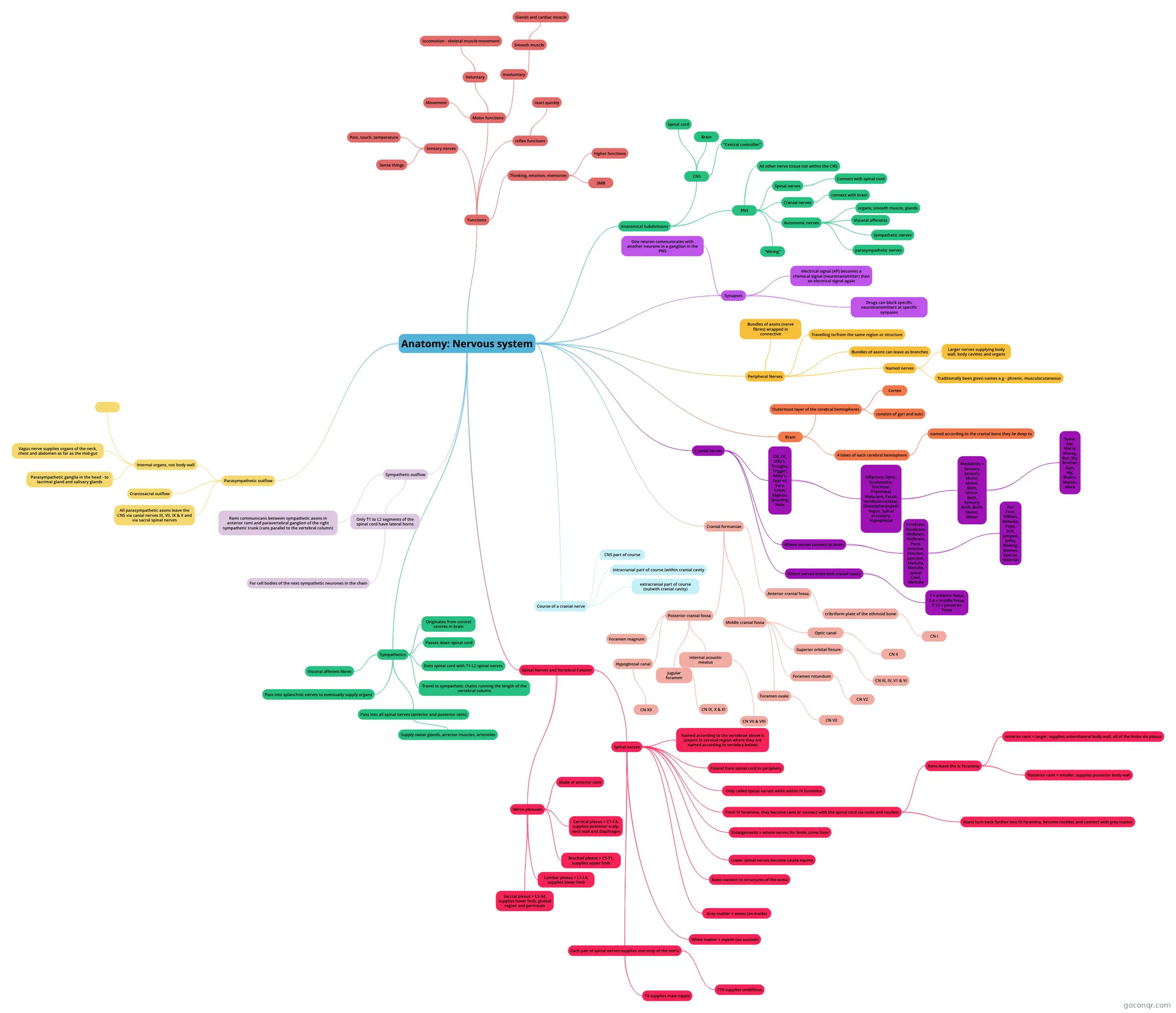
Mind map: Anatomy: Nervous system
Inhalt der Mindmap
Anatomy: Nervous system
Functions
- Sensory nerves
- Sense things
- Pain, touch, temperature
- Motor functions
- Movement
- Voluntary
- locomotion - skeletal muscle movement
- Involuntary
- Smooth muscle
- Glands and cardiac muscle
- Smooth muscle
- reflex functions
- react quickly
- Thinking, emotion, memories
- higher functions
- 3MB
- Sensory nerves
Anatomical Subdivisions
- CNS
- Brain
- Spinal cord
- "Central controller"
- PNS
- All other nerve tissue not within the CNS
- Spinal nerves
- Connect with spinal cord
- Cranial nerves
- connect with brain
- Autonomic nerves
- organs, smooth muscle, glands
- Visceral afferents
- Sympathetic nerves
- parasympathetic nerves
- "Wiring"
- CNS
Synapses
- One neuron communicates with another neurone in a ganglion in the PNS
- electrical signal (AP) becomes a chemical signal (neurotransmitter) then an electrical signal again
- Drugs can block specific neurotransmitters at specific synpases
Peripheral Nerves
- Bundles of axons (nerve fibres) wrapped in connective
- Travelling to/from the same region or structure
- Bundles of axons can leave as branches
- Named nerves
- Larger nerves supplying body wall, body cavities and organs
- Traditionally been given names e.g - phrenic, musculocutaneous
Brain
- Outermost layer of the cerebral hemispheres
- Cortex
- consists of gyri and sulci
- 4 lobes of each cerebral hemisphere
- named according to the cranial bone they lie deep to
- Outermost layer of the cerebral hemispheres
Cranial nerves
- Oll, Of, Olly's, Troughs, Trigger, Abby's, Fear of, Very, Great, Vaginas, Spouting, Hate
- Olfactory, Optic, Oculomotor, Trochlear, Trigeminal, Abducent, Facial, Vestibulocochlear, Glossopharyngeal, Vagus, Spinal accessory, Hypoglossal
- Modalities = Sensory, Sensory, Motor, Motor, Both, Motor, Both, Sensory, Both, Both, Motor, Motor
- Some, Say, Marry, Money, But, My, Brother, Says, Big, Brains, Matter, More
- Modalities = Sensory, Sensory, Motor, Motor, Both, Motor, Both, Sensory, Both, Both, Motor, Motor
- Olfactory, Optic, Oculomotor, Trochlear, Trigeminal, Abducent, Facial, Vestibulocochlear, Glossopharyngeal, Vagus, Spinal accessory, Hypoglossal
- Where nerves connect to brain
- Forebrain, Forebrain, Midbrain, Midbrain, Pons, Junction, Junction, Junction, Medulla, Medulla, spinal Cord, Medulla
- For, Four, Million, Millenia, Pepe, Just, Jumped, Jollily, Making, Memes, Special, Material
- Forebrain, Forebrain, Midbrain, Midbrain, Pons, Junction, Junction, Junction, Medulla, Medulla, spinal Cord, Medulla
- Where nerves enter/exit cranial cavity
- 1 = anterior fossa, 2-6 = middle fossa, 7-12 = posterior fossa
- Oll, Of, Olly's, Troughs, Trigger, Abby's, Fear of, Very, Great, Vaginas, Spouting, Hate
Cranial formaniae
- Anterior cranial fossa
- cribriform plate of the ethmoid bone
- CN I
- cribriform plate of the ethmoid bone
- Middle cranial fossa
- Optic canal
- CN II
- Superior orbital fissure
- CN III, IV, V1 & VI
- Foramen rotundum
- CN V2
- Foramen ovale
- CN V3
- Optic canal
- Posterior cranial fossa
- internal acoustic meatus
- CN VII & VIII
- jugular foramen
- CN IX, X & XI
- Hypoglossal canal
- CN XII
- Foramen magnum
- internal acoustic meatus
- Anterior cranial fossa
Course of a cranial nerve
- CNS part of course
- Intracranial part of course (within cranial cavity
- extracranial part of course (outwith cranial cavity)
Spinal Nerves and Vertebral Column
- Spinal nerves
- Named according to the vertebrae above it (expect in cervical region where they are named according to vertebra below)
- Extend from spinal cord to periphery
- Only called spinal nerves while within IV foramina
- From IV foramina, they become rami or connect with the spinal cord via roots and rootlets
- Rami leave the Iv foramina
- Anterior rami = larger, supplies anterolateral body wall, all of the limbs via plexus
- Posterior rami = smaller, supplies posterior body wall
- Roots turn back further into IV foramina, become rootlets and connect with grey matter
- Rami leave the Iv foramina
- Enlargements = where nerves for limbs come from
- Lower spinal nerves become cauda equina
- Rami connect to structures of the soma
- Grey matter = axons (on inside)
- White matter = myelin (on outside)
- Each pair of spinal nerves supplies one strip of the soma
- T10 supplies umbillicus
- T4 supplies male nipple
- Nerve plexuses
- Made of anterior rami
- Cervical plexus = C1-C4, supplies posterior scalp, neck wall and diaphragm
- Brachial plexus = C5-T1, supplies upper limb
- Lumbar plexus = L1-L4, supplies lower limb
- Saccral plexus = L5-S4, supplies lower limb, gluteal region and perineum
- Spinal nerves
Sympathetics
- Originates from control centres in brain
- Passes down spinal cord
- Exits spinal cord with T1-L2 spinal nerves
- Travel to sympathetic chains running the length of the vertebral column
- Pass into all spinal nerves (anterior and posterior rami)
- Supply sweat glands, arrector muscles, arterioles
- Pass into splanchnic nerves to eventually supply organs
- Visceral afferent fibres
Sympathetic outflow
- Only T1 to L2 segments of the spinal cord have lateral horns
- For cell bodies of the next sympathetic neurones in the chain
- Rami communicans between sympathetic axons in anterior rami and paravertebral ganglion of the right sympathetic trunk (runs parallel to the vertebral column)
- Only T1 to L2 segments of the spinal cord have lateral horns
Parasympathetic outflow
- All parasympathetic axons leave the CNS via canial nerves III, VII, IX & X and via sacral spinal nerves
- Craniosacral outflow
- Internal organs, not body wall
- Parasympathetic ganglia in the head - to lacrimal gland and salivary glands
- Vagus nerve supplies organs of the neck, chest and abdomen as far as the mid-gut
Informationen
- Beschreibung
- Mindmap am Anatomy: Nervous system, erstellt von Joanne McEwan am 30/10/2017.
- Erstellt von
- Joanne McEwan
- Aufrufe
- 0
- Bewertung