
Mindmap
Pleural Effusion

Inhalt der Mindmap
Pleural Effusion
Sign and symptoms
- pleural manifestation depends on the causes and size of the effusion.
- Asmptomatic in patient less than 300ml of fluid in the pleural cavity.
- Shortness of breath
- General features dyspea
- Pleuritic pain sharp and worsen in inspiration
- Dry cough
- Decreased cest wall movement
- Absence of breath sound
- Dullness of percussion
- Decreased tactile fremitus over the affected areas
- Absence of breath sound
- Decreased cest wall movement
- Dry cough
- pleural manifestation depends on the causes and size of the effusion.
Treatment/ Nursing intervension
- Directed to the underlying causes of the effusion
- ABCs first consideration
- Give enough oxygen for the body to function, by nasal cannula
- prevention of accumulation of pleural effusion
- ABCs first consideration
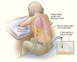
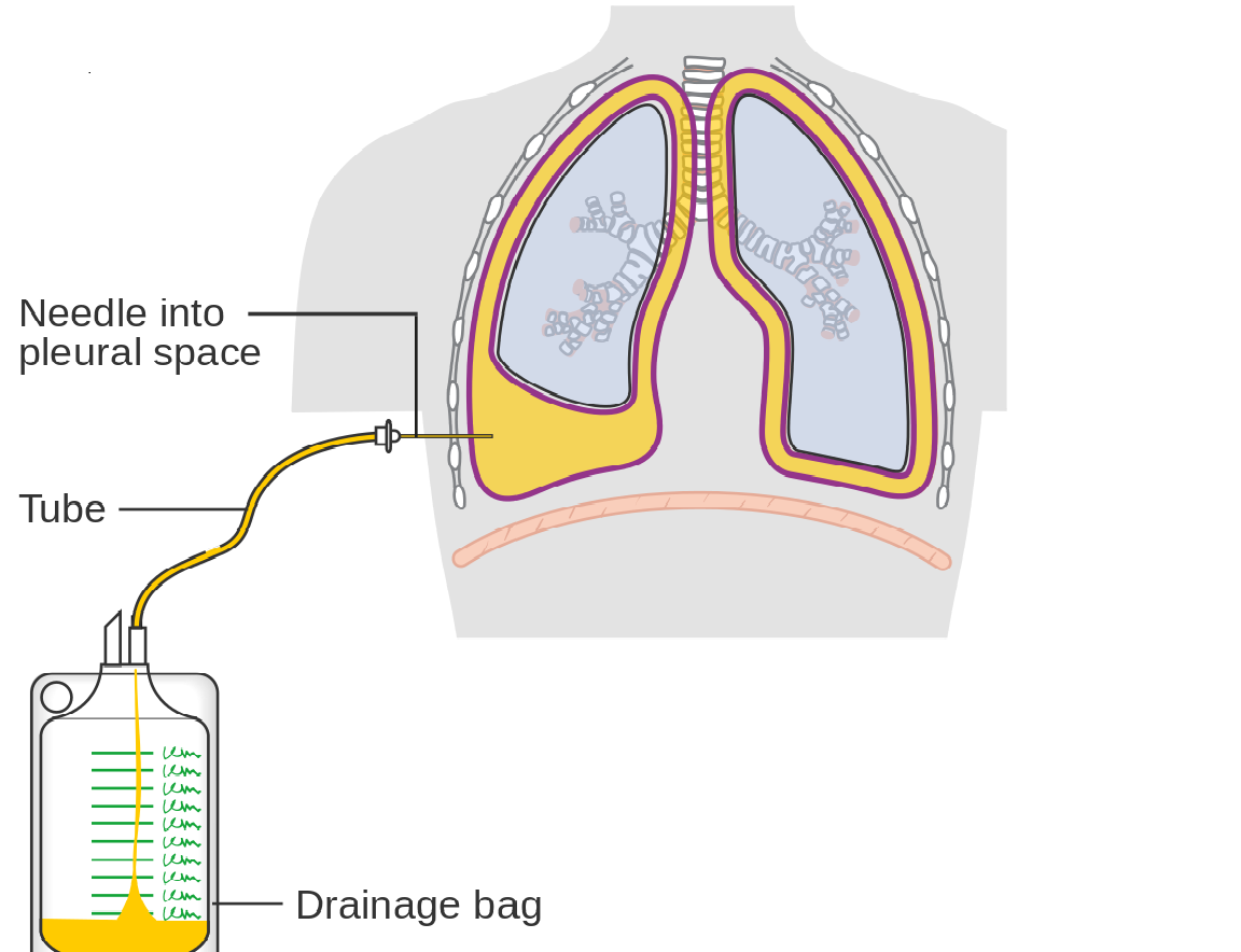
- Thoracentesis, if effusion is large
- Chest tube by inserting a plastic tube into the pleural space for days
- Patient teaching
- pursed lip and diaphragmatic breathing.
- Always take your medication
- Pleurodesis
- injecting an irritating substance through the chest wall tube into the pleural space. This substance inflame the pleura and the chest wall, healing will start. It prevent pleural effusion from coming back
- pleural decortication
- This is surgical method inside the pleural space removing unhealthy tissue. This result to thoracotomy.
- Medication
- Lasix
- Anti-inflammable
- Analgestic
- Treatment using Yoga -- it was found that implementing Yoga and deep breathing exercises after aspiration of fluid resulted in a faster recovery than in those who did not participate in yoga. Yoga is a complementary therapy that could be initiated by nurses and administered to patients suffering with pleural effusions.
- Quote: "The measures of lung function like FVC, PEFR and CE showed faster rise in the experimental group as compared to the control group indicating that lung expansion was faster in patients practising pranayama than in those who had the routine chest physiotherapy. However, the aim of the study was not to prove that one method is superior to another but to reveal the potential of other techniques" (Prakasamma & Bhaduri, 1984, p. 132
- Directed to the underlying causes of the effusion
Complications
- Significant inflammation of the lining of the lungs
- Increased hydrostatic pressure or decreased oncotic pressure.
- Transudates
- Lung compromise its full expansion for breathing
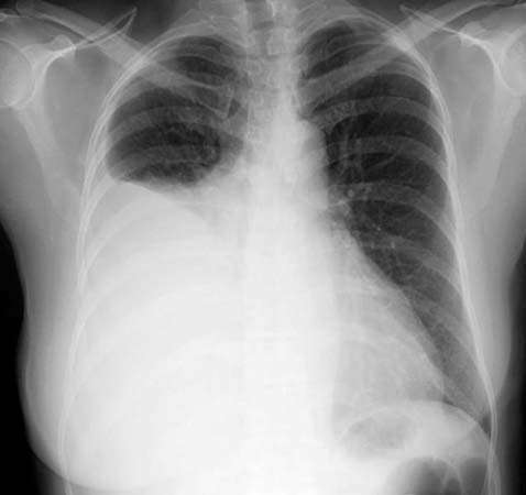
- Massive collection leads to contralateral tracheal shift,
- Lung compromise its full expansion for breathing
- Transudates
- Increased in permeability of the pleural membrane
- Exudate
- Abscess called empyema
- Exudate
- Lung scarring
- Result to permanent decreased in lungs functions
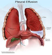
What is a Pleural Effusion?
- not a disease in itself but is a sign of a serious disease
- When this space becomes filled with fluid the lungs can no longer expand properly making it difficult to breathe
- when breathing is impaired this results in detrimental effects to the body such as a lack of oxygen which is essential for the body
- It occurs when the pleural space becomes filled with fluid
- pleural space: which is a small space filled with thin lubricating fluid between the lung and chest wall
- Transudative pleural effusions occurs in non inflammatory conditions usually protein/ cell poor fluid due to pressure in the capillary either from increased hydrostatic pressure or decreased oncotic pressure
- Eg1. of increased hydrostatic pressure : experienced in CHF where the heart is pumping hard and the pressure on the blood vessels causes fluid to be pushed out of the capillaries and into the pleural space. Eg2. of decreased oncotic pressure: When the body loses protein during kidney/ liver failure simply osmosis kicks in where fluid moves from an area of low solute to high, thus the low level of protein in the capillaries caused by the pathological loss draws fluid to move into the pleural space.
- Exudative pleural effusions Occurs during inflammation causing an accumulation of exudate and cells from the capillaries to for they will be more permeable due to the immune response to inflammation
- Eg1. malignancies or infections of the lungs, pulmonary embolisms, perforations of the esophagus.
- Types :1)Empyema 2) Hemothorax 3)Chylothorax
- Empyema : fluid inside the lung is pus - can be caused by lung infections such as pnuemonia, TB, infected wounds Hemothorax: when the pleurl space fills with blood - caused by blunt trauma to chest Chylothorax : results in leakage of lymph fluid into the pleural space due to insufficiency of the thoracic duct which transports lymph fluid - can be caused by tumors that press on the thoracic duct, blunt trauma to duct
- Helpfull video to aide your understanding https://www.youtube.com/watch?v=gASiQ2I_4KY
Bilder und Dateien der Mindmap
Informationen
- Beschreibung
- Mindmap am Pleural Effusion, erstellt von Gabe Igbinosa am 04/03/2018.
- Erstellt von
- Gabe Igbinosa
- Aufrufe
- 28
- Bewertung