410430
Beschreibung
Mindmap von melian.yates, aktualisiert more than 1 year ago
|
|
Erstellt von melian.yates
vor mehr als 10 Jahre
|
|
Pathology of Joints
- Types of Joints
- Fibrous
- Osseous
- Cartilaginous
- Synovial
- Most clinically
significant
- Anatomy
- Composed of two bone ends
bound together by a fibrous
capsule & ligaments
- Inner surface of capsule is
lined by synovial cells & the
joint space contains synovial
fluid
- Composed of two bone ends
bound together by a fibrous
capsule & ligaments
- Functions
- Absorb force of impact
(Cartilage & synovial fluid)
- Allow a variable degree
of movement
- Articular cartilage
- Minimizes friction
- Transmits force to
underlying bone
(Distributes weight)
- Composition:
- Proteoglycan (PG)
- Collagen
- Cells
- Water
- Water
- Cells
- Provides turgor &
elasticity to articular
cartilage
- Traps water
(lubrication, flow of
nutrients - joint flushing)
- Traps water
(lubrication, flow of
nutrients - joint flushing)
- Collagen
- Proteoglycan (PG)
- Minimizes friction
- Absorb force of impact
(Cartilage & synovial fluid)
- Causes of Injury:
- Trauma
- Joint instability
- Lubrication failure
- Infection
- Immune mediated
- Immune mediated
- Infection
- Lubrication failure
- Joint instability
- Trauma
- Response to Injury:
- 1st line of defense:
Articular cartilage
- Limited
response to injury
- Avascular,
reinforced gel
- Little reparative
capacity
- Chrondrocytes can't move very far
- Little remodeling occurs
- Few cells which can actually respond
- Few cells which can actually respond
- Little remodeling occurs
- Chrondrocytes can't move very far
- Only proteoglycan is
continually turned over
- Limited
response to injury
- 1st line of defense:
Articular cartilage
- Most clinically
significant
- Fibrous
- Degenerative Joint Disease (DJD)
- = Osteoarthritis
- Destructive disease leading to
loss of articular cartilage in one
or multiple joints
- Incorporates a variety of
diseases with a common
end stage
- Initiating changes may be caused by:
Trauma, Joint instability, synovial
inflammation, ageing, etc.
- Gross lesions identical:
Cartilage breakdown & loss
- Pathogenesis (Not
entirely clear):
- Primary event:
Proteoglycan loss
- => Loss of lubrication
- => Collagen disruption
- Fibrillation of cartilage, Frays, clefts,
Ulcers, Exposure of Bone, Eburnation, +/-
Osteophytes & joint mice, brown/yellow
discoloration, linear grooves, synovial
hypertrophy & capsular fibrosis
- Fibrillation of cartilage, Frays, clefts,
Ulcers, Exposure of Bone, Eburnation, +/-
Osteophytes & joint mice, brown/yellow
discoloration, linear grooves, synovial
hypertrophy & capsular fibrosis
- => Collagen disruption
- => Decrease in Tugor &
Elasticity of Articular
Cartilage
- => Loss of lubrication
- Failure of chrondrocytes to
maintain balance b/w matrix
break down & repair
- Degradative enzymes play a role:
- Matrix metalloproteinases (MMPs)
- Inhibitors normally control these enzymes
- Inhibitors are decreased in DJD => Activation
- Results in decreased proteoglycan =>
adverse effect on elastic properties of cartilage
- Release of cartilage breakdown products
induces inflammation via local irritation
- Release of cartilage breakdown products
induces inflammation via local irritation
- Results in decreased proteoglycan =>
adverse effect on elastic properties of cartilage
- Inhibitors are decreased in DJD => Activation
- Enzymes include:
- Gelatinases (digest type I &
BM collagens)
- Collagenases (digest collagen)
- Stromelysins (digest
non-collagenous proteins)
- Stromelysins (digest
non-collagenous proteins)
- Collagenases (digest collagen)
- Gelatinases (digest type I &
BM collagens)
- Inhibitors normally control these enzymes
- Produced by
chondrocytes,
synoviocytes &
inflammatory cells
- Matrix metalloproteinases (MMPs)
- Degradative enzymes play a role:
- Primary event:
Proteoglycan loss
- = Osteoarthritis
- Joint Inflammation
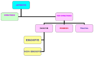
- Arthritis
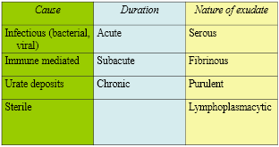
- Classifications:
- Infectious
- Farm animals & Horses
(esp. young)
- Portals of entry include navel & GI
tract => Bacteraemia
- Bacteria reach joints Haematogenously
=> Polyarthritis
- Multiple joints
- Other portals of entry:
- Traumatic inoculation
- Extension from bone or
periarticular soft tissue
- Traumatic inoculation
- Multiple joints
- Bacteria reach joints Haematogenously
=> Polyarthritis
- (Dogs & Cats -
One joint - trauma
related)
- Acute
- Fibrinous
- Fibrinogen in blood
vessels -> leaks -> fibrin
- Oedema, synovial villi thickened
- Early resolution is common, but fibrin
may be converted to fibrous tissue
(enlarged joint w/ restricted movement)
- Early resolution is common, but fibrin
may be converted to fibrous tissue
(enlarged joint w/ restricted movement)
- Oedema, synovial villi thickened
- Fibrinogen in blood
vessels -> leaks -> fibrin
- Suppurative
- Fibrinosuppurative
- Outcomes:
- Infection cleared -> complete resolution
- Infection cleared -> healing by fibrosis
- Continued inflammation:
- Persistent, subclinical
or intermittent
- Re-seeding of joint
- Failure to get rid of
irritant bacterial products
- Failure to get rid of
irritant bacterial products
- Re-seeding of joint
- Persistent, subclinical
or intermittent
- Infection cleared -> complete resolution
- Fibrinous
- Chronic
- Lymphoplasmacytic
- Proliferative
- Proliferative
- Lymphoplasmacytic
- Farm animals & Horses
(esp. young)
- Non-infectious
- Dogs & Cats
- Immune mediated disease
- Trauma
- Foreign material (urate
crystals = gout (birds))
- Foreign material (urate
crystals = gout (birds))
- Often
polyarthritis
- Two forms:
- 1) Erosive
- Immune process is JOINT centered
- Instability & luxation of multiple joints
- Ex. Rheumatoid arthritis:
Small & toy breed Dogs (rare
in Cats)
- Polyarthritis: Grey hounds
- Feline progressive polyarthritis
- Feline progressive polyarthritis
- Polyarthritis: Grey hounds
- Pathogenesis:
- Animal forms Abs against
endogenous (unknown) Ag
- Abs IgG & IgM =
Rheumatoid factor
- Complexes form & are
depositied on articular surfaces
- Pannus formation (granulation tissue)
=> Cartilage erosion
- Pannus formation (granulation tissue)
=> Cartilage erosion
- Complexes form & are
depositied on articular surfaces
- Abs IgG & IgM =
Rheumatoid factor
- Animal forms Abs against
endogenous (unknown) Ag
- Immune process is JOINT centered
- 2) Non-Erosive
- Joint NOT the primary target
- Immune complexes form
elsewhere & settle in the joint
- Ex. Systemic lupus
erythematosus (SLE)
- Dogs
- Anaemia,
thrombocytopaenia,
polymyositis,
glomerulonephritis
- Dogs
- Chronic diseases (such as pyometra,
otitis externa, IBD, endocarditis, UTI,
fungal infections) can lead to immune
complex deposition in joints
- No pannus & no
villous hyperplasia of
lining
- No cartilage destruction
- Idiopathic
- Drug associated
- Joint NOT the primary target
- 1) Erosive
- Trauma
- Results from:
- Persistent antigenic
material in the synovium
- Deposition of Ag/Ab
complexes in the synovium
- Persistent antigenic
material in the synovium
- Dogs & Cats
- Classifications:
- Arthritis
- Developmental Disorders
- Athrogryposis
- Persistent congenital flexure of a joint
in conjunction w/ muscle contraction
- Causes:
- Inactivity or paralysis (in utero)
- Spinal dysraphism
- Intrauterine viral infections
- Toxic plants (poison hemlock)
- Toxic plants (poison hemlock)
- Intrauterine viral infections
- Spinal dysraphism
- Inactivity or paralysis (in utero)
- Persistent congenital flexure of a joint
in conjunction w/ muscle contraction
- Hip dysplasia
- Common in Dogs
(large/giant breeds)
- Inherited disease in which joint
laxity (instability) results in secondary
degenerative joint disease (DJD)
- Joint laxity -> Subluxation -> flattening of
dorsal rim of acetabulum -> modeling of the
acetabulum & femoral head
- Contributing factors:
- Heredity, weight,
over-exercise, low
pelvic mass
- Heredity, weight,
over-exercise, low
pelvic mass
- Gross lesions:
- Articular cartilage (femoral
head & acetabulum)
- Erosion &/or
Ulceration
- Erosion &/or
Ulceration
- Joint capsule/synovium
- Subchondral bone
- Shallow, wide acetabulum,
Eburnation: Osteophyte
formation
- Shallow, wide acetabulum,
Eburnation: Osteophyte
formation
- Capsule stretched & thickened, w/
cartilage & bone formation w/in; round
ligament may be ruptured
- Subchondral bone
- Articular cartilage (femoral
head & acetabulum)
- Common in Dogs
(large/giant breeds)
- Osteochondrosis
- A disorder of growth cartilage
occurring in growing animals
- Most species
- Failure of
Endochondral
Ossification
- Really a defect in cartilage
growth (i.e. chrondrodysplasia)
- Growth cartilage is not
mineralized so is focally or
multifocally retained
- An area of growth cartilage fails to
undergo matrix calcification or
vascular invasion, & therefore does
not become converted to bone
- An area of growth cartilage fails to
undergo matrix calcification or
vascular invasion, & therefore does
not become converted to bone
- Manifestations include thickened articular
cartilage/cartilage flaps (osteochondritis
dissecans or OCD), retained cartilage cores,
bone cysts, angular limb deformity & DJD
- Endochrondral
ossification takes
place at the:
- Physis
- Subarticular epiphyseal
growth cartilage
- Subarticular epiphyseal
growth cartilage
- Physis
- Multifactorial: Trauma, genetic,
rapid growth, ischaemia, nutritional
- Idiopathic
- Giant breeds: humoral head
( 4-8 months old)
- Horses: Distal tibia
- Horses: Distal tibia
- Lesions bilateral in 70% of cases (But
lameness often unilateral)
- A disorder of growth cartilage
occurring in growing animals
- IVDD (Intervertebral Disk Disease)
- Chrondrodystrophic Dogs:
- Predisposed to
degenerative disk
change from early age
- The nucleus pulpsus is
replaced by chondroid tissue
which mineralizes & fragments
- Annulus fibrosus
secondarily
degenerates
- Annulus fibrosus
secondarily
degenerates
- Predisposed to
degenerative disk
change from early age
- Non - Chondrodystrophic Dogs:
- Degeneration begins in the
Annulus fibrosis
- Fibrosis of the nucleus (vs.
chondroid degneration)
- Middle-aged Dogs affected &
thoracolumbar area predisposed
- Middle-aged Dogs affected &
thoracolumbar area predisposed
- Fibrosis of the nucleus (vs.
chondroid degneration)
- Degeneration begins in the
Annulus fibrosis
- Disk can herniate (if Annulus is intact - more common
Non-chondrodystrophics) or Rupture through the Annulus
(More likely in Chondrodystrophics)
- Extruded material is gritty. haemorrhagic or "cheesy"
- Extruded material is gritty. haemorrhagic or "cheesy"
- Chrondrodystrophic Dogs:
- Athrogryposis
Medienanhänge
Möchten Sie kostenlos Ihre eigenen Mindmaps mit GoConqr erstellen? Mehr erfahren.