
Notizen
Topic 6A: Sensory processing
Inhalt der Notizen (8)
Seite 1
stimulus: any signal from the environment that can impact the sense
sensation: transmission of sensory stimuli signal to sensory organs
perception: integration of sensations into the brain and their interpretation
although perceptions are built from sensations, not all sensation result in perceptions. This is d/t sensory adaption; if a stimuli has been constant over a long period of time, you become decreasingly aware of it. Although you are still sensing the stimuli, you are not perceiving it.
perceptual process:
observation of objects in the world
perception is used to select objects
perception is organized in the brain
perception is interpreted
person responds to interpretations
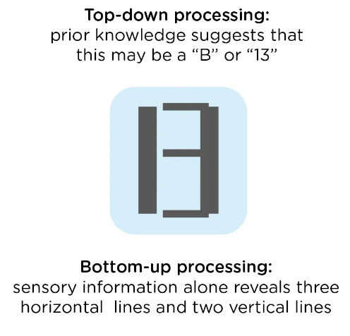
bottom-up processing: sensory receptors pick up signals that the brain processes and interprets
top-down processing: the interpretation of sensations is influenced by our knowledge, experiences, and thoughts
context effect: the bias of environmental factors on a personal perception of the stimulus
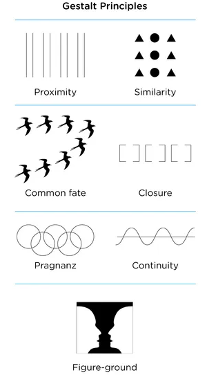
gestalt principles: psychological principles explaining how humans perceive stimuli in organized forms
laws of grouping: brain groups elements together whenever possible instead of keeping them as separate elements
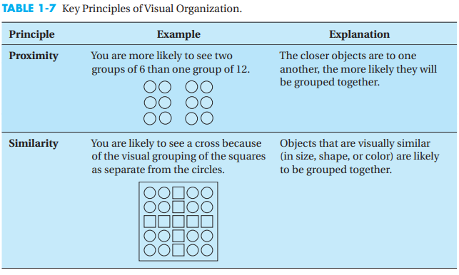
law of proximity: when we perceive a collection of objects, objects that are close to one another will be perceived as a group; this allows for faster processing, instead of focusing on several small stimuli. However this also can lead to misperception.
law of similarity: similar elements will be grouped; allows distinction b/w adjacent and overlapping objects
figure-ground law: a visual field can be separated into components of figures (prominent objects) and ground (objects in the background)
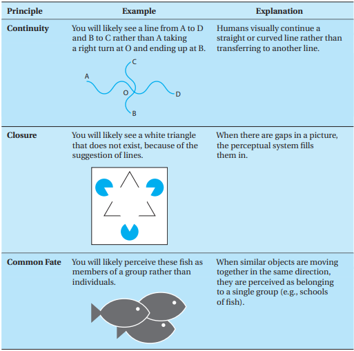
law of closure: our perception will complete incomplete objects
law of continuity: lines are seen as following the smoothest path
JND: just noticeable difference, the amount by which a stimulus must change for human perception 50% of the time
Also subject to sensory adaption (a stimulus is no longer perceived because of prolonged contact of sensory receptors)
Weber’s law: states the JND increases in proportion to the intensity of an initial stimulus.
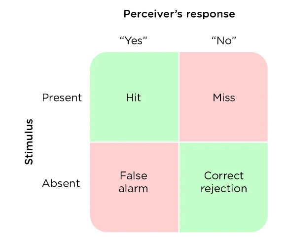
signal detection theory: detection of a stimulus depends on the intensity of stimulus and person’s physical and psychological state.
describes how people make decisions; the brain receives signals from the environment and using internal (person’s history, perceptions, and interpretations) and external (interferes with a person’s ability to interpret the information) noise, it makes decisions.
These decisions, especially simple true or false assumption type decisions, may be correct or incorrect.
It is more likely that an incorrect decision will be made when comparison stimuli are quite similar to one another.
Sensitivity: ability to detect a true signal; useful as screening tests, the results of which can be confirmed with more precise testing later prn (ex: screening mammogram vs diagnostic)
Selectivity: ability to correctly reject a negative signal; useful for confirmatory tests (ex: biopsy)
Mathematically difficult for both qualities to be present in high levels in a single test.
sensory interaction: one sense affects another; ex: turning down the music in the car to see street signs better
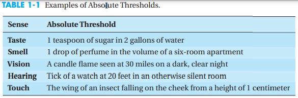
sensory threshold: the weakest sensation or stimulus an organism can detect
absolute threshold: the minimum intensity of a stimulus needed for human senses to percept it 50% of the time; each sense has its own threshold
Seite 2
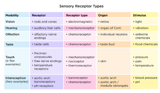
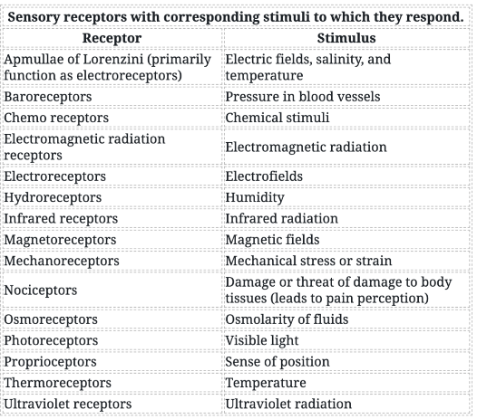
sensory receptors: transform environmental signals into stimuli that are transmitted to CNS via neurons
5 main types of receptors:
Chemoreceptors ---> sense chemicals
Thermoreceptors ---> sense temperature
Mechanoreceptors ---> sense pressure
Photoreceptors ---> sense light
Proprioceptors ---> sense position of limbs/body parts
Force sensitive mechanoreceptors are an example of how the placement of a sensory receptor affects brain processing of sensory inputs
Cutaneous touch receptors in the skin and muscle spindles are both mechanoreceptors, however have different functions.
Cutaneous receptors provide info about external environment, while spindle receptor provide info about internal environment.
Both detect physical forces arising from movement f local tissue
Most sensory information is processed cross-laterally
refractory period: neurons are not able to fire during this time, recovery for the neuron, no sensation will trigger a firing in this time.
Refractory and recovery period grows longer the more a receptor fires, and only a change in the stimulus or a new stimulus will trigger a response from the neuron.
Sensory pathways: stimuli trigger a cascade of information from flow from receptors via nerve cells to the CNS.
Once reaching the brain, all sensory information is routed through the thalamus, except for smell.
Then sent for processing to opposite side of brain as follows --->
Auditory---> temporal
Visual---> occipital
Tactile---> somatosensory cortex (parietal lobe)
Homunculus diagram: visual representation of sensory information processing
Vestibular sensors: monitor bodily response to gravity and motion, maintenance of balance, located in semicircular canals
Kinesthetic sensors: monitor position of body parts in space, Receptors located mostly in joints and muscles, send info to somatosensory cortex in parietal lobe
Seite 3
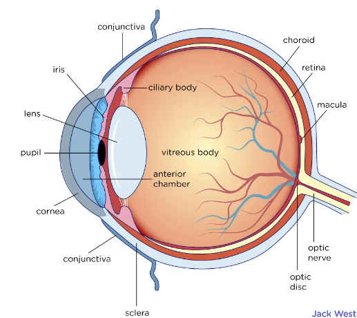
Structure of the eye:
light hits the cornea (front transparent layer of the eye)
lens (transparent convex structure behind the cornea) refracts light onto the retina so an image can be brought into focus
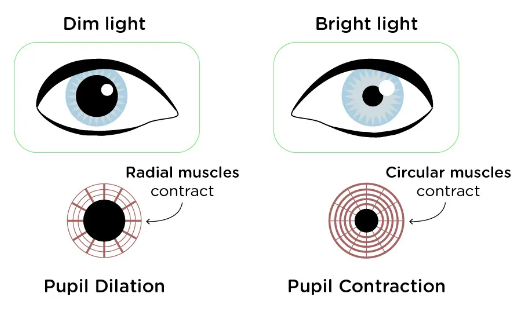
iris (colored part of the eye, circular muscular ring lying between the lens and cornea), regulates amount of light entering the eye by controlling the size of the pupil.
photoreceptive cells of the eye are in the retina, transduction of light to nervous impulses is done here
lens operated by muscles, which control the thickening or flattening of the lens. This changes the focal length of the entering light rays
presbyopia: d/t loss of flexibility of the lens, the image focuses behind the retina, results in farsightedness
hyperopia: farsightedness arising from elongated eyeball so the image nearsightedness, eyeball is elongated so the image falls in front of the retina
fovea: region in the back of the eye, responsible for acute vision, high density of cones
aqueous humor: maintains convex shape of the cornea
vitreous humor: supports the lens and maintains shape of entire eye
Vision:
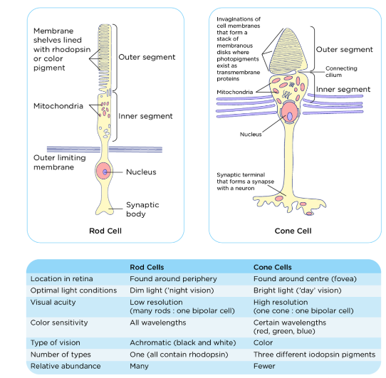
Light receptors: capture photons and signal info to bipolar cells
Rods:
more sensitive, scotopic vision (lower light)
low in acuity.
Not able to sense color differences because they have only 1 photopigment (rhodopsin)
located in outer edges of retina
Cones:
photopic vison (active in high levels of light), low sensitivity
higher acuity
source of color vision
located in center of retina
From the eye, to the brain:
optic nerves are made up of myelinated ganglion cells
some of the axons make up the magnocellular pathway, which carries info about form, movement, depth, and differences in brightness
others make up the parvocellular pathway, which carries info about color and fine details
optic chiasma: the crossing of optical pathways at the base of the brain, allows for coordination of info from both eyes
signals then go to the thalamus, which also has the magnocellular and parvocellular pathways separated into distinct parts of the thalamus.
From thalamus, visual signals enter the primary visual cortex at the back of the brain.
Then they can ether go to the parietal lobe, which carries magnocellular information. Or the temporal lobe which carries parvocellular info in addition to magnocellular.
pathway from retina to superior colliculus (in the midbrain): eye movements are coordinated and integrated with auditory information
pathway from retina to suprachiasmatic nucleus (in the hypothalamus): the SCN controls our circadian cycle and sends information to the pineal gland, which is important in sleep/wake patterns and annual cycles.
bottom-up processing of visual info:
Parallel processing: the use of multiple pathways (i.e. the magnocellular and parvocellular pathways) to process the same stimulus
feature detection: cells in the visual cortex respond to visual stimuli. They provide info about basic features of the image, which are then integrated to produce a perception
processing of light: the light itself is the proximal stimulus that excites photoreceptors of the eyes, while any physical object or event reflecting light is the distal stimulus
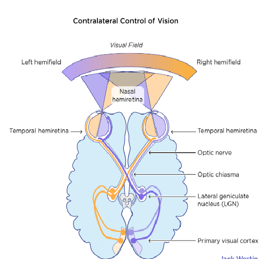
certain parts of the brain are specialized for specific visual tasks
fusiform gyrus: processes information needed to identify faces
prosopagnosia: deficit in identification of human faces, as a result of damage to the gyrus
binocular cues: differences between the view of the 2 eyes allow for things to be determined from the environment
retinal disparity: the 2 retinas register greater differences between their views if an object is close to the eyes.
Depth perception is a result of the brain putting together both pictures
convergence: incorporates physical movement of eyes; eyes will turn towards each other more if an object is closer to the body, resulting in 2 images being seen when you’re cross eyed
monocular cues:
linear perspective: parallel lines in the distance appear closer together
the height of an object in a image correlates with its distance (objects taller in an image are assumed to be farther away)
objects larger than other similar objects are assumed to be closer to the viewer
light and shadow interplay create the image of 3 dimensions in a 2d image
if an object is blocking the view of another object, the blocking object is presumed to be closer
texture becomes less distinct the further an object is
visual cues are believed to be innate or developed early on in life (visual cliff study)
constancy: processing system allows for you to continue to view objects as having a constant shape and size, no matter the distance or angle you view them from
Theories on visual processing:
Opponent process theory: 3 opposing color dichotomies (black-white, red-green, and yellow-blue) provide the basis for all color vision
No single cone can provide info about both opposite colors at the same time
Vision fatigue: after looking at an object with a single color for a certain time, after looking away, you may see an after-image of the opposing color
Trichromatic color theory: cones are divided into 3 types (1 type responds to short-wave (blue) light, 1 type to middle-wave (green), and 1 type to long-wave (yellow) light), with precise shades arising from activity from different cones.
color blindness: genes for mid-and long-wave cones are on X chromosome and they are recessive, resulting in males being more likely to be color blind as they only have one X chromosome. Complete color-blindness is quite rare.
Seite 4
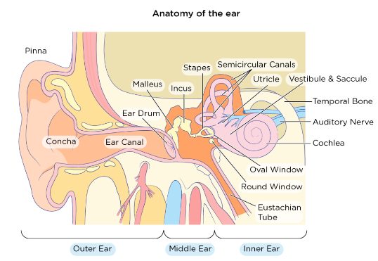
Hearing: the transduction of sound waves into a neural signal.
overview of auditory processing (from the ears to the brain):
sound waves travel through the auditory canal and hit the tympanic membrane.
TM vibrates, resulting in movement of the ossicles
The stapes presses into the oval window (cochlea)
Fluid inside the cochlea moves, which stimulates hair cells (basilar membrane of the cochlea in the inner ear)
As hair cells are activated, they generate neural impulses that travel along cranial nerve VIII to the brain.
in the brain, auditory info goes to the inferior colliculus (midbrain), then to the thalamus, then to the auditory cortex (temporal lobe).
Believed that auditory information also is processed in parallel pathways (like magnocellular and parvocellular pathways for visual info).
Localization of sound is analogous to depth perception; involves monaural and binaural cues (the way monocular and binocular cues are for vision)
Monaural cues provide information on where the sound is coming from (front, behind, above, or below you).
Binaural cues provide information on the location of a sound along a horizontal axis (relies on differences in patterns of vibration of the eardrum b/w both ears)
Interaural level differences: the intensity of sound heard from one side is greater if the sound is coming from that side
Interaural timing differences: there is a small difference in the time at which a sound wave arrives at each ear.
Specific brain areas monitor these cues to glean the origin of the sound
Frequency and pitch are directly related
ear structure:
outer ear:
the auricle (C shaped curve of the outer ear) guides sound waves into the ear canal.
The auditory canal has the tympanic membrane at end of it which vibrates with the movement of air d/t sound waves.
ceruminous glands all along the canal produce cerumen (ear wax) which helps prevent small particles from getting into the TM, also waterproofs the canal and prevents bacterial infection
middle ear: a space containing 3 small bones—the ossicles—and also is connected to the pharynx via the auditory tube.
malleus: aka hammer, attached to TM, transfers vibrations to the--->
incus: aka anvil, transfers vibrations to --->
stapes: aka stirrup, attached to inner ear at the oval window, transfers vibrations to inner ear
The auditory (Eustachian) tube:
functions in balance, by equalizing air pressure across the TM.
This is why chewing gum or drinking fluids while a plane takes off, helps with pressure related sxs, as the auditory tube opens with swallowing or yawning, and this will help equalize internal pressure to rising external pressure.
Eustachian tube also provides drainage pathway for fluids that accumulate with otitis media. The tubes themselves can be a cause for otitis media as the microbes can use this pathway to move from the pharynx to the middle ear
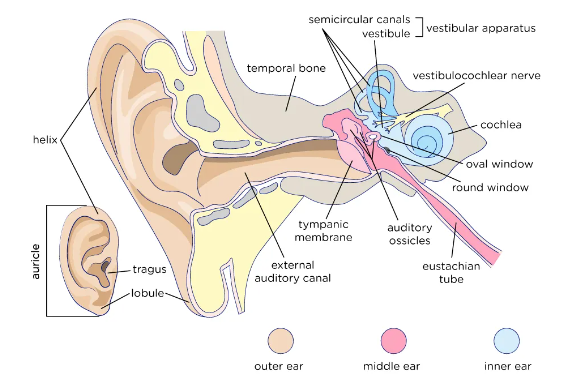
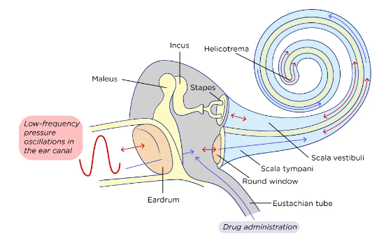
inner ear: entirely enclosed within the temporal bone. Made of 3 separate regions --->
cochlea: involved in hearing, connects middle and inner ear at the cochlea).
Membranous area at the entrance of cochlea).
Allows vibrations transmitted from ossicles into the cochlea
made up of 3 chambers separated from one another by membranes (scala vestibuli ---> cochlear duct ---> scala tympani)
the chambers are continuous and connected to one another by the helicotrema, which contains a fluid within it. When the oval window is pushed out by the vibrations from the middle ear, the round window at the end of the cochlea is pushed out by the fluid.
spiral organ of corti: organ for hearing, located throughout the cochlea, made of lower basilar membrane against the scala tympani and an upper membrane. The hair cells containing stereocilia which are receptors for sound waves are located b/w the 2 membranes.
the movement of the stereocilia simulate the nerve impulse created by the organ of Corti. The impulse is then sent to the brain via cranial nerve VIII.
when the stereocilia bend, this causes a gated ion channel to open, which depolarizes the membrane, which sends a signal to the nerve
vestibule and semicircular canals: involved in balance and equilibrium
semicircular canals: provide information about rotational movements
otoliths: indicate linear acceleration
vestibulo-ocular reflex: signals from the vestibular system are sent to neural structures that control eye movements and vision
other signals are sent to the muscles that control posture and keep us upright
both proprioception and vestibular system contribute to balance, however vestibular system provides information about where the entire body is in space as opposed to specific body parts.
the vestibulocochlear nerve (cranial near VIII) carries neural signals from the inner ear via separate fiber bundles, so the varying signals can go to appropriate centers in the brain.
the vestibular apparatus within the inner ear provides info about equilibrium --->
static (gravitational) equilibrium: movement of the head with respect to gravity
dynamic (rotational) equilibrium: acceleration of the head in rotation, in horizontal and vertical movements, mostly sensed by semicircular canals
use hair cells with stereocilia that detect the movement of fluid to determine bodily position; the stereocilia bend when otoliths (ear stones) move d/t gravity
the maculae are oriented 90° to one another, in x-y-z axes, to respond to positions in each of the different planes. Specific pattern of hair-cell activity across the maculae helps determine head position and direction of acceleration.
outer ear collects sound waves, sends to inner ear, which interprets as pressure, and the inner ear converts signals to electrical signals that the brain further interprets.
Inner hair cells, which are the primary auditory receptors, are the most important for conveying auditory information to the brain.
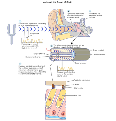
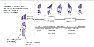
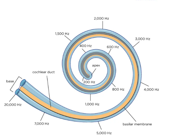
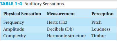
sound waves that reach the ear stimulate the vestibular nerve and the cochlear nerves, which combine into cranial nerve VIII.
Cranial nerve VIII goes into the brain stem, then the midbrain, then the thalamus.
From thalamus, signal is relayed to the primary auditory cortex in the temporal lobe.
Theories about sensory perception by hair cells in the cochlea:
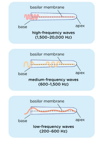
Place theory: pitch is perceived d/t specific vibrations along the basilar membrane of the inner ear. The brain interprets vibration at the specific frequency as a specific pitch.
Problem with this theory is that hair cells vibrating at a specific location are not independent of other nearby hair cells
Frequency (aka temporal) theory: the brain interprets pitch based on the vibration frequency of the entire basilar membrane (as opposed to the vibrations at specific locations like the place theory)—a 2000 Hz vibration would cause the basilar membrane to vibrate 2000x/second, which would be interpreted as a 2000 Hz pitch
Problem with this theory is that neurons struggle to fire at speeds greater than 1000x/second
Volley theory: unification of the above theories. Sounds <1000 Hz follow frequency theory, sounds 1000-5000 Hz follow a combo of theories, and sounds >5000 Hz follow place theory only.
Auditory localization: determination of sound location based on comparison of how sound is perceived by each ear.
Binaural disparity: comparison of the timing and strength of the same sound in each ear.
How the sound strikes each ear depends on the distance b/w ears and the ability to turn the head.
Seite 5
Somatization: skin is the largest bodily organ, and has 6 types of sensory receptors at least (touch, temperature, posture, limb position, etc.)
uses both sensory receptor neurons in the periphery and deeper neurons in the CNS
mechanoreceptors:
these are pressure receptors: have a receptive field like most nerve cells, which depending on the body part, is of a different shape and size.
The field has an excitatory center circled by an inhibitory part.
Once the stimulus activates the nerve fibers, the nerves travel up the spinal cord ascending pathways to the brain stem
the signal crossed sides of the brain via thalamus, landing into the parietal lobe containing the somatosensory cortex.
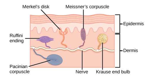
in the skin there are 4 types of tactile mechanoreceptors:
Merkel’s disks:
in the upper layers of skin
dense distribution in the fingertips and lips
slow adapting
respond to light touch
Meissner’s corpuscles:
skin of eyelids and fingertips
respond to fine touch/pressure and low-frequency vibration or flutter
Ruffini endings:
slow adapting
detect skin stretch and deformations w/in joints
Pacinian corpuscle:
deep in the dermis
rapid adapting
sense deep, transient pressure and high-frequency vibration when they are compressed
Proprioceptors:
Relies on 2 main stretch receptors
Golgi tendon organs: located at the insertion of skeletal muscle fibers into tendons
Muscle spindles: detect changes in the length of a muscle
kinesthesia: awareness of the position and movement of body parts using proprioceptors
key in muscle memory and hand-eye coordination
proprioception vs kinesthesia: awareness of movements vs the actual movement, proprioception is more cognitive while kinesthesia is behavioral.
both rely on vestibular system
thermoreceptors:
mechanism not known exactly, however mammals believed to have at least 2 types of sensors—those that detect heat and those that detect cold (temps below body temperature)
nociception: respond to potentially damaging stimuli, which usually causes the sensation of pain, 2 pathways of transmission
fast pathway: uses A-delta fibers (large, myelinated neurons allowing for quacking signal transmission).
This is the sharp fast pain you feel with a cut
slow pathway: C-fiber neurons, nonmyelinated, lag of 1-2 seconds in signal transmission as compared to fast pathway.
Muscle pain is an example
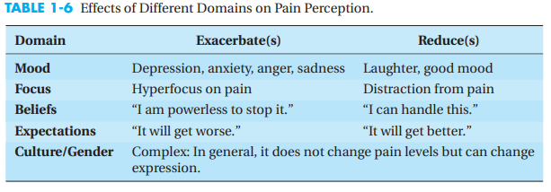
pain is perceived differently by various individuals, even if the stimulus is the same, as a result of emotion and cognition, personality, drug abuse hx, and endorphins.
Seite 6
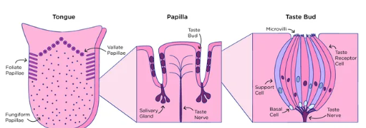
Taste: A chemical sense uses chemoreceptors (which is why smell and taste are closely related as they both use chemoreceptors), varies across individuals
each taste has 1 corresponding receptor; therefore, each receptor is specific to respond to only 1 of the 5 tastes.
salty: salty tastants provide Na+ ions that directly excite the corresponding receptor neuron by entering it
sour: sour tastants are typically acids, which trigger a change in the ion channel they bind to, ultimately increasing the [H+ ion] in the taste neurons which depolarizes them
sweet, bitter, and umami:
these above tastants bind to their receptors, and via GPCR mechanism, they excite the associated neurons
Primary mechanism: taste buds, brief life cycle of 10-60 days.
taste buds are on the tongue, contain clusters of gustatory receptors on papillae (bumps containing 1-8 taste buds, and receptors of pressure and temperature)
Density of buds on the tongue (how many you have on your tongue) is genetically variable.
Supertasters have 10,000 taste buds on their tongue, and experience greater sensitivity of and stronger tastes; nontasters have ~500 taste buds only.
Cranial nerves VII and IX collect data from the tongue
Signal sent up the thalamus to the gustatory area (insular cortex of frontal lobe)
Emotional aspects of food and taste are a result of close relationship of limbic system and gustatory area.
Taste adaptation: acute overstimulation of taste buds, after which the neurons have to recharge before firing again, can occur after a few bites of the same food or after a longer time.
Seite 7
smell: chemical sense uses chemoreceptors (which is why smell and taste are closely related as they both use chemoreceptors), much still unknown about mechanisms.
Main mechanism:
ligands (odor particles) bind to specific receptors on the surface of cilia in the nose
olfactory receptors signal using GPCR mechanism
binding of cAMP to its receptor causes opening of nucleotide-gated ion channel, which allow influx of Na+ and Ca2+ ions into cell, which depolarizes it.
Ca2+ activates Cl- channels, leading to outflux of Cl- ions, which further depolarizes the cell and sends signal to brain
Humans can differentiate b/w ~10,000 different scents as a result of AA variations, the proteins in the cilia are variable
The only sense NOT routed through the thalamus; instead goes into cerebral cortex
Olfactory nerve fibers (cranial nerve I) converge in the olfactory bulb in the brain
Olfactory system has projections into the amygdala, which is part of the limbic system and the hippocampus (emotional and memory centers respectively)
Anosmia: lack of smell, can be caused by injury or disease (COVID for example)
Smell used as a communication tool by animals ---> Pheromones
Seite 8
Cerebral cortex: controls voluntary movement, cognitive functions, and memory storage, higher-order cognitive processing
Frontal lobe: attention, decision making
Temporal lobe: memory, language
Parietal lobe: movement, sensation
Occipital lobe: vision
Perception: processing of sensory stimuli so they can be understood
Bottom-up processing: sensory stimuli from the sense receptors is sent in bits and pieces up to the brain, where they are put together into a complete sensation (like a whole image, or identification of a sound or smell)
Top-down processing: the brain uses a priori knowledge to form a quick impression, which it then breaks apart to confirm whether this impression is correct
Gestalt perception: idea that the whole is greater than the sum of its parts.
Law of Pragnanz: when things can be visually grouped together, they will be
5 principles --->
Cultural influences on visual perception:
People who grew up in round houses were less susceptible to the following illusion than those who grew up in more rectangular, European style homes.
Informationen
- Beschreibung
- Notiz am Topic 6A: Sensory processing, erstellt von 000 000 am 01/08/2022.
- Erstellt von
- 000 000
- Aufrufe
- 0
- Bewertung