
Karteikarten
Histology Cell Structure and Function
Karten in diesem Stapel (33)
- 1What is histology?the study of microscopic structure of cells, tissues, and organs
*It is central to biological and medical science to understand pathological situations - 2What is pathology?the study of ABNORMAL microscopic structures
- 3Units of measurements used in histology 10 angstroms (Å) = 1 nanometer (nm) 10,000 angstroms = 1 micrometer (μm) 1,000 micrometers = 1 millimeter (mm) 10 millimeters = 1 centimeter (cm) 100 centimeters = 1 meter (m)We will mostly use:
1,000 micrometers = 1 millimeter (mm)
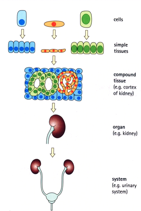
*RBC's = 7.5-8 microns - 4From Cells to Systems cells --> tissues --> organsCells:
are basic structural and functional units
Cells differentiate to perform specialized tasks
Tissues:
are functional arrangements of similarly specialized cells
Compound tissues contain a mixture of cells with different functions
Tissues form organs and systems
An organ is an anatomically distinct group of tissues that perform specific functions (examples: heart, liver, kidney)
The term system may be used to
1. describe cells with a similar function but widely distributed in several anatomical sites (example: diffuse endocrine system)
2. describe a group of organs which have similar or related functional roles (example: digestive system, urinary system) - 5How to Make a Slide with TissueTissue is harvested and fixed, usually in 4% paraformaldehyde
After fixation, tissue is serially transferred through alcohol and finally xylene
prior to embedding in wax (paraffin)
Sectioning of sample via a Microtome (machine cuts incredibly thin pieces of the tissue embedded in wax into ribbons)
Mounting and Staining (for contrast) - 6The four basic tissues1. Epithelium
2. Connective Tissue
3. Muscle Tissue
4. Nerve Tissue - 71. Epithelium

- 82. Connective Tissue

- 93. Muscle Tissue

- 104. Nerve Tissue

- 11Fundamentals of histological stainsMost (but not all) stains are based on ionic interactions:
negatively charged (“acidic”) dyes bind to positively charged tissue components, and vice versa.
Tissue components may be acidophilic or basophilic. - 12Acidophilic“acid-loving binding”
binds acidic (-) charged dyes.
Usually red or pink.
Example: PROTEINS (+ charge)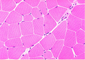
- 13Basophilic“base loving binding”
binds basic (+) charged dyes,
usually BLUE
Example: nucleic acids & DNA, (- charge)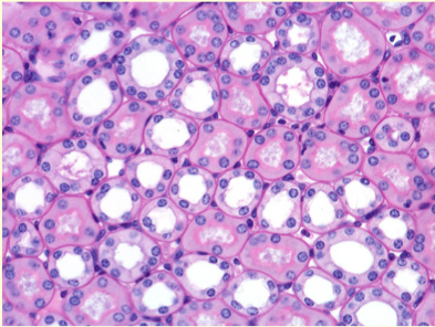
- 14Frequently used histological stains1. Hematoxylin and Eosin (H&E)
2. Toluidine Blue (TBO)
3. Periodic Acid-Schiff (PAS) method - 151. Hematoxylin and Eosin (H&E)The combination of two dyes, hematoxylin (dark blue, basic) and eosin (red, acidic).
Nucleic acids (e.g., in cell nuclei) stain dark blue, proteins and most other tissue components stain pink/red.
*one of the BEST stains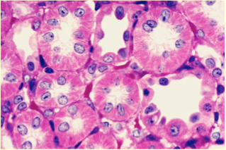
- 162. Toluidine Blue (TBO)a basic BLUE dye that binds to nucleic acids.
It also stains mast cell granules, mucins, and cartilage metachromatically (purple).
(strong dye)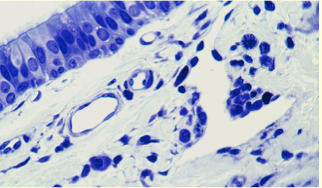
- 173. Periodic Acid-Schiff (PAS) methoddemonstrates complex CARBOHYDRATES either alone (e.g., glycogen)
or combined with proteins (glycoproteins), mucin, and some basement membranes,
which all stain characteristic PURPLE.
Often combined with hematoxylin to label cell nuclei.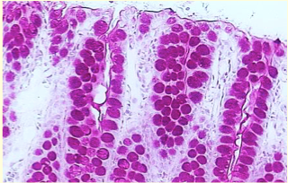
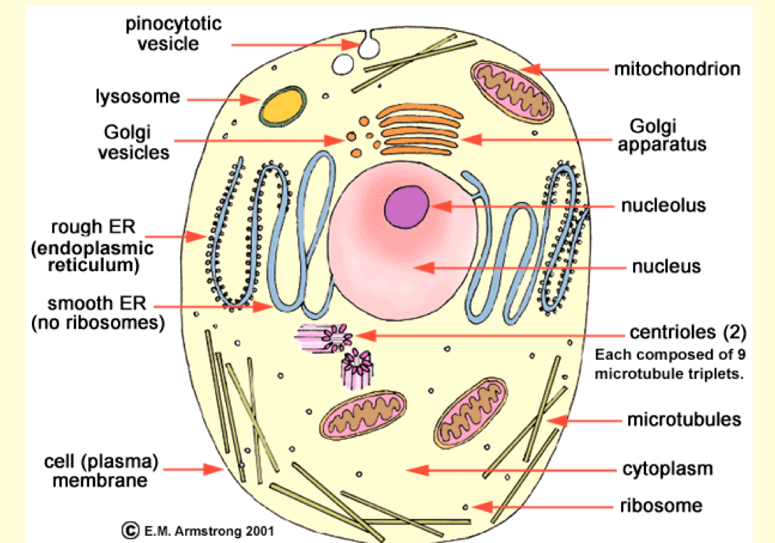
- 18CELL KNOW difference b/w the nucleus of a cell & the cell itself!

- 19The cell membrane (or plasma membrane)Main components:
1. Phospholipids (amphipathic)
2. Proteins (inserted or loosely attached to surface)
(G: glycocalyx)
Main functions:
1. Maintains STRUCTURAL integrity of the cell (MAIN GOAL)
2. Interface between cytoplasm and
external environment
3. Controls movements of substances
in and out of cells
4. Regulates cell-cell interactions
5. Transduces extracellular signals into
intracellular events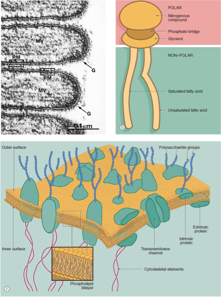
- 20Nucleus (N), nuclear envelope (NE), and rough endoplasmic reticulum (RER) Note: Appearance of nuclei is often characteristic of cell type and functional state **NE and RER tend to surround the nucleus

- 21NEURON *RER: stains darker than the nucleus

- 22The Golgi ApparatusComposed of several flattened,
slightly curved cisternae, which
widen at the periphery to form vesicles.
It has an entry (cis) face and an exit (trans) face.
It serves as a transit station where proteins synthesized in the RER are modified, packaged, and sorted into vesicles according to their destination.
(POST-translational)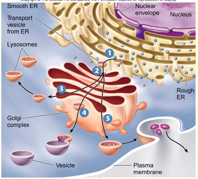
- 23Golgi Apparatus (major functions?)Major functions:
Modifications:
e.g., glycosylation; adding specific “addressing” molecules
Packaging:
enzymes for lysosomes, proteins for secretion
Sorts all materials for lysosomes, secretion, and incorporation into the plasma membrane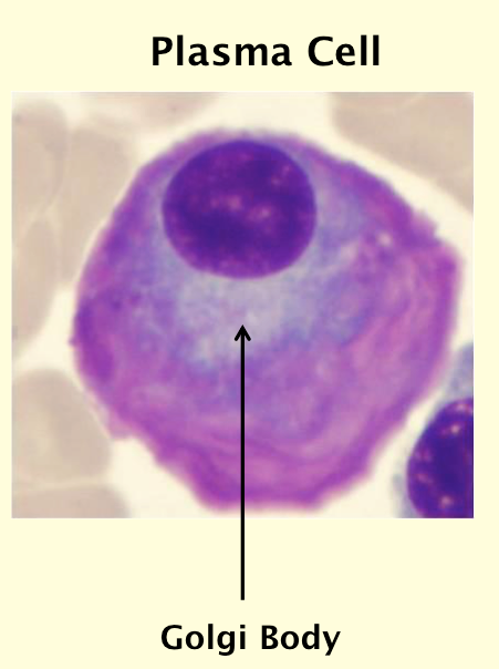
- 24Protein synthesis, targeting, and secretion: Erythroblast (General Pathway: Nucleus --> RER --> Golgi --> Other cells)

- 25Protein synthesis, targeting, and secretion: Eosinophil leukocyte

- 26Protein synthesis, targeting, and secretion: Plasma Cell

- 27Protein synthesis, targeting, and secretion: Pancreatic Acinar Cell

- 28Mitochondria (Greek: mitos=thread, chondrion=small cartilage)Mitochondria produce energy in the form of ATP or, in brown fat, as heat
("power house of the cell")
They consist of an outer membrane, an inner membrane, which forms the cristae, a central space, the matrix, and the intermembranous space
They are visible with routine LM as acidophilic (pink) DOTS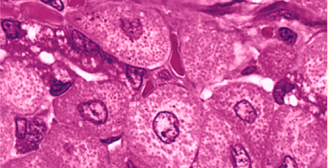
- 29Lysosomes (degrade intracellular things)Membrane-bound vesicles containing “digestive” enzymes
The enzymes are synthesized in the RER and packaged and sorted in the Golgi apparatus
In a few special cases, the enzymes are secreted into the extracellular space
Residual bodies contain undigested remnants, usually lipid-containing pigments such as lipofuscin.
They tend to accumulate in aging cells (old stuff).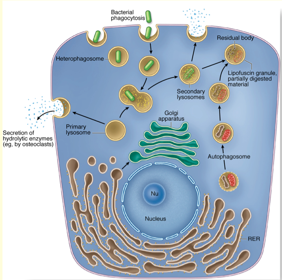

- 30Other Cellular Components: CytoskeletonThin Filaments (actin and myosin)
Intermediate Filaments
Microtubules
- 31Other Cellular Components: GlycogenGlucose storage

- 32Other Cellular Components: LipidsStored in the cytosol or the smooth ER
Cytosolic storage in form of non-membrane
bound LIPID DROPLETS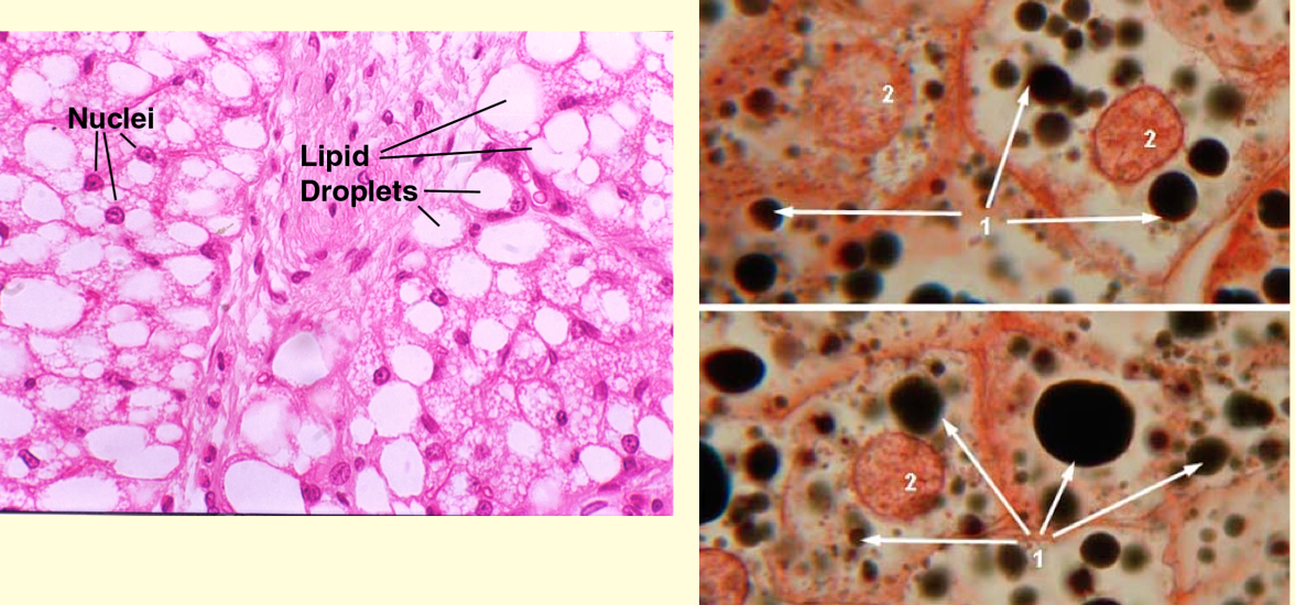
- 33Exam Question The Lysosome is: A. An organelle for storage of glycogen B. Non-organelle that stores intracellular lipids C. Digestive membrane bound vesicle of cells containing enzymes D. Responsible for protein synthesis in cellsC. Digestive membrane bound vesicle of cells containing enzymes
Informationen
- Beschreibung
- Histology (Alb) Lecture 1 (for Exam 1)
- Erstellt von
- Marissa Alvarez
- Aufrufe
- 47
- Bewertung