
Fichas
Visual Neglect
Fichas del conjunto (49)
- 1Unilateral visual neglect is a special case of ---- Most often associated with ---- , but not always.visuospatial agnosia.
right parietal lesions - 2what activities does visual neglect effect?Drawing
Reporting
Reading
Writing
Eating
Dressing
Grooming - 3what tasks are used in studies of neglect?Crossing out tasks
Bisection tasks
Copying tasks
Reading tasks - 4Define hyperkinesia?patient has difficulty initiating limb movements into the neglected field
- 5Define anosagnosia?Patient neglects one whole side of the body and may fail to dress one side.
- 6What did Marshall and Halligan (1988) find?showed neglect patients two houses, one with fire down the neglect side of the house.
Patients did not see any fire but claimed that they would rather live in the house that wasnt on fire - 7one characteristic of neglect is that Detection of objects is a function of ------their importance
- 8define extinction/repetition?Little problem when one object presented on the left, but when one object on left and one on right, left object is ignored, especially if both objects are the same (repetition).
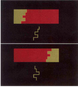
- 9OUtline experiment 1 of Driver et al (1992)Patient CC with Hemispatial neglect
Aim: Determine whether deficit is in spatial or object-attention
Method: Match dividing contours to probe line
Results: Significantly worse when the green figure was on the right hand side
Discussion: Supports object-attention - 10Outline experiment 2 by Driver et al (1992)Aim: Testing where deficit occurs in attentional process
Method: Reported colour of figure
Results: Showed same effect as controls - that symmetrical shapes are seen as the figure
Did not report any symmetry seen unlike controls
Discussion: Preattentive stage likely intact - 11Outline experiment 3 of Driver et al (1992)Aim: Can CC explicitly judge symmetry?
Method: judge shape symmetry
Results: Vertical at chance and horizontal good
Discussion: Neglected information may still be represented implicitly. - 12Give evidence that Hemispatial neglect isn’t purely based on retinal coordinatesKooistra and Heilman (1989) who found that changes in body and head position as well as fixation can affect whether an object is seen
Studies show cells in monkey PPC code both retinal and extra-retinal information - explain why lesions don’t result in retinotopic problem - 13----- SEGMENTATION CAN BE PRESERVED IN NEGLECTFIGURE-GROUND
- 14Whether stimuli seen depends on?VISUAL FIELD POSITION and SEGMENTATION
- 15Object based neglect can occur, what is this?Affects the contralateral side of the object rather than spatial area
- 16What was found by Mattingley et al (1997)when stimuli in both hemisphere seen as one object, unaffected by neglect
- 17Outline Driver and Mattingley (1998)Compared the effects of semantic priming in Blindsight and Neglect
Greater effect of priming in Neglect patients
Can identify colour and shape in neglected field/objects
Ventral stream is mostly intact
Visual neglect is NOT purely retinotopic
Some aspects of neglected stimuli are IMPLICITLY processed (colour, semantics, shape)
Parietal lobe is involved in visual experience and motor planning (ventral and dorsal stream)
Neglect is an ATTENTIONAL deficit - 18Early selection is based on low-level features such as?the sensory and physical attributes of the stimulus.
- 19Late selection is based on?high-level features such as stimulus identity and meaning.
- 20There is mixed evidence of whether early or late selection occurs in visual neglect, outline the predictions of both theories?Early selection theory predicts stimuli presented contralesionally are not semantic elaborated whereas late selection theory predicts that they are.
- 21What visual neglect patient did Ladavas et al. (1993) look at?B.P. 63 year old
large damaged area in the fronto-temporo- parietal region of the right cerebral hemisphere.
Severe left visual neglect
Control group: 2 participant matched on age and education. - 22Outline the method of Ladavas et al (1993)A prime was presented to the neglect visual field followed by a word or non-word and participant had to indicate if it was a word
Results: B.P. was faster to response to the target when it was preceded by a related word.
Information in the neglect field can still be processed semantically or lexically.
B.P. was asked to read a word or non-word presented to the neglected visual field. He was not able to read it
B.P. was asked to decide whether the stimuli presented to the neglected visual field was words or non-words. Performed at chance level
B.P. was asked to decide whether stimuli presented in the neglected visual field was living or non-living.
Performed at chance level
B.P. had to detected the present or absent of the stimuli in the neglected visual field. Performed at chance level. - 23outline the discussion of Ladavas et al (1993)Semantic information processing demonstrated - show both subjective and objective lack of awareness - Visual information presented in the neglected space is processed sufficiently to activate semantic representation.
Supports the late selection theory - 24Outline the three main theories regarding visual neglect?Stimulus-bound theories- the syndrome is the result of an interruption, attenuation, or lack of synthesis in the flow of information conveyed to one hemisphere
The one-sided breakdown of mechanisms subserving the 'orienting response' to incoming stimulations. These theories evolved contemporaneously with a second approach, which took into account the concept of 'body schema'
Psychodynamic line of reasoning suggesting motivational explanations for some phenomena of the syndrome - 25The three main groups differ in the level of -----functioning which is disordered and causes the behavioural manifestations
- 26Outline the method of Bisiach et al (1979)19 right brain damaged patients - unilateral neglect. All showed unilateral neglect via a circle task
30 Control Subjects
Subjects were required to detect differences occurring within pairs of patterns successively presented at intervals. These were random, white cloud-like shapes lying on a blue background. Central sector was common to all patterns, left and right sectors were varied.
Experiment 1: Static
Participants required to detect differences occurring within pairs of stimuli
Experiment 2: Dynamic
Pattern moved from left →right or right → left
Screen was blacked out with the exception of a central vertical slit which allowed partial view of the stimulus
Participants required to detect differences - 27describe the results of Bisiach et al (1979)Static Patterns
Patients were significantly worse at detecting differences on the left side of the pattern
Right side performance similar to controls
Dynamic Patterns
Looked at the effect of Side (Left/Right side of pattern), Recency (which side of a pattern was last seen), Direction of Movement (Left/Right)
Only effect of Side was found to be significantly different in patients - 28Outline the discussion of bisiach et al (1979)?Patients were worse at detecting left-side differences in both static and dynamic conditions
Representational Disorder
Dynamic images are also affected as the mental representation of the cloud pattern suffers from unilateral neglect - 29What was the aim of Young et al (1992)?To further explore the phenomenon of neglect by investigating the object, face & word recognition abilities of a patient with a stable left-sided spatial neglect
- 30describe patient B.Q (tested by Young et al 1992)64 year old female
Suffered stroke in parietal region of right hemisphere
Persistent Unilateral Neglect
Left Hemiplegia
Left Hemianopia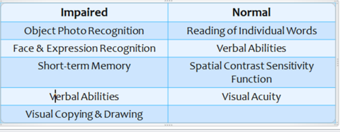
- 31What stimuli were used by Young et al (1992)?Chimaerics
Left/right halves of everyday objects and famous faces, juxtaposed together
Mirror reversal of image halves
Pretest performed to establish recognition - 32OUtline the methods and results of Young et al (1992)?Stimuli presented one at a time in random order
Had to identify each half-object & both halves of the chimaeric objects & famous faces
Further experiments conducted on:
Orientation
Different arrangements of chimaeric faces
Position in visual field
Chimaeric expressions
Insight and awareness
Defining features
Reading
B.Q. showed a marked problem in identifying the left side of
chimaeric objects, but had barely any errors in the right
visual field. - 33what was found when using chimaeric faces with different expressions?BQ shown chimaerics consisting of two half-faces; one smiling, one neutral.
Each was accompanied by a mirror-reversed chimaeric placed either vertically or horizontally.
BQ asked to choose which chimaeric looked happier.
Significant right-side bias, especially in horizontal trials. - 34Outline what Young et al (1992) found about BQ's insight and awarenessBQ showed a lack of insight into the nature of chimaerics shown to her.
She could trace the outline of face and object chimaerics but would identify them using their right-side only.
Suggests that BQ could ‘see’ the left-side of image but wouldn’t take it into account for identification.
Sometimes also ‘made-up’ the left-side of the image e.g. tracing the rim of a bowl across the boundary. - 35Outline BQ's performance on tasks involving defining features?BQ shown line-drawings of objects, each with a defining feature on the left or right which allowed recognition.
BQ made errors in 4/16 drawings with the defining feature on the left, and 2/16 with defining features on the right.
When shown faces with the right-side internal features obscured, BQ made errors in 8/20 cases.
BQ seems to be able to use left-sided information if it is critical to identification. - 36How was BQ found to perform on reading tasks?When reading, BQ often omitted words on the left side of the page.
However, when shown words whose first letter could be substituted or deleted to make different words (e.g CLOVE -> GLOVE/LOVE), BQ only misread 1/57.
BQ also correctly read both words in compound and non-compound word pairs (e.g. EGG CUP vs. TIP CAP).
Performance was worse for non-words and letter strings, suggesting pre-existing representations are important.
Consistent with the idea that BQ uses left-side information when it is crucial for identification. - 37Outline the discussion of Young et al (1992)B.Q. was unable to recognise the left identity of chimaeric faces and objects regardless of visual field position, object type, or orientation.
Not due to her left visual field defect or defective control of eye movements
However found that features on the left side do influence interpretations if they contribute to essential information.
Reason for findings may be that the information from the dominant right visual field is being excited so strongly that left visual field information is being further inhibited.
Higher order information that is still intact can therefore replace this left field information, sometimes causing confabulation. - 38what is visual extinction related to?lesions to the right inferior parietal cortex
- 39What did Rees et al (2000) investigate?Do extinguished stimuli produce striate or extrastriate cortical activation in the damaged hemisphere?
- 40Describe the patient of Rees et al (2000)Patient G.K. – damage to the right inferior parietal lobe
- 41What did Rees et al (2000) find in the extinction study?fMRI data showed right hemisphere activation to the extinguished left stimulus in both striate and early extrastriate areas.
- 42What was the conclusion of Rees et al (2000)Why isn’t striate/extrastriate activation enough to cause awareness?
fMRI has poor temporal resolution. Neuronal Synchrony?
(EEG suggests a difference between extinguished and bilateral trials)
Maybe visual cortex and parietal/frontal cortex interact to produce awareness - 43Describe cancellation tests?patient has to detect a target amidst distractors and to place a mark through the
target objects (e.g., stars), presented on a sheet of paper aligned
with the patient’s own body midline
Danckert and Ferber (2006) - 44What issue did Mort et al (2003) intend to address?The brain regions that are critically associated with
visual neglect have become intensely disputed. In particular,
one study of middle cerebral artery (MCA)
stroke patients has claimed that the key brain region
associated with neglect is the mid portion of the
superior temporal gyrus (STG), on the lateral surface
of the right hemisphere, rather than the posterior parietal
lobe. Such a result has wide-ranging implications for
both our understanding of the normal function these
cortical areas and the potential mechanisms underlying
neglect. - 45Outline the method of Mort et al (2003)Here, we use novel high resolution MRI protocols
to map the lesions of 35 right-hemisphere patients
who had suffered either MCA or posterior cerebral
artery (PCA) territory stroke. - 46What did Mort et al (2003) conclude?For patients with MCA
territory strokes, the critical area involved in all neglect patients was the angular gyrus of the inferior parietal
lobe (IPL). Although the STG was damaged in half of
our MCA neglect patients, it was spared in the rest.
For PCA territory strokes, all patients with neglect had
lesions involving the parahippocampal region, on the
medial surface of the temporal lobe. PCA patients without
neglect did not have damage to this area. We conclude
that damage to two posterior regions, one in the
IPL and the other in the medial temporal lobe, is associated
with neglect. Although some neglect patients do
have damage to the STG, our ®ndings challenge the
recent in¯uential proposal that lesions of this area are
critically associated with neglect. Instead, our results
implicate the angular gyrus and parahippocampal
region in this role. - 47What factors challenge researchers looking at the neural basis of visual neglect?Several factors challenge clinicians and researchers investigating the neural basis of VN. First, patients with VN often manifest extensive brain damage, which hinders precise anatomical correlation. Second, pure assessment of VN is problematic in the stroke patient who has co-existing cognitive impairment, causing difficulty in recruiting “ideal” VN patients for further investigation. Also, the lack of detailed classification of the different neglect subtypes can lead to over-generalization of the neural basis of VN. For example, the distinction between egocentric VN and allocentric VN, which can occur independently,58 and 109 has not been made in many studies.
Ting et al (2011) - 48What did Ting et al (2011 review) report regarding the amounts of left/right hemispheric stroke patients who get VN?A US study141 of 1,281 stroke patients reported a prevalence rate of VN of 43% and 20% following right and left hemispheric stroke, respectively. This asymmetry in prevalence of VN is consistent with other literature4, 13 and 21 and probably occurs because the right hemisphere tends to allocate attention to both hemispaces, but the left hemisphere accords attention more selectively to right hemispace. Also, VN patients with right hemispheric stroke generally have a worse prognosis than those with left hemispheric stroke.
- 49Outline Ting et al's (2011) thoughts on the dorsal/ventral stream dichotomy as proposed by Milner and Goodalethe dorsal stream, which connects the visual cortex with the superior parietal lobe and the intraparietal sulcus, subconsciously assimilates incoming visual information in order to bring about immediate visually guided movements. The ventral stream, which bridges the visual cortex and the inferior temporal lobe, is accountable for our conscious, object-based visual perception and recognition. Dysfunction of dorsal stream results in optic ataxia (impaired visual guidance of movement), and dysfunction of ventral stream causes visual agnosia (impaired recognition of visual stimuli), respectively. Although this model provides a comprehensive explanation for various components of visuospatial perception and visuomotor function, the manifestation of VN, which which normally follows the damage of inferior parietal lobe, has yet to be fully considered in the context of this dorsal–ventral dichotomy. Also, the double dissociation between VN and optic ataxia or visual agnosia has suggested separate underlying disorders of the neural substrates
Información
- Descripción
- Fichas sobre Visual Neglect, creado por Jo Gledhill el 06/05/2015.
- Creado por
- Jo Gledhill
- Visitas
- 5
- Etiquetas
- Valoración