
7554652


| Pregunta | Respuesta |
| Arteries | * Deliver oxygenated blood to the tissues * Thick-walled. with elastic tissue and smooth muscle * Under high pressure * Blood volume is called stressed volume |
| Arterioles | * Smallest branches of the arteries * Site of highest resistance in the cardiovascular system * Resistance regulated by the ANS * Alpha1 adrenergic receptors found on arterioles of the skin, splanchnic and renal circulations * Beta2 adrenergic receptors found on arterioles of skeletal muscle |
| Capillaries | * Have largest total cross-sectional and surface area * Single layer of endothelial cells surrounded by basal lamina * Thin-walled * Site of exchange of nutrients, water, and gases |
| Venules | * Formed from merged capillaries |
| Veins | * The largest vein is the vena cava which returns blood to the heart * Thin-walled * Low pressure * Contain highest proportion of blood in the cardiovascular system * Blood volume contained is called unstressed volume * Have Alpha1 adrenergic receptors |
| Velocity | v = Q/A Q = blood flow A = cross-sectional area * directly proportional to blood flow * inversely proportional to the cross-sectional area |
| Velocity is _________ in the aorta than in the sum of all the capillaries. | Blood velocity is higher in the aorta (small-cross-sectional area) than in the sum of all the capillaries (large cross-sectional area). The lower velocity in the capillaries optimizes conditions for exchange of substances across the capillary wall. |
| Cardiac Output = | Q = ΔP/R CO = (MAP - Right arterial pressure)/Total peripheral resistance * Blood flow is inversely proportional to the resistance of the blood vessels |
| Resistance = | R = (8ηl)/(πr^4) η = viscosity of blood l = length of vessel * Resistance is directly proportional to viscosity and the length of the vessel * It is inversely proportional to the 4th power of the vessel radius. Ex: If vessel decreased by a factor of 2 then resistance increases by a factor of 16 and blood flow decreases by a factor of 16. |
| Parallel Resistance | * Systemic circulation: each organ is supplied by an artery that branches off the aorta. R(total) = 1/R1 + 1/R2 + 1/R3 + 1/R4, etc. * Total resistance is less than the resistance of any of the individual arteries. * The pressure in each parallel artery is the same. |
| Series Resistance | * Illustrated by the vessels within an organ. Each organ is supplied by a large artery, smaller arteries, arterioles, capillaries, and veins. R(total) = R(artery) + R(arterioles) + R(capillaries) * The largest portion of resistance is from the arterioles. * As blood flows through the series of blood vessels, pressure decreases. |
| Reynold's Number | * Predicts whether blood flow will be laminar or turbulent. * The higher the number, the greater the chance for turbulent flow. * Increased by a decrease in blood viscosity * Increased by an increase in blood velocity |
| Capacitance (Compliance) | * Describes the dispensability of blood vessels * C = V/P (volume/pressure) * Is much greater for veins than arteries * A decrease in venous capacitance decreases venous volume and increases arterial volume * Capacitance of the arteries decreases with age |
| The largest decrease in pressure... | Occurs across the arterioles because they are the site of highest resistance. |
| Mean pressures in the: Aorta Arterioles Capillaries Vena cava | Aorta: 100 Arterioles: 50 Capillaries: 20 Vena cava: 4 |
| Systolic Pressure | * Highest arterial pressure during a cardiac cycle * Measured after the heart contracts (systole) and blood is ejected into the arterial system |
| Diastolic Pressure | * Lowest arterial pressure during a cardiac cycle * Measured when the heart is relaxed (diastole) and blood is returned to the heart via the veins |
| Pulse Pressure | * Difference between the systolic and diastolic pressures |
| The most important determinant of pulse pressure is | Stroke volume. As blood is ejected from the left ventricle into the arterial system, arterial pressure increases because of the relatively low capacitance of the arteries. |
| Decreases in capacitance, cause ________ in pulse pressure. | Increases |
| Mean Arterial Pressure (MAP) | Average arterial pressure with respect to time. ~ diastolic pressure + 1/3 pulse pressure |
| P wave | * Represents atrial depolarization |
| PR interval | * The interval from the beginning of the P wave to the beginning of the Q wave * Varies with conduction velocity through the AV node. If conduction decreases, the PR interval increases |
| An increase in the conduction velocity by the ___________ nervous system, ___________ the PR interval. | An increase in the conduction velocity by the sympathetic nervous system, decreases the PR interval. |
| A decrease in the conduction velocity by the ___________ nervous system, ___________ the PR interval. | A decrease in the conduction velocity by the parasympathetic nervous system increases the PR interval. |
| QRS complex | Represents depolarization of the ventricles. |
| QT interval | * The interval from the beginning of the Q wave to the end of the T wave * Represents the entire period of depolarization and repolarization of the ventricles. |
| ST segment | * From the end of the S wave to the beginning of the T wave * Represents the period when the ventricles are depolarized * Is isoelectric |
| T wave | Represents ventricular repolarization |
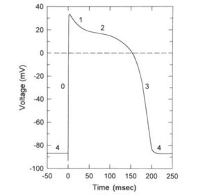
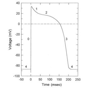
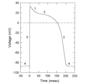
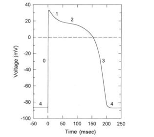
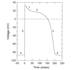
| Phase O: | * Caused by the transient increase in the Na+ conductance which results in an inward Na+ current that depolarizes the membrane. * At the peak of the action potential, the membrane potential approaches the Na+ equilibrium potential. |
| Phase 1 | * Brief period of initial repolarization * Caused by an outward current, in part by the movement of K+ ions out of the cell and a decrease in Na+ conductance. |
| Phase 2 | * Plateau of the action potential * Caused by a transient increase in Ca++ conductance which results in an inward Ca++ current and by an increase in K+ conductance. * Outward and inward currents are approximately equal to membrane potential is stable. |
| Phase 3 | * Repolarization * Ca++ conductance decreases, K+ conductance increases * High K+ conductance results in large outward K+ current which hyperpolarizes the membrane back toward the K+ equilibrium potential. |
| Phase 4 | * The resting membrane potential * Period during which the inward and outward currents are equal and the membrane potential approaches the K+ equilibrium potential. |
| Sinoatrial (SA) node | * The pacemaker of the heart * Has an unstable resting potential |
| AV node | * The upstroke of the action potential in the AV node is the result of an inward CA++ current. |
| Conduction Velocity | * Reflects the time required for excitation to spread throughout cardiac tissue * The larger the inward current during the upstroke, the higher the velocity |
| Conduction velocity is fastest in the | Purkinje system |
| Conduction velocity is slowest in the | AV node (PR interval) allowing time for ventricular filling. If conduction velocity through the AV node is increased, ventricular filling may be compromised. |
| Excitability | The ability of cardiac cells to initiate action potentials in response to inward, depolarizing current |
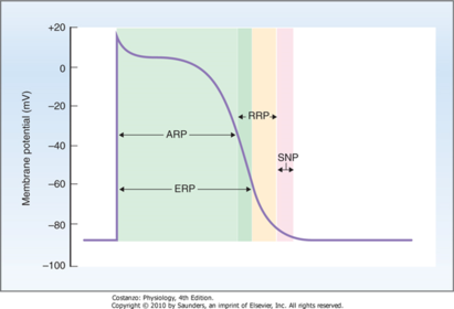
| Absolute refractory period | Reflects the time during which no action potential can be initiated regardless of how much inward current is supplied |
| Effective refractory period | Reflects the time during which a conducted action potential cannot be elicited. |
| Relative refractory period | Period during which an action potential can be elicited, but more than the usual inward current is required. |
| Chronotropic Effects | * Produce changes in heart rate * A negative effect decreases heart rate by decreasing the firing rate of the SA node * A positive effect increases heart rate by increasing the firing rate of the AV node |
| Dromotropic Effects | * Produce changes in conduction velocity primarily through the AV node * A negative effect decreases conduction velocity --> slowing the conduction of action potentials from the atria to the ventricles and increasing the PR interval. |
| The SA node, atria, and AV node have __________ _________ innervation, but the ventricles do not. | The SA node, atria, and AV node have parasympathetic vagal innervation, but the ventricles do not. |
| The parasympathetic neurotransmitter, ACh | * Acts on muscarinic receptors * Has a negative chronotropic effect: decreases HR * Has a negative dromotropic effect: decreases conduction velocity through the AV node and increases the PR interval. |
| The sympathetic nervous system neurotransmitter is __________ and acts on ___________ receptors. | The sympathetic nervous system neurotransmitter is norepinephrine and acts on Beta-1 receptors. |
| Norepinephrine has ____________ chronotropic and ___________ dromotropic effects on the heart. | * Positive chronotropic effect: Increase HR * Positive dromotropic effect: Increase conduction velocity, decrease PR interval |
| Which organs have ONLY sympathetic innervation? | Sweat glands, vascular smooth muscle, pilomotor muscles of skin, liver, adipose tissue, and kidneys (adrenal medulla) |
| Where are α1 adrenergic receptors located and what is their mechanism of action? | *Vascular smooth muscle (Skin, renal, splachnic) *GI tract sphincter muscles *Badder sphincter *Radial muscle (iris) *MOA: IP3 increases intracellular Ca++ |
| Where are the α2 adrenergic receptors and what is their mechanism of action? | *GI wall muscles *Presynaptic adrenergic neuron *MOA: Inhibit adenylyl cyclase, decreasing cAMP |
| Where are the β1 adrenergic receptors located and what is their mechanism of action? | *Heart *Salivary glands *Adipose tissue *Kidney *MOA: Stimulates adenylyl cyclase, increase cAMP |
| Where are the β2 adrenergic receptors located and what is their mechanism of action? | *Vascular smooth muscle *GI tract wall muscle *Bladder wall muscle *Bronchioles *MOA: Stimulates adenylyl cyclase, increases cAMP |
| Where are the nicotinic (colinergic) receptors located and what is their mechanism of action? | *Post-ganglionic neurons of the SNS/PNS *Adrenal medulla *Skeletal muscle *MOA: Opening Na+ and K+ channels leading to depolarization |
| Where are the muscarinic (cholinergic) receptors located and what is their mechanism of action? | *Effector organs of PNS *Sweat glands of SNS *MOA: IP3 increases intracellular Ca++ Inhibits adeylyl cyclase, decreasing cAMP Direct K+ channel activation |
¿Quieres crear tus propias Fichas gratiscon GoConqr? Más información.