
Fichas
Sin título
Fichas del conjunto (45)
- 1LIMBS ILOSDescribe limb compartments and their contents
Describe muscle groups, their function and innervation
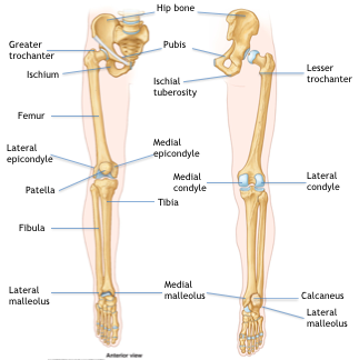
Illustrate knowledge of the main bony features of the limbs
Understand and give examples of joint types
Start to consider what can go wrong with the limbs - 2Limb regionsGluteal region
Anterior thigh
Posterior thigh
Anterior knee
Posterior knee
Anterior leg
Posterior leg
Anterior ankle
Posterior ankle
Foot - 3Flexiondecreases angle of a joint
- 4Extensionincreases angle of a joint
- 5AbductionMove region away from midline
- 6AdductionMove region back to midline
- 7Condylerounded articular area (e.g. femoral condyles)
- 8Epicondylebony region either superior or next to a condyle (epi = upon, adjacent to)
- 9Facetflat, smooth formation where articulation occurs (e.g. facets of thoracic vert. for articulation with rib tubercle)
- 10fossadepression or concave found in bone (e.g. iliac fossa)
- 11malleolusrounded process (e.g. malleoli of the ankle)
- 12trochanterlarge blunt elevation (e.g. greater & lesser trochanters of femur)
- 13tuberclesmall raised eminence (e.g. greater and lesser tubercles of humerus)
- 14tuberositylarge round elevation (e.g. ischial tuberosity of pelvis)
- 15Joint typesFibrous
Cartilaginous ( 1 and 2 )
Synovial - 16Fibrous2 bones united by fibrous tissue
e.g. skull sutures - 171º cartilaginousunited by hyaline cartilage
e.g. costochondrial - 182º cartilagelike 1º but disc of fibrocartilage between
e.g. all midline symphyses - intervertebral, pubis - 19synovial atypicalarticular surface covered with fibrocartilage
e.g. temporomandibular, sternoclavicular, 2-7 sternochondral - 20synovial typicalarticular covered with hyaline cartilage
e.g. all synovial not atypical - condyloid between lower arm and wrist - 21typical synovial joints characteristicsTypical synovial (including all limb joints) are characterised by 6 features:
The bone ends involved are covered by hyaline cartilage
Surrounded by a joint capsule
Encloses a joint cavity
Capsule reinforced externally/internally by ligaments
Lined by synovial fluid
Joints are capable of varying degrees of movement - 22Lower limbExtensions from the trunk required to support body mass, allow for propulsion and maintenance of balance.
Two hip bones articulate with one another at the pubic symphysis.
Each firmly attached to the sacro-iliac joint
Rigid structure allows transmission of body weight AND propulsive force. - 23Thighabove knee
- 24Thigh compartmentsanterior or extensor (of the knee)
medial or adductor (of the hip)
posterior or flexor (of the knee). - 25gluteal regionThe gluteal region or buttock lies behind the pelvis and hip, above the posterior compartment.
- 26Leg compartmentsanterior or extensor (of the ankle and foot)
lateral or fibular
posterior or flexor (of the ankle and foot), the calf - 27Footdorsum - upper surface
sole - plantar surface - 28Lower limb - bone
- 29Gluteal regionP to pelvis
Iliac crest to gluteal fold
Important region owing to important vessels and nerves which emerge - 30Muscles of gluteal regionGluteus maximus
Gluteus medius
Gluteus minimus
Piriformis
Tensor fascia lata
Iiliotibial tract
S gemellus
Obturator interns
Quadratus femoris - 31Gluteus maximuship extensor
- 32Gluteus mediusabductor
- 33Piriformislateral rotator
- 34Lateral rotationIf the tendon of the muscle passes posteriorly to the joint, then the muscle confers lateral rotation
- 35Medial rotationIf the tendon of the muscle passes anteriorly to the joint, then the muscle confers medial rotation
- 36Neurovascular structuresinternal pudendal a and nerve
I gluteal a and nerve
S gluteal a and nerve
Sciatic nerve - 37Thigh
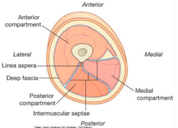
- 38Thigh prosection
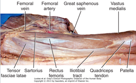
- 39Anterior compartment on thighQuadriceps femoris - group of 4 muscles - knee extensors
Satorius - hip flexion, lateral rotation
Innervated by femoral nerve (L2-L4) - 40Quadriceps femorisRectus femoris
Vastus medialis
Vastus intermedius
Vastus lateralis
Quadriceps insert (via quadriceps tendon)
into the patellar ligament
ALL muscles of this compartment
Innervated by femoral nerve (L2-L4) - 41Femoral triangleS - inguinal ligament
M - adductor longus
L - satorius
Floor - pectinous, iliopsoas
Structures L - M : Femoral NAV and lymph nodes - 42Medial (adductor) compartment of thighGracilis
Adductor brevis
Adductor longus
Adductor magnus
obturator nerve (L2-L4), except hamstring part of adductor magnus. - 43Posterior compartment of thighSemitendinous
Semimembranosus
Biecps femoris (long head)
- Hamstrings - 44Common features of 3/4 muscles in P compartmentOG - ischial tuberosity
Insert to bones of leg
Span and act on 2 joints (hip extension and knee flexion)
Innervation = tibial division of sciatic nerve
Short head of BF does not meet any of these criteria (except insertion point) - 45Innervation of thighL4 - S3
Sciatic nerve
Información
- Descripción
- Fichas sobre Sin título, creado por anna jones el 22/05/2018.
- Creado por
- anna jones
- Visitas
- 1
- Valoración