
Diagrama de flujo
Burn Managment
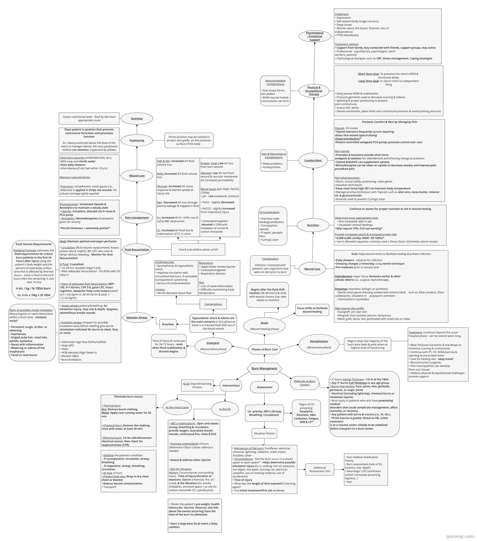
Flowchart: burn management
Contenido del diagrama
Burn Managment
Phases of Burn Care
Assessment
Referrals to Burn Centers
• 2° burns Partial Thickness: >10 % of the TBSA
• Any 3° burns Full Thickness in any age group
• Burns that involve: Face, joints, feet, genitalia, perineum, or major joints
• Electrical (including lightning), chemical burns or inhalation injuries
• Burn injury in patients who also have preexisting medical
disorders that could complicate management, affect
mortality, or recovery.
• Any patient with burns & trauma (i.e., fx, etc.).
*If the trauma is greater threat to life, initial treatment
is at a trauma center initially to be stabilized before transport to a burn center.
Interventions
1st priority: ABC’s (Airway, Breathing, Circulation)!
Situation History
• Mechanism of the burn: fire/flame, electrical, chemical, lightning, radiation, scald, steam, frostbite, other
• Circumstances: "Did the burn occur in a closed space vs open space?" - Helps determine possible inhalation injury (Ex.’s; cooking, hot oil, explosion, hot object, hot water, burning car, electrical, campfire, use of smoking material, use of accelerants)
• Time of injury
• What was the length of time exposed to burning agent?
• Any initial treatment/first aid rendered.
• Past medical /medication history
• Burn assessment (rule of 9’s, location, size, depth)
• Neurologic: LOC (confusion-carbon monoxide poisoning, hypoxia,..)
• Pain
Addtional Assessment Info
Goal: Stop the burning Process
At the Initial Scene
In the ED
Eliminate burn source
• Thermal burn
Dry: Remove burnt clothing,
Moist: Apply cool running water for 20 min
• Chemical burn: Remove the clothing, rinse with water at least 20 min
• Electrical burn: 1st be safe/disconnect electrical source, then check for responsiveness (CPR)
• Stabilize the patient’s condition:
- If unresponsive: circulation, airway, breathing
- If responsive: airway, breathing, circulation
• ID type of burn
• Prevent heat loss: Wrap in dry clean sheet or blanket
• Reduce wound contamination
• Transport!
• ABC’s (stabilization): Open and assess airway, breathing & circulation, provide oxygen, auscultate breath sounds, continuous Pox, vitals & ECG
• Estimate extent/depth of burn: Determine if Burn Center referral is needed
• Assess & address other injuries
• Get the Situation History: Circumstances surrounding injury - Time of injury/duration of exposure, Source (chemical, fire, oil, scald), & the Situation (Ex; smoke inhalation, enclosed space = at risk for carbon monoxide CO, cyanide pois)
• Obtain the patient’s pre-weight, health history (Ex: Vaccine: Tetanus), and info about the events occurring from the time of the burn to admission
• Start 2 large bore IVs & insert a foley catheter
*Signs of CO poisoning: Headache, Dizziness, N&V, Confusion, Fatigue, SOB & CP*
Emergent
(Resuscitative phase)
Acute
(Wound healing phase)
Rehabilitative
(Restorative phase)
Time of injury & continues for 24-72 hours - ends when fluid mobilization & diuresis begins.
Hypovolemic shock & edema are the main concerns in this phase as there is a massive fluid shift out of the blood vessels
Priorities
• Assess airway patency/breathing: for inhalation injury, resp rate & depth, dyspnea, adventitious breath sounds
• Establish airway: Prepare for possible intubation early before swelling gets worse (intubation indicated for burns to chest, face, or neck)
• Administer high flow O2/humidified
• Keep NPO
• Vitals
• HOB elevated (high fowler’s)
• Monitor ABGs
• Bronchodilators
Maintain Airway
Signs of possible smoke inhalation - (May progress to rapid obstruction within a short time - Intubate early!)
• Persistent cough, stridor, or wheezing
• Hoarseness
• Singed scalp hair, nasal hair, eyelids, eyelashes
• Nares with inflammation
• Blistering or edema of the oropharynx
• Facial or neck burns
Fluid Resuscitation
Goal: Maintain optimal end-organ perfusion
• Circulation (fluid volume replacement): Assess pulses-rate & rhythm, BP, CRT, skin color & temp; obvious bleeding - Monitor for Over Resuscitation!
IV fluid: Crystalloid
• 1st 24 hrs: lactated ringer’s (LR)
• After adequate resuscitation - D5.45NS with 20 mEq K+
• Signs of adequate fluid resuscitation: SBP> 100, P<110/min, CVP 3-8, good CRT, intact cognition, adaquete foley urine output (adult 0.5-1 mL/kg/hr or 30-50 mL/hr & peds 1-1.5 mL/kg/hr)
Fluid Volume Requirements
Parkland Formula: estimates the fluid requirements for critical burn patients in the first 24 hours after injury using the patient's body weight and the percent of total body surface area that is affected by thermal burns - (Give ½ fluid in the1st 8 hours then the remaining ½ over next 16 hrs)
4 mL / kg / % TBSA burn
Ex: 4 mL x 70kg x 20 TBSA
Pain management
Pharmacologic: Scheduled Opioids & Anxiolytics to maintain a steady state
• Opioids: morphine, dilaudid via IV route & PCA pump
• Anxiolytics: Benzodiazepines (lorazepam) given for anxiety
*Partial thickness = extremely painful*
Wound care
Clean burn wounds (embedded bits, etc.):
•Mild soap and sterile water
•Start daily showers
•Give tetanus (if not had within 10 yrs)
Remove ruptured blister
•Dressings: nonadherent, mesh gauze (i.e., Telfa) that is applied in strips not around, f/b tubular bandage lightly applied
Positioning
Place patient in position that prevents contracture formation and promotes function
Ex: Head positioned above the level of the heart to manage edema, the neck positioned midline (no rotation), supported by pillows, etc.
Nutrition
Assess nutritional need - feed by the most appropriate route
Check Labs before admin of IVF
Hgb & Hct: Increased d/t fluid volume loss
BUN: Increased d/t fluid volume loss
Glucose: Increased d/t stress response & altered uptake at injury site
Na+: Decreased d/t loss through plasma leakage & trapped in fluid
K+: Increased d/t K+ shifts out of cell & RBC destruction
Cl-: Increased d/t fluid loss & reabsorption of Cl- in urine
Protein, total: Low d/t loss from burn wound
Albumin: Low d/t lost from wound & vascular membrane d/t increased permeability
Blood Gases (pH, Pa02, PaCO2, COHb):
• pH - Low (metabolic acidosis)
• PaO2 - slightly decreased
• PaCO2 - slightly increased from respiratory injury
• Carboxyhemoglobin - elevated (>10%) with inhalation of smoke & carbon monoxide
Cardiovascular:
• Dysrhythmias & hypovolemic shock
• Impaired circulation with circumferential burn, if untreated (compartment syndrome)
• Venous thromboembolism
Urinary
• AKI d/t decrease blood flow
Respiratory
• Upper/lower airway injuries
• Carboxyhemoglobin
• Respiratory distress
Skin
• Loss of water/electrolytes
• Difficulty maintaining body temperature
Complications
Begins after the fluid shift resolves (36-48 hours) & ends with wound closure (can take weeks to months).
Focus shifts to facilitate wound healing
Wound Care
Goal: keep wound moist to facilitate healing & prevent infection
•Daily showers - observe for infection
•Dressing changes 2 times/day using sterile technique
•Pre-medicate prior to wound care!
Debridement-major focus: Removes eschar & other
cellular debris (i.e., surgical, hydrotherapy).
Dressings (standard, biologic or synthetic):
•Sterile mesh gauze dressing soaked with antimicrobial such as Silver product, Silver sulfadiazine, Silvadene or polysporin ointment
• PermeaDerm (synthetic)
May require skin grafts:
•Autograft: pt’s own skin
•Allograft: from another person; temporary
•Mesh graft: donor skin perforated with small slits or holes
Continue to assess for proper nutrition to aid in wound healing
Determine most appropriate route
• Non-intubated: able to eat
• Intubated: enteral feedings
*May require TPN, if GI not working*
Provide increased calorie & increased protein diet
*4,000-6,000 cal/day (RMR-↑50-100%)*
• Harris–Benedict equation x Activity Level x Stress factor (Estimates calorie needs)
Nutrition
Comfort/Rest
Promote Comfort & Rest by Managing Pain
Opioids (IV route):
*Opioid tolerance frequently occurs requiring
doses that exceed typical dosing
(hypermetabolism)*
•Patient-controlled analgesia PCA pumps promote control over care
Non-opioid:
•Precedex & ketamine provide short-term
analgesia & sedation for debridement and dressing change procedures
•Tylenol &NSAIDS can supplement opiates.
•Benzodiazepine can be taken w/ opiods to decrease anxiety and improve post procedure pain
Non-pharmacologic:
•Music, virtual reality, positioning, video game,
relaxation techniques
•Keep room temp high (85°) to maintain body temperature
•Manage pruritus (itchiness): with Topicals such as aloe vera, cocoa butter, mineral oil, & glucocorticoids
•Antacids used to prevent Curling’s ulcer
Physical & Occupational
Therapy
:
Short Term Goal: To preserve the client’s ROM &
functional ability
Long Term Goal: to return client to independent
living
• Early passive ROM & mobilization
• Pressure garments used to decrease scarring & edema
• Splinting & proper positioning to prevent
joint contractures
• Assess ADL ability
• Elevate extremities, place limbs anti-contracture positions & avoid prolong pressure.
Prone position may be utilized to protect skin grafts on the posterior surface of the body.
Psychological
/ Emotional
Support
Challenges:
• Depression
• Self esteem/body image concerns
• Sleep issues
• Worries about the future, finances, loss of
independence
• PTSD (flashbacks)
Treatment options:
• Support from family, stay contacted with friends, support groups, stay active
• Professional - psychiatrists, psychologist, social
workers, pastoral
• Psychological therapies such as CBT, Stress management, Coping strategies
Complication
Infection: Colonized with patient’s own organisms that were on skin prior to burn
GI Complication:
• Diarrhea: tube feedings/antibiotics
• Constipation: opioids
• If septic: paralytic ileus
• Curling’s ulcer
Pain & Neurological Complications
• Sleep problems
• Anxiety/stress
Musculoskeletal Complications
• Scar tissue forms, less pliable
• ROM may be limited. Contractures can form
Begins when the majority of the burn area heals & ends when at highest level of functioning
Treatment: continues beyond the acute hospital phase - can be several years long.
• Wear Pressure Garments & Ace Wraps to minimize scarring & contractures
• Continue with PT, OT, ROM,exercise & splinting at prescribed times
• Care for healing skin - keep moist!
• Reconstructive surgeries
• Pain (neuropathies can develop
from scar tissue)
• Address physical & psychosocial challenges - provide support
Información
- Descripción
- NCLEX H&I5 Diagrama sobre Burn Managment, creado por MacKenzie Allen el 29/10/2022.
- Creado por
- MacKenzie Allen
- Visitas
- 2
- Valoración
Más sobre Burn Assessment and Management