
Mapa mental
Spinal Cord & Tracts 2018
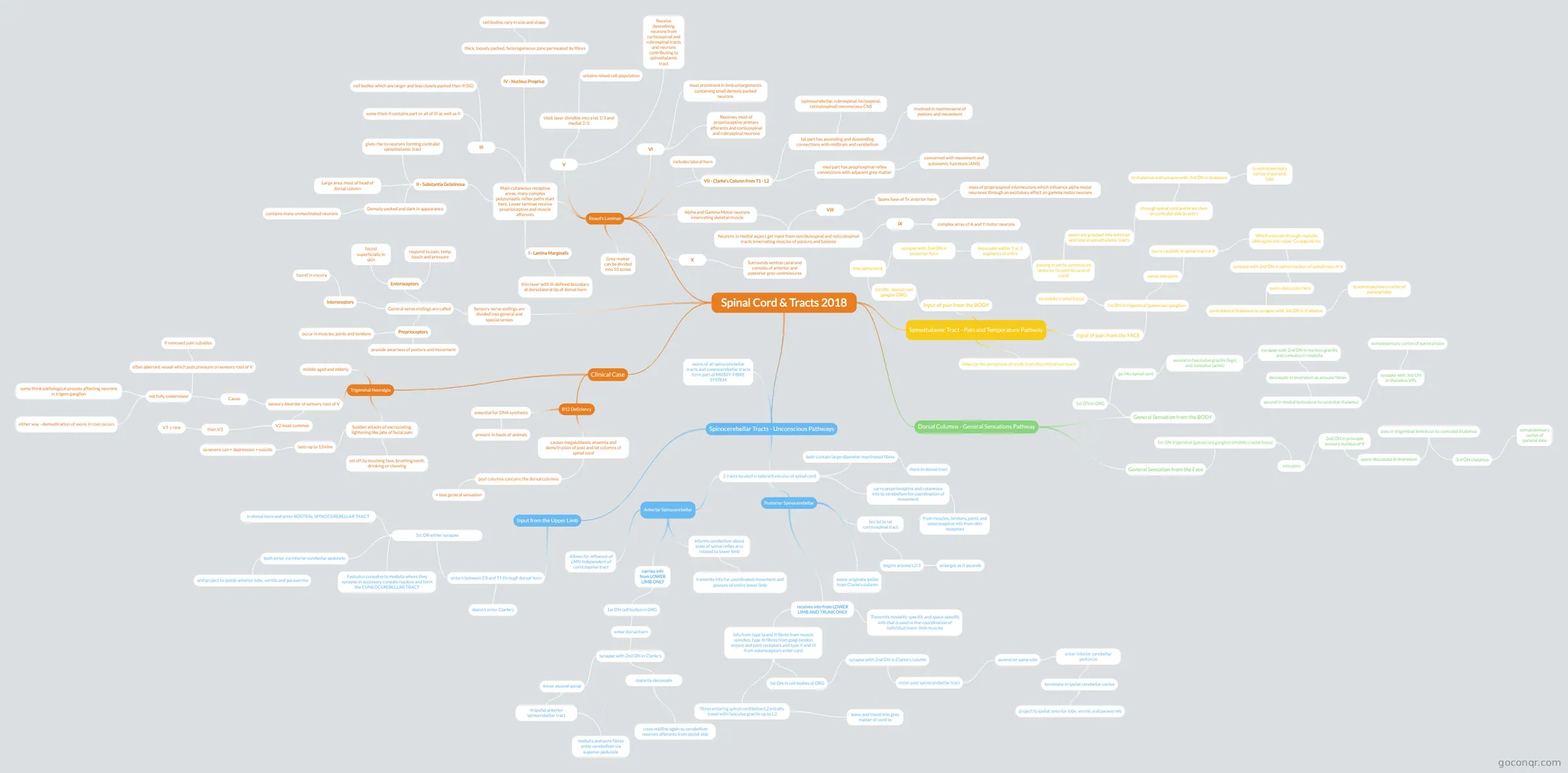
Mind map: Spinal cord & tracts
Contenido del mapa
Spinal Cord & Tracts 2018
Sensory nerve endings are divided into general and special senses
- General sense endings are called
- Enteroceptors
- found superficially in skin
- respond to pain, temp, touch and pressure
- Interoceptors
- found in viscera
- Proprioceptors
- occur in muscles, joints and tendons
- provide awarness of posture and movement
- Enteroceptors
- General sense endings are called
Rexed's Laminae
- Grey matter can be divided into 10 zones
- Main cutaneous receptive areas, many complex polysynaptic reflex paths start here. Lower laminae receive proprioceptive and muscle afferents
- I - Lamina Marginalis
- thin layer with ill-defined boundary at dorsolateral tip of dorsal horn
- II - Substantia Gelatinosa
- Large area, most of head of dorsal column
- Densely packed and dark in appearance
- contains many unmyelinated neurons
- gives rise to neurons forming contralat spinothelamic tract
- III
- cell bodies which are larger and less closely packed then II (SG)
- some think II contains part or all of III as well as II
- IV - Nucleus Proprius
- thick, loosely packed, heterogeneous zone permeated by fibres
- cell bodies vary in size and shape
- thick, loosely packed, heterogeneous zone permeated by fibres
- I - Lamina Marginalis
- V
- Receive descedning neurons from corticospinal and rubrospinal tracts and neurons contributing to spinothalamic tract
- thick layer divisible into a lat 1/3 and medial 2/3
- ontains mixed cell population
- VI
- Receives most of proprioceptive primary afferents and corticospinal and rubrospinal neurons
- most prominent in limb enlargements containing small densely packed neurons
- VII - Clarke's Column from T1 - L2
- includes lateral horn
- lat part has ascending and descending connections with midbrain and cerebellum
- (spinocerebellar, rubrospinal, tectospinal, reticulospinal) unconscious CNS
- involved in maintenacne of posture and movement
- med part has propriospinal reflex connections with adjacent grey matter
- concerned with movement and autonomic functions (ANS)
- Alpha and Gamma Motor neurons innervating skeletal muscle
- Neurons in medial aspect get input from vestibulospnal and reticulospinal tracts innervating muscles of posture and balance
- VIII
- Spans base of Tx anterior horn
- mass of propriospinal interneurons which influence alpha motor neurones through an excitatory effect on gamma motor neurons
- Spans base of Tx anterior horn
- IX
- complex array of A and Y motor neurons
- VIII
- Neurons in medial aspect get input from vestibulospnal and reticulospinal tracts innervating muscles of posture and balance
- X
- Surrounds ventral canal and consists of anterior and posterior grey commissures
Spinothalamic Tract - Pain and Temperature Pathway
- Input of pain from the BODY
- 1st ON - dorsal root ganglia (DRG)
- into spinal cord
- synapse with 2nd ON in posterior horn
- decussate within 1 or 2 segments of entry
- passing in white commissure (anterior to central canal of cord)
- axons are grouped into anteirior and lateral spinothalamic tracts
- through spinal cord and brain stem on contralat side to entry
- to thalamus and synapse with 3rd ON in thalamus
- to somatosensory cortex of parietal lobe
- to thalamus and synapse with 3rd ON in thalamus
- through spinal cord and brain stem on contralat side to entry
- axons are grouped into anteirior and lateral spinothalamic tracts
- passing in white commissure (anterior to central canal of cord)
- decussate within 1 or 2 segments of entry
- synapse with 2nd ON in posterior horn
- into spinal cord
- 1st ON - dorsal root ganglia (DRG)
- Input of pain from the FACE
- 1st ON in trigeminal (gasserian) ganglion
- (in middle cranial fossa)
- axons into pons
- turns caudally in spinal tract of V
- Which extends through medulla oblongata into upper Cx segements
- synpase with 2nd ON in spinal nucleus of spinal tract of V
- axons decussate here
- contralateral thalamus to synapse with 3rd ON in thalamus
- to somatosensory cortex of parietal lobe
- contralateral thalamus to synapse with 3rd ON in thalamus
- axons decussate here
- synpase with 2nd ON in spinal nucleus of spinal tract of V
- Which extends through medulla oblongata into upper Cx segements
- turns caudally in spinal tract of V
- 1st ON in trigeminal (gasserian) ganglion
- (Also carries sensations of crude (non-discriminative) touch
- Input of pain from the BODY
Clinical Case
- Trigeminal Neuralgia
- sensory disorder of sensory root of V
- V2 most common
- then V3
- V1 = rare
- then V3
- Cause
- not fully understood
- often aberrant vessel which puts pressure on sensory root of V
- if removed pain subsides
- some think pathological process affecting neurons in trigem ganglion
- either way - demyelination of axons in root occurs
- often aberrant vessel which puts pressure on sensory root of V
- not fully understood
- V2 most common
- middle aged and elderly
- Sudden attacks of excruciating, lightening like jabs of facial pain
- lasts up to 15mins
- so severe can = depression + suicide
- set off by touching face, brushing teeth, drinking or chewing
- lasts up to 15mins
- sensory disorder of sensory root of V
- B12 Deficiency
- essential for DNA synthesis
- present in foods of animals
- causes megaloblastic anaemia and demylination of post and lat columns of spinal cord
- post columns contains the dorsal columns
- = lose general sensation
- post columns contains the dorsal columns
- essential for DNA synthesis
- Trigeminal Neuralgia
Dorsal Columns - General Sensations Pathway
- General Sensation from the BODY
- 1st ON in DRG
- go into spinal cord
- ascend in fasciculus gracilis (legs) and cuneatus (arms)
- synapse with 2nd ON in nucleus gracilis and cuneatus in medulla
- decussate in brainstem as arcuate fibres
- ascend in medial leminsicus to contralat thalamus
- synapse with 3rd ON in thalamus VPL
- somatosensory cortex of parietal lobe
- synapse with 3rd ON in thalamus VPL
- ascend in medial leminsicus to contralat thalamus
- decussate in brainstem as arcuate fibres
- synapse with 2nd ON in nucleus gracilis and cuneatus in medulla
- ascend in fasciculus gracilis (legs) and cuneatus (arms)
- go into spinal cord
- 1st ON in DRG
- General Sensation from the Face
- 1st ON trigeminal (gasserian) ganglion (middle cranial fossa)
- into pons
- 2nd ON in principle sensory nucleus of V
- axons decussate in brainstem
- pass in trigeminal lemniscus to contralat thalamus
- 3rd ON thalamus
- somatosensory cortex of parietal lobe
- 3rd ON thalamus
- pass in trigeminal lemniscus to contralat thalamus
- axons decussate in brainstem
- 2nd ON in principle sensory nucleus of V
- into pons
- 1st ON trigeminal (gasserian) ganglion (middle cranial fossa)
- General Sensation from the BODY
Spinocerebellar Tracts - Unconscious Pathways
- 2 tracts located in lateral funiculus of spinal cord
- carry proprioceptive and cutaneous info to cerebellum for coordination of movement
- from muscles, tendons, joints and exteroceptive info from skin receptors
- both contain large-diameter myelinated fibres
- more in dorsal tract
- Posterior Spinocerebellar
- lies lat to lat corticospinal tract
- begins around L2/3
- enlarges as it ascends
- begins around L2/3
- axons originate ipsilat from Clarke's column
- receives info from LOWER LIMB AND TRUNK ONLY
- Transmits modality-specific and space-specific info that is used in fine coordination of individual lower limb muscles
- info from type Ia and II fibres from muscle spindles, type Ib fibres from golgi tendon organs and joint receptors and type II and III from extereceptors enter cord
- 1st ON in cell bodies of DRG
- synapse with 2nd ON in Clarke's column
- enter post spinocerebellar tract
- ascend on same side
- enter inferior cerebellar peduncle
- terminate in ipsilat cerebellar cortex
- project to ipsilat anterior lobe, vermis and paravermis
- terminate in ipsilat cerebellar cortex
- enter inferior cerebellar peduncle
- ascend on same side
- enter post spinocerebellar tract
- synapse with 2nd ON in Clarke's column
- fibres entering spinal cord below L2 initially travel with fasiculus gracilis up to L2
- leave and travel into grey matter of cord to
- 1st ON in cell bodies of DRG
- lies lat to lat corticospinal tract
- Anterior Spinocerebellar
- informs cerebellum about state of spinal reflex arcs related to lower limb
- transmits info for coordinated movement and posture of entire lower limb
- carries info from LOWER LIMB ONLY
- 1st ON cell bodies in DRG
- enter dorsal horn
- synapse with 2nd ON in Clarke's
- majority decussate
- cross midline again so cerebellum receives afferents from ipsilat side
- minor ascend ipsilat
- in ipsilat anterior spinocrebellar tract
- medulla and pons fibres enter cerebellum via superior peduncle
- in ipsilat anterior spinocrebellar tract
- majority decussate
- synapse with 2nd ON in Clarke's
- enter dorsal horn
- 1st ON cell bodies in DRG
- Allows for influence of LMN independent of corticospinal tract
- informs cerebellum about state of spinal reflex arcs related to lower limb
- carry proprioceptive and cutaneous info to cerebellum for coordination of movement
- Input from the Upper Limb
- enters between C8 and T1 through dorsal horn
- doesn't enter Clarke's
- 1st ON either synapse
- in dorsal horn and enter ROSTRAL SPINOCEREBELLAR TRACT
- Fasiculus cuneatus to medulla where they synapse in accessory cuneate nucleus and form the CUNEOCEREBELLAR TRACT
- both enter via inferior cerebellar peduncle
- and project to ipsilat anterior lobe, vermis and paravermis
- enters between C8 and T1 through dorsal horn
- axons of all spinocerebellar tracts and cuneocerebellar tracts form part of MOSSY-FIBRE SYSTEM
- 2 tracts located in lateral funiculus of spinal cord
Información
- Descripción
- Mapa Mental sobre Spinal Cord & Tracts 2018, creado por Harriet Colquhoun el 20/04/2018.
- Creado por
- Harriet Colquhoun
- Visitas
- 0
- Valoración
Más sobre Neural Pathways and Spinal Tracts