
Mapa mental
Cardiac Action Potential
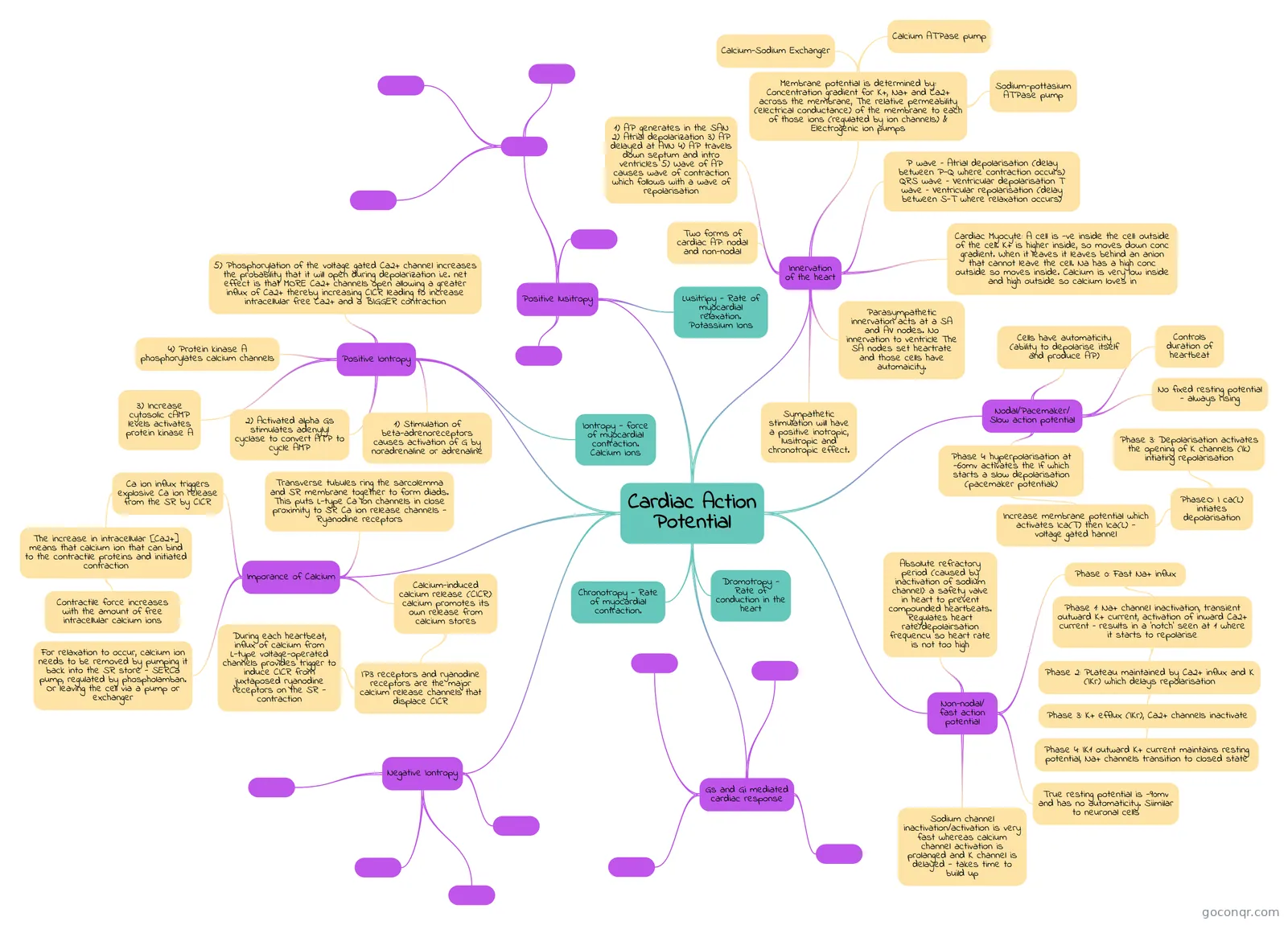
Mind map: Cardiac action potential
Contenido del mapa
Cardiac Action Potential
Positive Iontropy
- Iontropy - force of myocardial contraction. Calcium ions
- 5) Phosphorylation of the voltage gated Ca2+ channel increases the probability that it will open during depolarization i.e. net effect is that MORE Ca2+ channels open allowing a greater influx of Ca2+ thereby increasing CICR leading to increase intracellular free Ca2+ and a BIGGER contraction
- 1) Stimulation of beta-adrenoreceptors causes activation of G by noradrenaline or adrenaline
- 2) Activated alpha Gs stimulates adenylyl cyclase to convert ATP to cycle AMP
- 3) Increase cytosolic cAMP levels activates protein kinase A
- 4) Protein kinase A phosphorylates calcium channels
Imporance of Calcium
- For relaxation to occur, calcium ion needs to be removed by pumping it back into the SR store - SERCa pump, regulated by phospholamban. Or leaving the cell via a pump or exchanger
- Ca ion influx triggers explosive Ca ion release from the SR by CICR
- The increase in intracellular [Ca2+] means that calcium ion that can bind to the contractile proteins and initiated contraction
- Contractile force increases with the amount of free intracellular calcium ions
- The increase in intracellular [Ca2+] means that calcium ion that can bind to the contractile proteins and initiated contraction
- Transverse tubules ring the sarcolemma and SR membrane together to form diads. This puts L-type Ca ion channels in close proximity to SR Ca ion release channels - Ryanodine receptors
- Calcium-induced calcium release (CICR): calcium promotes its own release from calcium stores
- IP3 receptors and ryanodine receptors are the major calcium release channels that displace CICR
- During each heartbeat, influx of calcium from L-type voltage-operated channels provides trigger to induce CICR from juxtaposed ryanodine receptors on the SR - contraction
- IP3 receptors and ryanodine receptors are the major calcium release channels that displace CICR
Negative Iontropy
Gs and Gi mediated cardiac response
Non-nodal/ fast action potential
- Absolute refractory period (caused by inactivation of sodium channel): a safety valve in heart to prevent compounded heartbeats. Regulates heart rate/depolairsation frequencu so heart rate is not too high
- Phase 0: Fast Na+ influx
- Phase 1: Na+ channel inactivation, transient outward K+ current, activation of inward Ca2+ current – results in a ‘notch’ seen at 1 where it starts to repolarise
- Phase 2: PLateau maintained by Ca2+ influx and K (IKr) which delays repolarisation
- Phase 3: K+ efflux (IKr), Ca2+ channels inactivate
- Phase 4: IK1 outward K+ current maintains resting potential, Na+ channels transition to closed state
- Phase 3: K+ efflux (IKr), Ca2+ channels inactivate
- Phase 2: PLateau maintained by Ca2+ influx and K (IKr) which delays repolarisation
- Phase 1: Na+ channel inactivation, transient outward K+ current, activation of inward Ca2+ current – results in a ‘notch’ seen at 1 where it starts to repolarise
- True resting potential is -90mv and has no automaticity. Siimilar to neuronal cells
- Sodium channel inactivation/activation is very fast whereas calcium channel activation is prolanged and K channel is delayed - takes time to build up
Nodal/Pacemaker/ Slow action potential
- Cells have automaticity (ability to depolarise itself and produce AP)
- No fixed resting potential - always rising
- Controls duration of heartbeat
- Phase 4: hyperpolarisation at -60mv activates the If which starts a slow depolarisation (pacemaker potentiak)
- Increase membrane potential which activates Ica(T) then Ica(L) - voltage gated hannel
- Phase:O: I ca(L) intiates depolarisation
- Phase 3: Depolarisation activates the opening of K channels (Ik) intiating repolarisation
- Phase:O: I ca(L) intiates depolarisation
- Increase membrane potential which activates Ica(T) then Ica(L) - voltage gated hannel
Positive lusitropy
- Lusitripy - Rate of myocardial relaxation. Potassium Ions
Innervation of the heart
- P wave – Atrial depolarisation (delay between P-Q where contraction occurs) QRS wave – Ventricular depolarisation T wave – Ventricular repolarisation (delay between S-T where relaxation occurs)
- 1) AP generates in the SAN 2) Atrial depolarization 3) AP delayed at AVN 4) AP travels down septum and intro ventricles 5) Wave of AP causes wave of contraction which follows with a wave of repolarisation
- Cardiac Myocyte: A cell is -ve inside the cell: outside of the cell. K+ is higher inside, so moves down conc gradient. When it leaves it leaves behind an anion that cannot leave the cell. Na has a high conc outside so moves inside. Calcium is very low inside and high outside so calcium loves in
- Membrane potential is determined by:: Concentration gradient for K+, Na+ and Ca2+ across the membrane, The relative permeability (electrical conductance) of the membrane to each of those ions (regulated by ion channels) & Electrogenic ion pumps
- Sodium-pottasium ATPase pump
- Calcium ATPase pump
- Calcium-Sodium Exchanger
- Two forms of cardiac AP: nodal and non-nodal
- Sympathetic stimulation will have a positive inotropic, lusitropic and chronotropic effect.
- Parasympathetic innervation acts at a SA and AV nodes. No innervation to ventricle The SA nodes set heartrate and those cells have automaicity.
Dromotropy - Rate of conduction in the heart
Chronotropy - Rate of myocardial contraction.
Información
- Descripción
- Mapa Mental sobre Cardiac Action Potential, creado por Priya Mistry el 24/05/2020.
- Creado por
- Priya Mistry
- Visitas
- 6
- Valoración