
Mapa mental
Acute Neurological Deficit
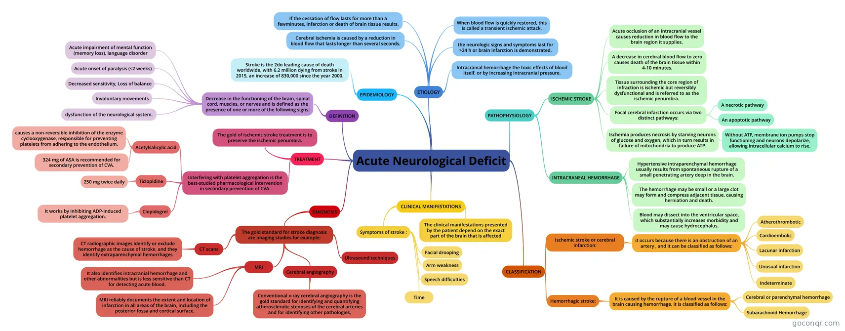
Mind map: Acute neurological deficit
Contenido del mapa
Acute Neurological Deficit
DEFINITION
- Decrease in the functioning of the brain, spinal cord, muscles, or nerves and is defined as the presence of one or more of the following signs:
- Acute impairment of mental function (memory loss), language disorder
- Acute onset of paralysis (<2 weeks)
- Decreased sensitivity, Loss of balance
- Involuntary movements
- dysfunction of the neurological system.
- Decrease in the functioning of the brain, spinal cord, muscles, or nerves and is defined as the presence of one or more of the following signs:
EPIDEMIOLOGY
- Stroke is the 2do leading cause of death worldwide, with 6.2 million dying from stroke in 2015, an increase of 830,000 since the year 2000.
ETIOLOGY
- Cerebral ischemia is caused by a reduction in blood flow that lasts longer than several seconds.
- If the cessation of flow lasts for more than a fewminutes, infarction or death of brain tissue results.
- When blood flow is quickly restored, this is called a transient ischemic attack.
- the neurologic signs and symptoms last for >24 h or brain infarction is demonstrated.
- Intracranial hemorrhage the toxic effects of blood itself, or by increasing intracranial pressure.
PATHOPHYSIOLOGY
- ISCHEMIC STROKE
- Acute occlusion of an intracranial vessel causes reduction in blood flow to the brain region it supplies.
- A decrease in cerebral blood flow to zero causes death of the brain tissue within 4-10 minutes.
- Tissue surrounding the core region of infraction is ischemic but reversibly dysfunctional and is referred to as the ischemic penumbra.
- Focal cerebral infarction occurs via two distinct pathways:
- A necrotic pathway
- An apoptotic pathway
- Ischemia produces necrosis by starving neurons of glucose and oxygen, which in turn results in failure of mitochondria to produce ATP.
- Without ATP, membrane ion pumps stop functioning and neurons depolarize, allowing intracellular calcium to rise.
- INTRACRANEAL HEMORRHAGE
- Hypertensive intraparenchymal hemorrhage usually results from spontaneous rupture of a small penetrating artery deep in the brain.
- The hemorrhage may be small or a large clot may form and compress adjacent tissue, causing herniation and death.
- Blood may dissect into the ventricular space, which substantially increases morbidity and may cause hydrocephalus.
- ISCHEMIC STROKE
CLASSIFICATION
- Ischemic stroke or cerebral infarction:
- it occurs because there is an obstruction of an artery , and it can be classified as follows:
- Atherothrombotic
- Cardioembolic
- Lacunar infarction
- Unusual infarction
- Indeterminate
- it occurs because there is an obstruction of an artery , and it can be classified as follows:
- Hemorrhagic stroke:
- It is caused by the rupture of a blood vessel in the brain causing hemorrhage, it is classified as follows:
- Cerebral or parenchymal hemorrhage
- Subarachnoid Hemorrhage
- It is caused by the rupture of a blood vessel in the brain causing hemorrhage, it is classified as follows:
- Ischemic stroke or cerebral infarction:
CLINICAL MANIFESTATIONS
- The clinical manifestations presented by the patient depend on the exact part of the brain that is affected
- Symptoms of stroke :
- Facial drooping
- Arm weakness
- Speech difficulties
- Time
DIAGNOSIS
- The gold standard for stroke diagnosis are imaging studies for example:
- CT scans
- CT radiographic images identify or exclude hemorrhage as the cause of stroke, and they identify extraparenchymal hemorrhages
- MRI
- MRI reliably documents the extent and location of infarction in all areas of the brain, including the posterior fossa and cortical surface.
- It also identifies intracranial hemorrhage and other abnormalities but is less sensitive than CT for detecting acute blood.
- Cerebral angiography
- Conventional x-ray cerebral angiography is the gold standard for identifying and quantifying atherosclerotic stenoses of the cerebral arteries and for identifying other pathologies,
- Ultrasound techniques
- CT scans
- The gold standard for stroke diagnosis are imaging studies for example:
TREATMENT
- The gold of ischemic stroke treatment is to preserve the ischemic penumbra.
- Interfering with platelet aggregation is the best-studied pharmacological intervention in secondary prevention of CVA.
- Acetylsalicylic acid
- causes a non-reversible inhibition of the enzyme cyclooxygenase, responsible for preventing platelets from adhering to the endothelium,
- 324 mg of ASA is recommended for secondary prevention of CVA.
- Ticlopidine
- 250 mg twice daily
- Clopidogrel
- It works by inhibiting ADP-induced platelet aggregation.
- Acetylsalicylic acid
Información
- Descripción
- Mapa Mental sobre Acute Neurological Deficit, creado por nikol moreno el 16/06/2021.
- Creado por
- nikol moreno
- Visitas
- 0
- Valoración