
Mapa mental
Cell death
Contenido del mapa
Cell death
~ 50% neurons lost in development
- Other cell types die too
- Most death is deliberate
Where does it occur?
- Everywhere
- E.g.DRG's
- DRG’s innervating limbs show less death
- Trophic support
- ?
- Trophic support
- DRG’s innervating limbs show less death
- E.g. motorneurons
- Still cell death even though target tissue is increasing
- Death occurring during synaptogenesis
- Reflects competition
- Death occurring during synaptogenesis
- Still cell death even though target tissue is increasing
- E.g.DRG's
- Pre and post synaptically
- e.g. somatosensory barrels
- e.g. chick auditory system
- nucleus magnocellularis
- nucleus laminaris
- Matching cell death
- Everywhere
How does it occur?
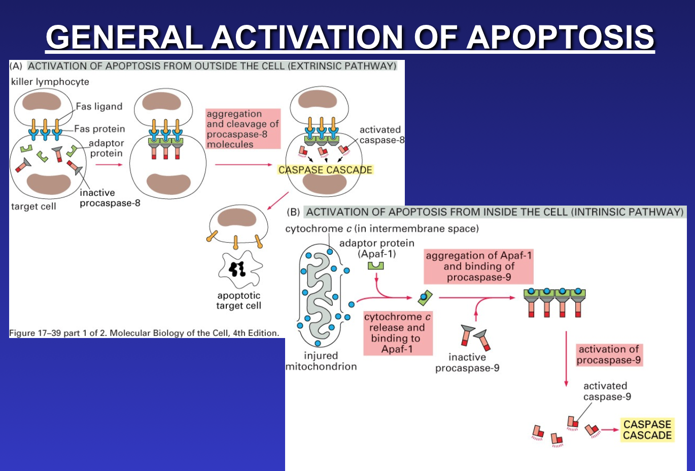
- Most cells die through apoptosis
- Programmed cell death
- Active process
- Inhibited by actinomycin D (transcription)
- Or cycloheximide (protein synthesis)
- Pro and anti apoptotic influences
- Multiple, opposing genes involved with apoptosis
- In C.elegans ced - 3 and ced - 4 required
- Anti-apoptotic genes e.g. ced-9 (bcl-2)
- Multiple, opposing genes involved with apoptosis
- Triggers for development apoptosis
- Lack of trophic support
- Neurotrophin supply (retrograde, anterograde, autocrine)
- Transmitters can be trophic factors
- Glutamate receptor (NMDA) activation
- MK801 injection P7 forebrain
- Other NMDA antagonists effective (e.g. paediatric anaesthetics)
- Other transmitter ligands not
- NMDA block – apoptosis ↑
- Where receptors are matters
- Hippocampus in vitro
- NMDA receptors synaptic and non-synaptic
- STAU evokes apoptosis
- Reduced by blocking GABA (bicucculine)
- Synaptic receptors are anti-apoptotic
- Extrasynaptic receptors are pro-apoptotic
- STAU evokes apoptosis
- NMDA receptors synaptic and non-synaptic
- Hippocampus in vitro
- Glutamate receptor (NMDA) activation
- Absence of depolarising stimulus
- e.g. anterograde transport of NT3 in retinal ganglion neurons
- e.g. early DRG’s in cell culture release NT3 and promote self differentiation
- NGF deprivation
- NGF withdrawal from sympathetic ganglion neurons in vitro
- SGN have p75NTR, TrkA, LOW TrkC
- Low NGF + KCl + BDNF (p75NTR, TrkB) = apoptosis
- NT’s give pro and anti-apoptotic signals
- An individual neuron needs both the “correct” NT’s and the absence of “wrong” NT’s
- An axon in the wrong hood is in danger
- Find the GIF
- Lack of trophic support
- Roles for proneurotrophins too?
- Synthesized in prepro form
- Pre portion is signal peptide
- Pro portion cleaved after release
- PRO NGF strongly activates p75NTR
- so the levels of enzymes to cleave PRO NGF are important too
- growth cones can release cleavage enzymes
- Release
- Basal release
- Stimulated release
- depends on Ca2+
- triggered by depolarization or ligand binding, e.g. other NT
- More efficient synapse, more stimulated release, more binding / uptake by membrane recycling, more pre-synaptic effect?
- More target more release.
- Synthesized in prepro form
- Most cells die through apoptosis
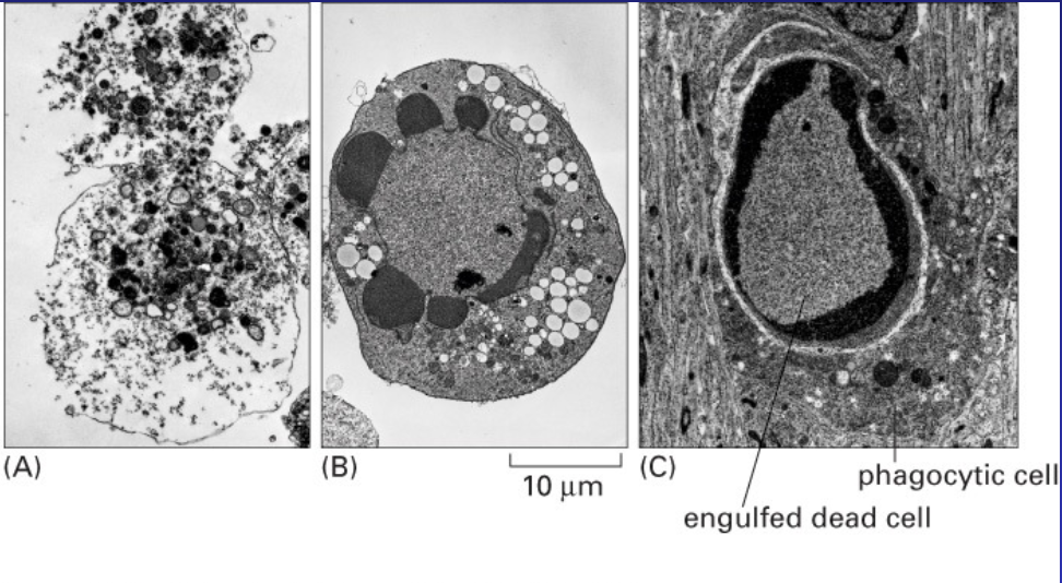
Cleaning up the mess
- Tidy removal of dying cells by macrophages
- Plus neuritic disintegration and removal
Imágenes y archivos del mapa
Información
- Descripción
- Undergraduate (MCD lectures) BMS236 Building Nervous Systems Mapa Mental sobre Cell death, creado por Kristi Brogden el 12/08/2014.
- Creado por
- Kristi Brogden
- Visitas
- 38
- Valoración