
Apuntes
Liver
Contenido del apunte (1)
Página 1
ACUTE HEPATITIS
-short-term, self-limiting inflammation of the liver lasting less than 6 months
Common infective causes of acute hepatitis:
Viral causes:
Hep A (faeco-oral transmission)
Hep B (parenteral transmission): acute in 15%, chronic in 5%
Hep C (parenteral transmission)
Hep D (requires Hep B infection concurrently) and Hep E, Epstein-Barr, varicella
Bacterial:
Leptospira
Syphilis
TB (miliary)
Protozoal:
Amoebiasis
Malaria
Leishmaniasis
Helminths:
Ascaris
Schistosoma
Liver flukes
Hydatid disease
Hepatotoxic drugs:
2 types of drugs:
intrinsic hepatotoxins: dose dependant, predictable, responsible for high incidence of liver damage
idiosyncratic hepatotoxins: due to hypersensitivity or abnormal drug metabolism
Types of damage:
Fatty change: methotrexate, tetracyclines, sodium valproate
Hepatic granulomas: sulphonamides, allopurinol
Acute hepatitis: isoniazid, rifampicin, halothane
Chronic hepatitis: isoniazid, methyldopa, NSAIDS
Cholestasis: steroids, chlorpromazine
Central vein occlusion: cytotoxic drugs (azothiaprine)
Tumours: oral contraceptives, anabolic steroids
Acute necrosis: PARACETAMOL
CHRONIC HEPATITIS
-inflammatory disease of the liver lasting for more than six months.
Common causes of chronic hepatitis:
Hep B (most common cause worldwide)
Hep C (25% of liver disease in UK)
Other viruses: CMV, Epstein-Barr
Autoimmune hepatitis (rare), Primary biliary cirrhosis, primary sclerosing cholangitis
Metabolic: non-alcoholic fatty liver disease (NAFLD), which may progress to non-alcoholic steatohepatitis (NASH): risk factors are metabolic syndrome, obesity, diabetes, and hyperlipidemia.
Other metabolic: haemochromatosis, Wilson's disease, alpha-1-antitrypsin deficiency.
Toxic and drugs: alcoholic liver disease, amiodarone, isoniazid, methyldopa, methotrexate, nitrofurantoin.
Sarcoidosis.
Morphology and Pathology:
Acute hepatitis:
commonly presents with: jaundice, nausea, anorexia, RUQ discomfort, fever and fatigue
LFT derangement
Fulminant liver failure: progression from normal function to liver failure within 8 wks, commonly caused by PARACETAMOL
encephalopathy (metabolic flap/asterixis)
jaundice
haemorrhage
Chronic hepatitis:
fibrosis: progressive decrease in liver cell mass and portal hypertension
symptoms: weakness, anorexia, muscle loss (esp at shoulders) and adipose tissue loss (cachexia)
GI bleeding from varices
Ascites: due to Na+ retention, esp with high portal pressure and low albumin
Jaundice
encephalopathy: reduced attention span, reversed sleep pattern, metabolic flap, hepatic coma
Coagulopathy
JAUNDICE
- due to increased bilirubin concentration (detectable clinically when >50 mcr mmol/L)
Bilirubin metabolism:-
SPLEEN: Unconjugated bilirubin (from RBC lysis)
LIVER: hepatic conjugation into water-soluble bilirubin glucoronide (conjugated bilirubin)
Excretion into bile
GUT: conversion into stercobilinogen (stool pigment) by gut bacteria
BLOOD: conjugated bilirubin from gut into blood (urobilinogen)
KIDNEY: Urobilinogen is excreted in urine
Classification:
Hepatocellular (hepatic): acute hepatitis, viral hepatitis, drugs, chronic liver disease, Hep B and C, alcoholic liver disease, udd-Chiari, autoimmun hep
Cholestatic/obstructive (post-hepatic): biliary obstruction (gallstones), secondary biliary disease, pancreatitis, pancreatic cancer, chronic biliary disease (PBC, PSC)
Relevant features:
Encephalopathy: liver failure only (not biliary)
acute or chronic presentation?
Pain? Gallstones most likely
risk factors for chronic liver disease: alcohol, travel, sexual practices, IV drug abuse etc
dark urine and pale stools: cholestatic!
Blood tests:
FBC: macrocytosis, thrombocytopenia, low urea, low sodium (chronic liver disease)
LFTs: Low albumin (nonspecific)
Conjugated bilirubin rise+LFT derangement
Hepatocellular (transaminases picture: AST or ALK> AlkPhos) vs cholestatic (AlkPhos or GGT > AST).
Unconjugated bilirubin rise: hemolysis or Gilberts (with normal LFTs)
Iron (haemochromatosis) and copper (Wilson's) studies
Viral hepatitis serology
Autoantibody profile and Ig
US : liver abnormalities (abscess, mets), biliary obstruction (dilated bile ducts), gallstones, pancreatic ca
MRCP, ERCP
Liver biopsy
Common primary sites for liver mets:
Colorectal, stomach, pancreas, breast, lung and eye.
In children, the most common primaries are neuroblastoma, Wilm's tumour and leukaemia
Primary hepatocellular carcinoma:
90-95% of people who develop HCC have underlying cirrhosis but non-cirrhotic HCC does occur.
Cirrhosis may be due to:
Hep B or Hep C infection (3-5% per year risk of developing HCC)
Alcoholism.
Genetic haemochromatosis.
Primary biliary cirrhosis.(PBC)
high concentration of aflatoxins, a group of mycotoxins produced by the fungi Aspergillus flavus and Aspergillus parasiticus in food. In sub-Saharan Africa and in Asia such high levels can occur due to fungal contamination of foodstuffs including peanuts and grains.
The metabolic syndrome, diabetes and smoking.
Rare associations include: androgenic steroids, primary sclerosing cholangitis, alpha-1-antitrypsin deficiency, oral contraceptives, porphyria cutanea tarda.
Cirrhosis:
characterised by fibrosis and the conversion of normal liver architecture into structurally abnormal nodules.
diffuse replacement of normal liver cells by nodules of regenerated liver cells separated by bands of collagenous fibrosis
Key Characteristics:
long-standing destruction of liver cells
chronic inflammation that stimulates fibrosis
regeneration of hepatocytes to cause nodules
Investigation
Alk Phos: greatly elevated in biliary obstruction
AST and ALT elevated transaminases proportional to activity of hepatocyte destruction
GGT: elevated in active alcoholics
Bilirubin: elevated, especially high in biliary obstruction
Serum albumin: low (failure of synthesis by liver)
Prothrombin time: prolonged (due to failure of synthesis of coagulation factors)
FBC: occult bleeding may produce anaemia; hypersplenism may cause thrombocytopenia; macrocytosis can suggest alcohol abuse.
Renal function tests and electrolytes: hyponatraemia may be present (due to increased activity of antidiuretic hormone)
Ferritin: low ferritin may indicate iron deficiency from diet or blood loss; ferritin is raised in haemochromatosis.
Viral antibody screen: to look for evidence of hepatitis B or C infection.
Pathophysiology:
Hypoproteinaemia: reduced hepatocyte function and reduced number of functional liver mass results in reduced synthesis of proteins
Oesophageal varices: fibrosis and alteration in liver architecture results in portal hypertensions, causing porto-systemic shunts to develop e.g oesophageal varices.
Ascites: accumulation of fluid due to increased portal pressure, increased plasma volume (salt and water retention in kidneys) and low protein levels. May lead to spontaneous bacterial peritonitis.
Encephalopathy; due to accumulation of toxic waste (e.g ammonia) leading to biochemical disturbance of brain function, mainly caused by liver failure and portosystemic shunting
Increase risk of Hepatocellular carcinoma
increase risk of portal vein thrombosis
anaemia (due to bleeding), thrombocytopenia (due to hypersplenism) and coagulopathies (reduced synthesis of clotting factors)
fibrinolysis and disseminated intravascular coagulation (DIC) may occur
Clinical features of complications:
-oesophageal varices: only sign may be on endoscopy or with variceal bleeding. Other signs of portal hypertension: caput medusae, spider naevi, haemorrhoids and periumbilical collateral veins
-ascites: distended abdomen, shifting dullness and fluid thrill on examination
-encephalopathy: metabolic flap/asterixis
(slow, clonic flexion-relaxation movement against your hand if asterixis is present.) Other signs: personality changes, intellectual impairment and reduced levels of consciousness
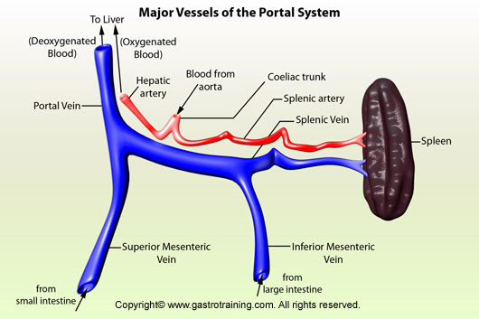
Portal hypertension: prolonged elevation of portal venous pressure (normal 2-5mmHg). To develop clinical features or complications, patients usually have pressures >12mmgHg
Mechanism:
Increased vascular resistance in the portal venous system - from various mechanical causes , and also as an active process in which liver damage activates stellate cells and myofibroblasts, contributing to the abnormal blood flow patterns.
Increased blood flow in the portal veins - from splanchnic arteriolar vasodilatation, caused by an excessive release of endogenous vasodilators.
Causes:
Prehepatic - blockage of the portal vein before the liver
Congenital atresia or stenosis.
Portal-vein thrombosis (idiopathic, umbilical and portal sepsis, malignancy, hypercoagulable states -pregnancy, The Pill)
Pancreatitis
Splenic vein thrombosis.
Abdo trauma (inc surgery)
Extrinsic compression, eg tumours.
Hepatic
Cirrhosis.
Chronic hepatitis.
Schistosomiasis.
Myeloproliferative diseases.
Idiopathic portal hypertension.
Drugs
Granulomata, eg sarcoid.
Nodular (nodular regenerative hyperplasia, partial nodular transformation).
Toxins (arsenic, vinyl chloride).
Fibropolycystic disease (including congenital hepatic fibrosis).
Posthepatic - blockage of hepatic veins or venules
Budd-Chiari syndrome (hepatic vein obstruction).
Constrictive pericarditis.
Right heart failure.
Veno-occlusive disease of the smaller hepatic veins/venules (due to ingestion of pyrrolizidine alkaloids; antileukaemic drugs, radiation).
Other causes Increased hepatic blood flow:
Increased splenic blood flow, eg massive splenomegaly.
Hepatoportal arteriovenous fistula.
Idiopathic (a diagnosis of exclusion)
Left-sided (sinistral) portal hypertension Rare. It is confined to the left side of the portal system. It may present as bleeding from gastric varices.Usually it is due to pathology involving the splenic vein or the pancreas
Clinical manifestations:
Symptoms of complications:
Haematemesis or melaena - suggest bleeding varices.
Lethargy, irritability and changes in sleep pattern - suggest encephalopathy.
Increased abdominal girth, weight gain - suggest ascites.
Abdominal pain and fever - suggest spontaneous bacterial peritonitis.
Signs:
Dilated veins in the anterior abdominal wall (periumbilical collaterals) and caput medusae (tortuous collaterals around the umbilicus).
Spider naevi
Hepatomegaly and splenomegaly
A venous hum, loudest during inspiration, is sometimes heard over large upper abdominal collaterals.
Splenomegaly.
Ascites.
Hepatitis
liver neoplasms
chronic liver disease
portal hypertension
Información
- Descripción
- Apunte sobre Liver, creado por bessimajamal el 14/06/2014.
- Creado por
- bessimajamal
- Visitas
- 457
- Etiquetas
- Valoración