
Test
A&P Lab Practical 2
Este test tiene tipos de pregunta que se abren en el reproductor de siempre.
Preguntas del test (48)
1Label the picture
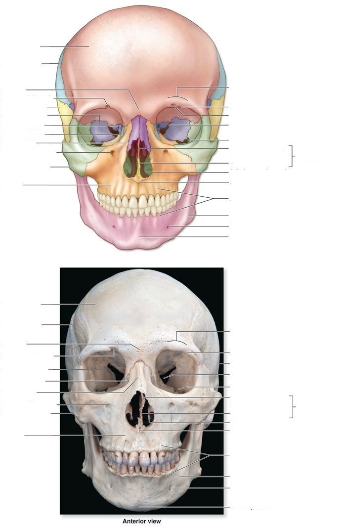
- Afrontal bone
- Bparietal bone
- Csupraorbital foramen
- Dtemporal bone
- Esphenoid bone
- Fnasal bone
- Gzygomatic bone
- Hmaxilla
- Imandible
- Jinfraorbital foramen
- Kmental foramen
- Lmental protuberance
- Malveolar process
- Ninferior nasal concha
- Ovomer
- Pperpendicular plate
- Qsuperior orbital fissure
2Label the picture
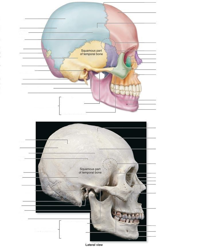
- Acoronal suture
- Bfrontal bone
- Cparietal bone
- Dsquamous suture
- Elambdoid suture
- Ftemporal bone
- Goccipital bone
- Hexternal acoustic meatus
- Imastoid process
- Jstyloid process
- Kmandibular ramus
- Ltemporal process of zygomatic bone
- Mmental protuberance
- Nmental foramen
- Omaxilla
- Pzygomatic bone
- Qethmoid bone
- Rlacrimal bone
- Snasal bone
- Tsphenoid bone
- Umandibular condyle
- Vcoronoid process
- Wmandibular notch
3Label the picture
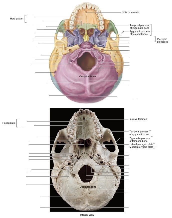
- Apalatine process
- Bhorizontal plate
- Cvomer
- Dforamen ovale
- Esphenoid bone
- Fjugular foramen
- Gcarotid canal
- Hlambdoid suture
- Iforamen magnum
- Joccipital condyle
- Kmastoid process
- Lmandibular fossa
- Mstyloid process
- Nlateral pterygoid plate
- Omedial pterygoid plate
- Pexternal occipital protuberence
4Label the picture
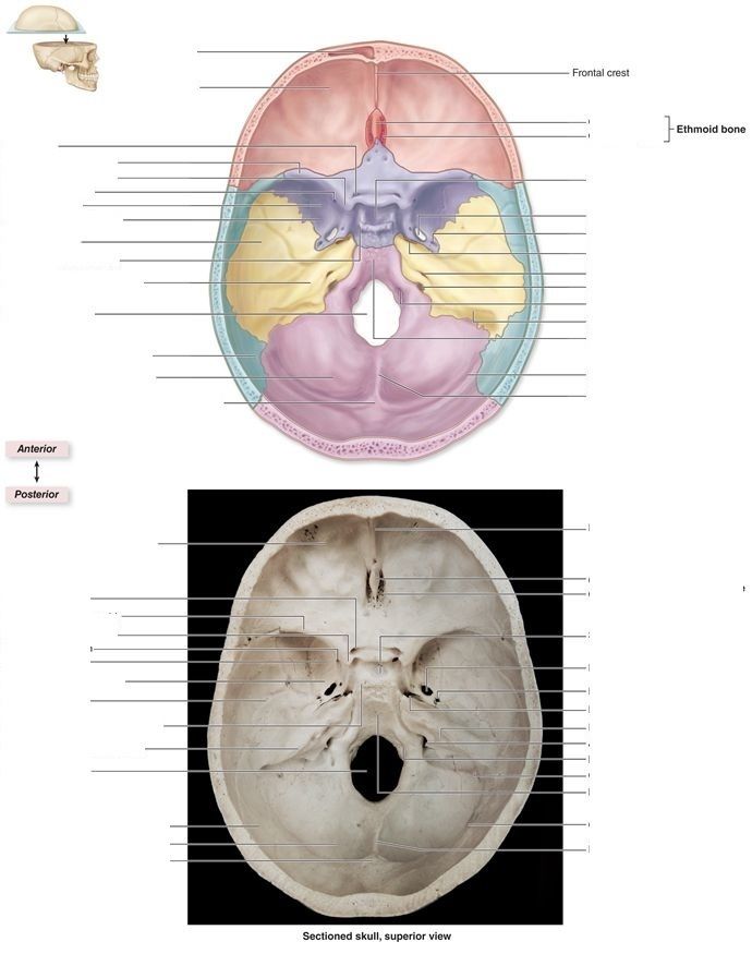
- Afrontal sinus
- Bcrista galli
- Ccribriform plate
- Doptic canal
- Elesser wing of sphenoid
- Fsella turcica
- Gforamen rotundum
- Hgreater wing of sphenoid
- Iforamen ovale
- Jinternal acoustic meatus
- Kjugular foramen
5Label the picture
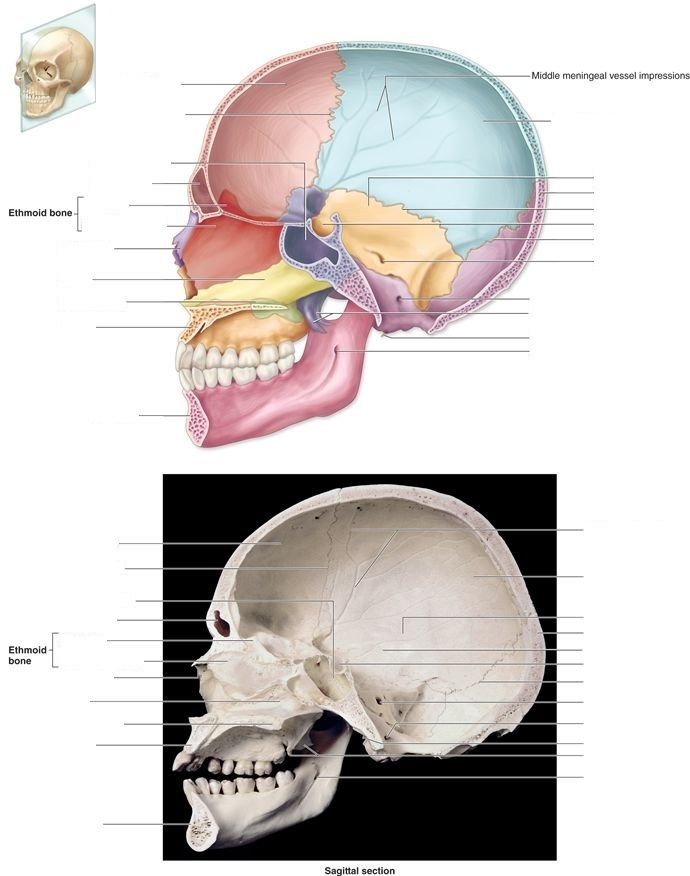
- Asphenoid sinus
- Bfrontal sinus
- Ccrista galli
- Dperpendicular plate
- Emandibular foramen
6Label the picture
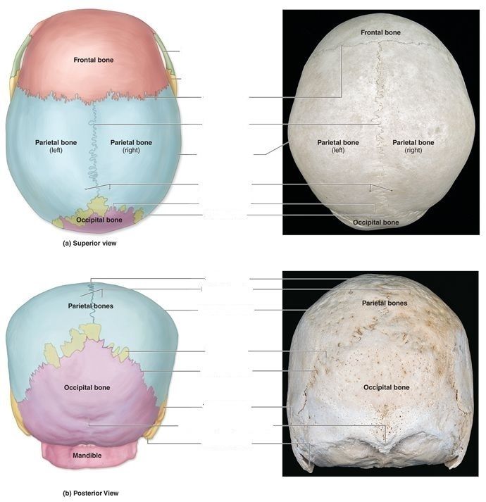
- Acoronal suture
- Bsagittal suture
- Clambdoid suture
7[blank_start]Long bones[blank_end] are longer tan they are wide and include the bones of the upper and lower limbs, excluding the ankle and wrist bones.
- ALong bones
8[blank_start]Flat bones[blank_end] are shaped exactly as they're named. These bones include the ribs, sternum, certain skull bones, and hip bones.
- AFlat bones
9[blank_start]Irregular bones[blank_end] are those whose shape doesn't fit into any of the other classes. These bones include the vertebrae, the sacrum, and certain bones of the skull, such as the sphenoid bone.
- AIrregular bones
10[blank_start]Sesamoid bones[blank_end] are usually small, round, and flat, and shaped somewhat like a sesame seed.
- ASesamoid bones
11[blank_start]Sutural bones[blank_end], or Wormian bones, are small, flat, irregularly shaped bones between the flat bones of the skull. There are individual variations in the number, shape, and position of the sutural bones.
- ASutural bones
12The axial skeleton consists of
- Askull
- Bupper and lower limbs
- Cthoracic cage
- Dpelvic girdle
- Ebones associated with the skull
- Fvertebral column
13The appendicular skeleton consists of
- AUpper and lower limbs
- BSkull
- CPectoral girdle
- DVertebrae
- EPelvic girdle
14The vertebral column consists of 24 unfused vertebrae and 5 fused vertabrae of the sacrum and coccyx. Of the 24 unfused vertebrae, [blank_start]7[blank_end] are cervical, [blank_start]12[blank_end] are thoracic, and [blank_start]5[blank_end] are lumbar.
- A7
- B12
- C5
15[blank_start]C1[blank_end], or [blank_start]atlas[blank_end], articulates with the occipital condyles, and holds up the head. It also allows us to nod our head yes.
- AC1
- Batlas
16[blank_start]C2[blank_end], or [blank_start]axis[blank_end], fuses with C1 and allows us to rotate our head left and right.
- AC2
- Baxis
17What type of vertebra is this?
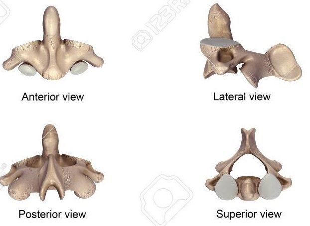
- Acervical vertebrae
- Bthoracic vertebrae
- Clumbar vertebrae
18What type of vertebra is this?
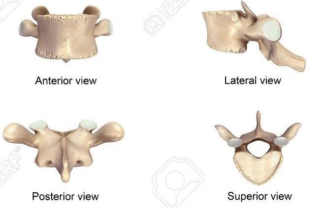
- Acervical vertebrae
- Bthoracic vertebrae
- Clumbar vertebrae
19What type of vertebra is this?
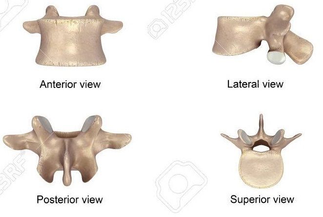
- Acervical vertebrae
- Bthoracic vertebrae
- Clumbar vertebrae
20Label the picture
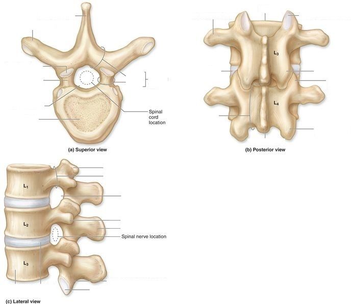
- Aspinous process
- Btransverse process
- Clamina
- Dpedicle
- Evertebral arch
- Fsuperior articular facet
- Gsuperior articular process
- Hvertebral foramen
- Ibody
- Jsuperior articular process
- Kpedicle
- Lintervertebral foramen
- Mtransverse process
- Nspinous process
- Oinferior articular process
- Pinferior articular facet
- Qbody
- Rtransverse process
- Ssuperior articular facet
- Tinferior articular process
- Usuperior articular process
- Vinferior articular process
- Wspinous process
- Xlamina
- Yintervertebral disc
- Zvertebral arch
21Label the picture
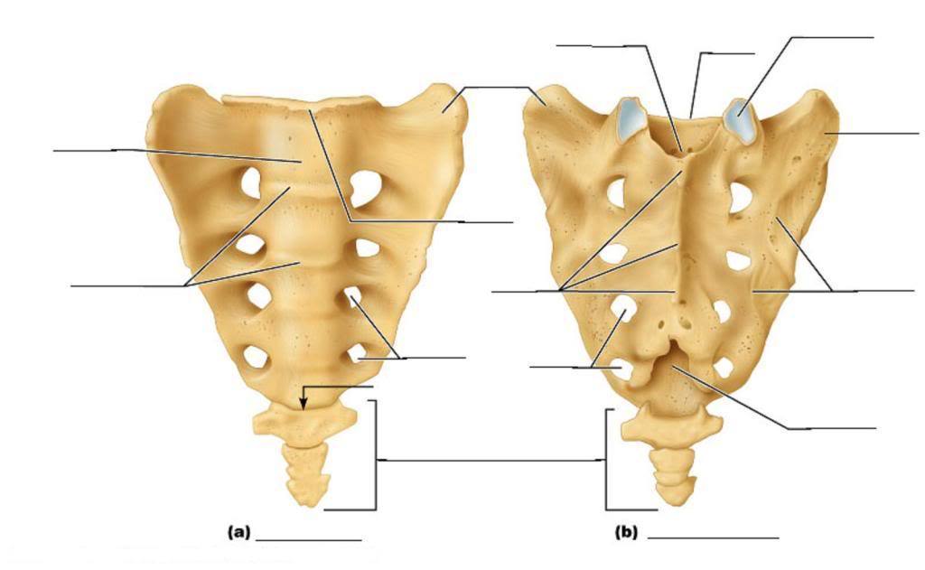
- Aauricular surface
- Bsacral foramina
22Label the picture
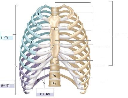
- Atrue ribs
- Bfalse ribs
- Cfloating ribs
- Dmanubrium
- Ebody
- Fxiphoid process
- Gsternum
- Hjugular notch
23Label the picture
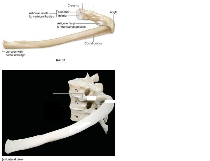
- Ahead
- Bneck
- Ctubercle
- Dbody
- Esternal end
24Ribs 1-7 are considered [blank_start]true ribs[blank_end], or vertebrosternal ribs, because they attach directly to the sternum by their own costal cartilage. Ribs 8-12, on the other hand, are classified as [blank_start]false ribs[blank_end] because they lack this direct attachment to the sternum. Ribs 8-10 are [blank_start]vertebrochondral ribs[blank_end] as they have an indirect attacment to the sternum, as their cartilage attaches to the costal cartilage of the true ribs. Ribs 11-12 have no attachment to the sternum at all, so they are often referred to as [blank_start]floating ribs[blank_end] or vertebral ribs.
- Afalse ribs
- Bvertebrochondral ribs
- Ctrue ribs
- Dfloating ribs
- Efloating ribs
- Fvertebrochondral ribs
- Gtrue ribs
- Hfalse ribs
- Ivertebrochondral ribs
- Jvertebrosternal ribs
- Kfloating ribs
- Ltrue ribs
- Mfalse ribs
- Nfloating ribs
- Overtebrochondral ribs
- Pvertebrosternal ribs
25Label the picture
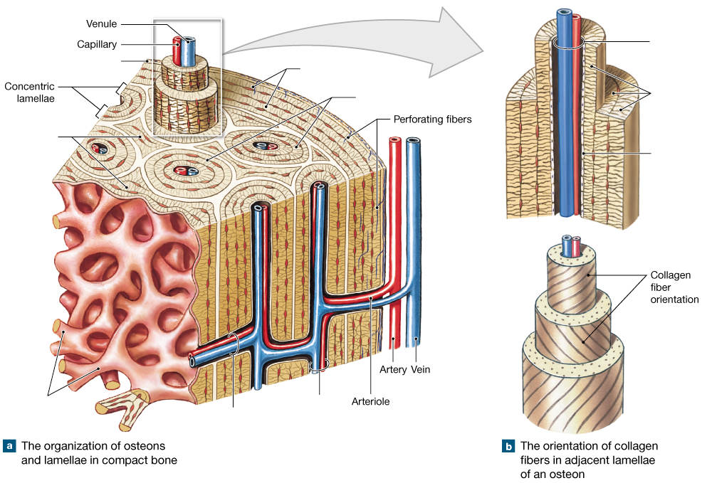
- Acircumferential lamellae
- Bperiosteum
- Cinterstitial lamellae
- Dtrabeculae of spongy gone
- Eosteons
- Fcentral canal
- Gperforating canal
- Hcentral canal
- Iconcentric lamellae
- Jendosteum
26Label the picture
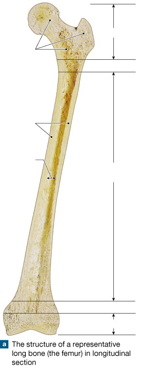
- Aepiphysis
- Bepiphysis
- Cmetaphysis
- Dmetaphysis
- Ediaphysis
- Fcompact bone
- Gmedullary cavity
- Hspongy bone
27Label the picture
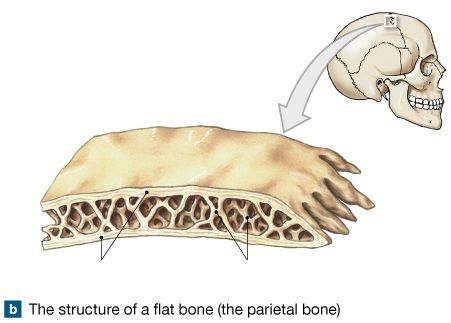
- Acompact bone
- Bdiploe
28Label the picture
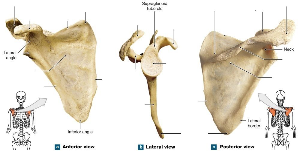
- Aacromion process
- Bacromion process
- Cacromion process
- Dcoracoid process
- Ecoracoid process
- Fcoracoid process
- Gsuperior border
- Hsuperior border
- Isuperior angle
- Jsupraspinous fossa
- Ksubscapular fossa
- Lspine
- Mspine
- Nglenoid cavity
- Oinfraspinous fossa
- Pmedial border
- Qlateral border
- Rinferior angle
- Slateral border
- Tmedial border
29Label the picture
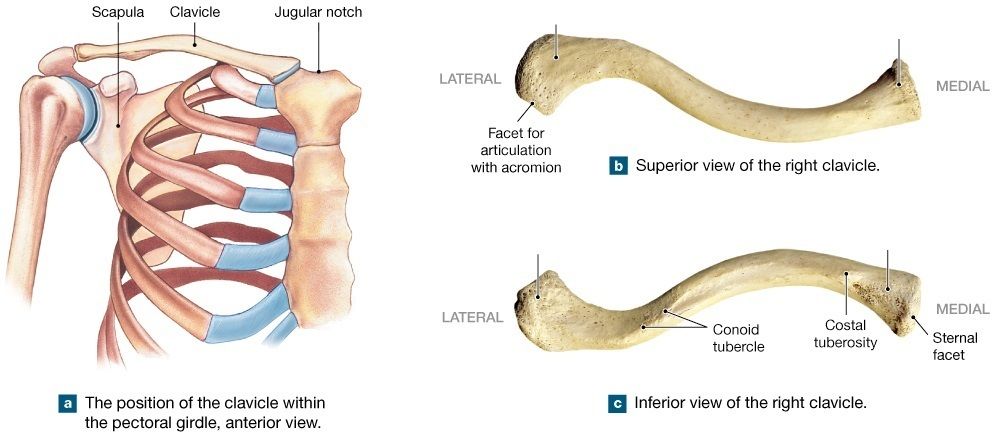
- Asternal end
- Bacromial end
30Label the picture
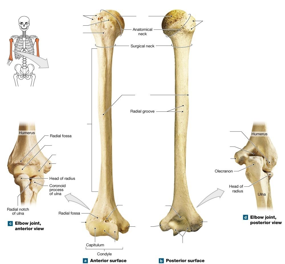
- Ahead
- Bgreater tubercle
- Cgreater tubercle
- Dlesser tubercle
- Eintertubercular groove
- Fshaft
- Gdeltoid tuberosity
- Hcapitulum
- Itrochlea
- Jmedial epicondyle
- Ktrochlea
- Lmedial epicondyle
- Mcoronoid fossa
- Nolecranon fossa
- Otrochlea
- Plateral epicondyle
- Qmedial epicondyle
- Rolecranon fossa
- Strochlea
31Label the picture
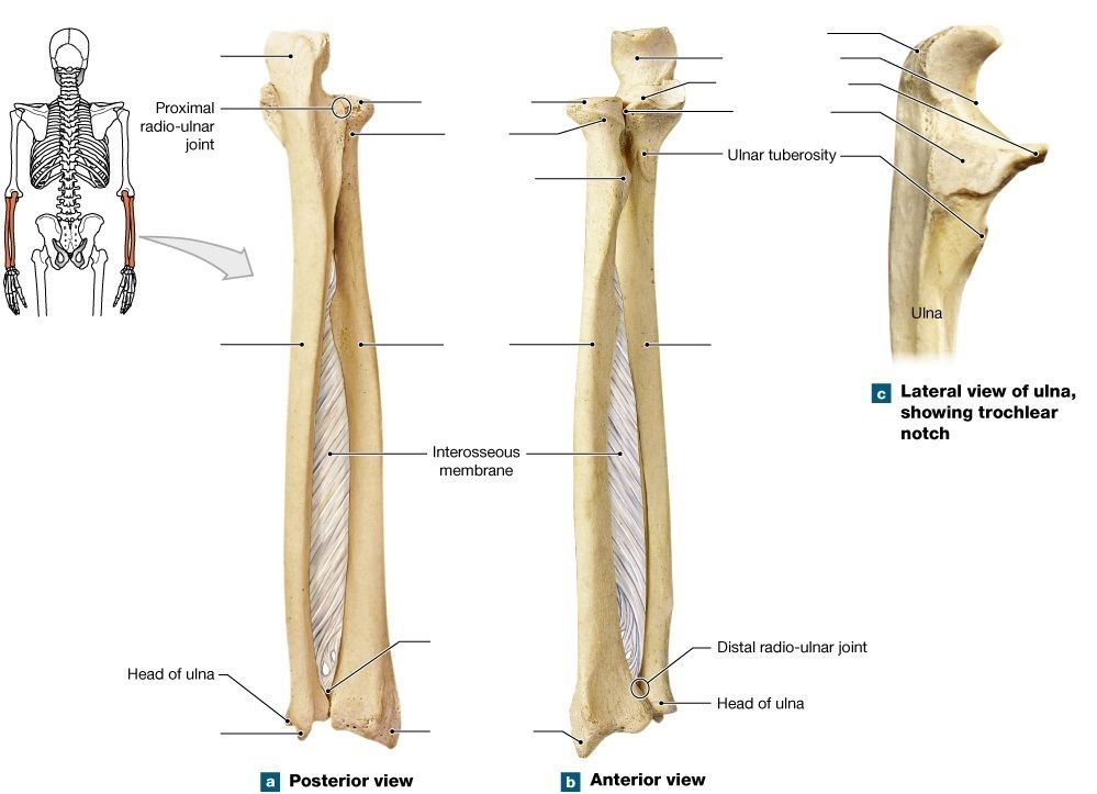
- Aolecranon process
- Bhead of radius
- Cneck of radius
- Dradial tuberosity
- Eradius
- Fulna
- Gulna
- Hulnar styloid process
- Iradial styloid process
- Julnar notch
- Kolecranon process
- Ltrochlear notch
- Mcoronoid process
- Nradial notch
32Label the picture
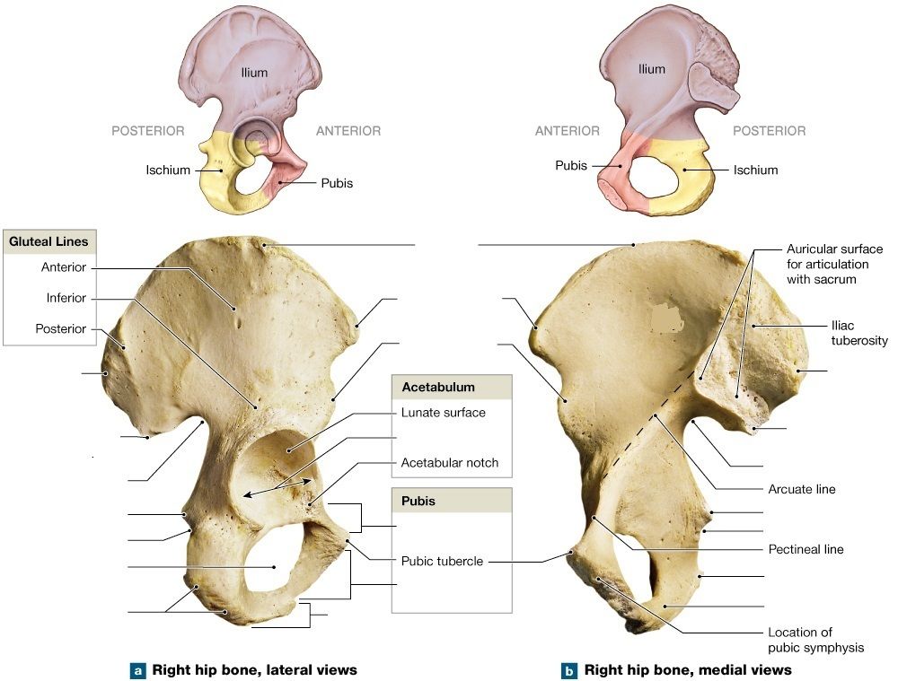
- Ailiac crest
- Banterior superior iliac spine
- Canterior inferior iliac spine
- Dacetabulum
- Esuperior pubic ramus
- Finferior pubic ramus
- Gischial ramus
- Hischial tuberosity
- Iobturator foramen
- Jlesser sciatic notch
- Kischial spine
- Lgreater sciatic notch
- Mposterior inferior iliac spine
- Nposterior superior iliac spine
- Oposterior superior iliac spine
- Pischial ramus
- Qischial tuberosity
- Rlesser sciatic notch
- Sischial spine
- Tgreater sciatic notch
- Uposterior inferior iliac spine
33Label the picture
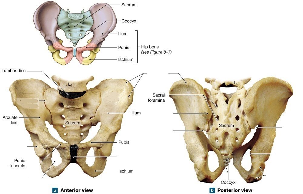
- Ailiac crest
- Bposterior superior iliac spine
- Cposterior inferior iliac spine
- Dsacro-iliac joint
- Epubic symphysis
- Fgreater sciatic notch
- Gischial spine
- Hischial tuberosity
34Label the picture
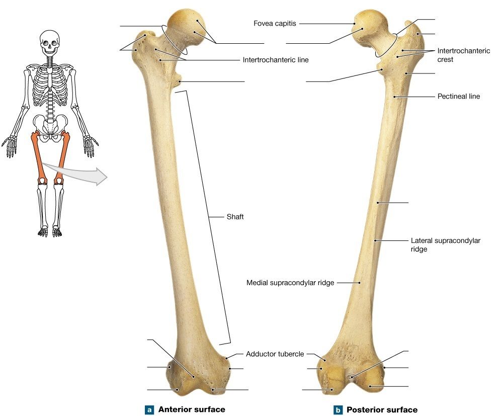
- Ahead
- Bneck
- Cneck
- Dgreater trochanter
- Egreater trochanter
- Flesser trochanter
- Ggluteal tuberosity
- Hlinea aspera
- Ipatellar surface
- Jlateral epicondyle
- Klateral condyle
- Lmedial epicondyle
- Mmedial condyle
- Nintercondylar fossa
- Olateral epicondyle
- Plateral condyle
35Label the picture
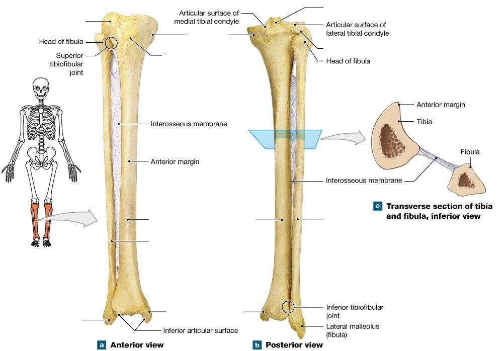
- Amedial tibial condyle
- Blateral tibial condyle
- Ctibial tuberosity
- Dtibia
- Efibula
- Ftibia
- Gfibula
- Hlateral malleolus
- Imedial malleolus
- Jintercondylar eminence
- Klateral tibial condyle
36[blank_start]Fibrous joints[blank_end] consists of two bones joined by short collagen fibers. Most [blank_start]fibrous joints[blank_end] allow no motion. [blank_start]Cartilaginous joints[blank_end] consist of bones united by cartilage rather than fibrous connective tissue. Most [blank_start]cartilagenous joints[blank_end] allow some motion. [blank_start]Synovial joints[blank_end] are freely movable joints.
- AFibrous joints
- BCartilaginous joints
- CSynovial joints
- Dfibrous joints
- Ecartilaginous joints
- Fsynovial joints
- GCartilaginous joints
- HFibrous joints
- ISynovial joints
- Jcartilagenous joints
- Kfibrous joints
- Lsynovial joints
- MSynovial joints
- NCartilaginous joints
- OFibrous joints
37Fibrous joints include
- Asuture
- Bsynchondrosis
- Csyndesmosis
- Dpivot
- Egomphosis
38Cartilaginous joints include
- ASuture
- BSynchondrosis
- CSyndesmosis
- DSymphysis
39Synovial joints include
- Aplane
- Bsuture
- Chinge
- Dsymphysis
- Epivot
- Fsynchondrosis
- Gellipsoid
- Hsyndesmosis
- Isaddle
- Jball and socket
40The cartilagenous connection between the ends of ribs 1 and 2 to the manubrium and sternum is an example of syndesmosis.
- AVerdadero
- BFalso
41Gomphosis is a type of joint that binds what to what?
- Ateeth to bony sockets
- Bribs to sternum
- Cskull bones to each other
- Dlong bones to each oher
42Bones connected by a ligament, such as the distal joint between the tibia and fibula is an example of syndesmosis.
- AVerdadero
- BFalso
43Drag and Drop the joint into the correct category

- Aintercarpal joints
- Bvertebrocostal joints
- Csacroiliac joint
- Dacromioclavicular joint
- Eclaviculosternal joint
- Felbow joint
- Gknee joint
- Hankle joint
- Iinterpharangeal joint
- Jatlanto-axial joint
- Kproximal radio-ulnar joint
- Lradiocarpal joint
- Mmetacarpophalangeal joint
- Nmetatarsophalangeal joint
- Ofirst carpometacarpal joint
- Pshoulder joint
- Qhip joint
44Label the picture
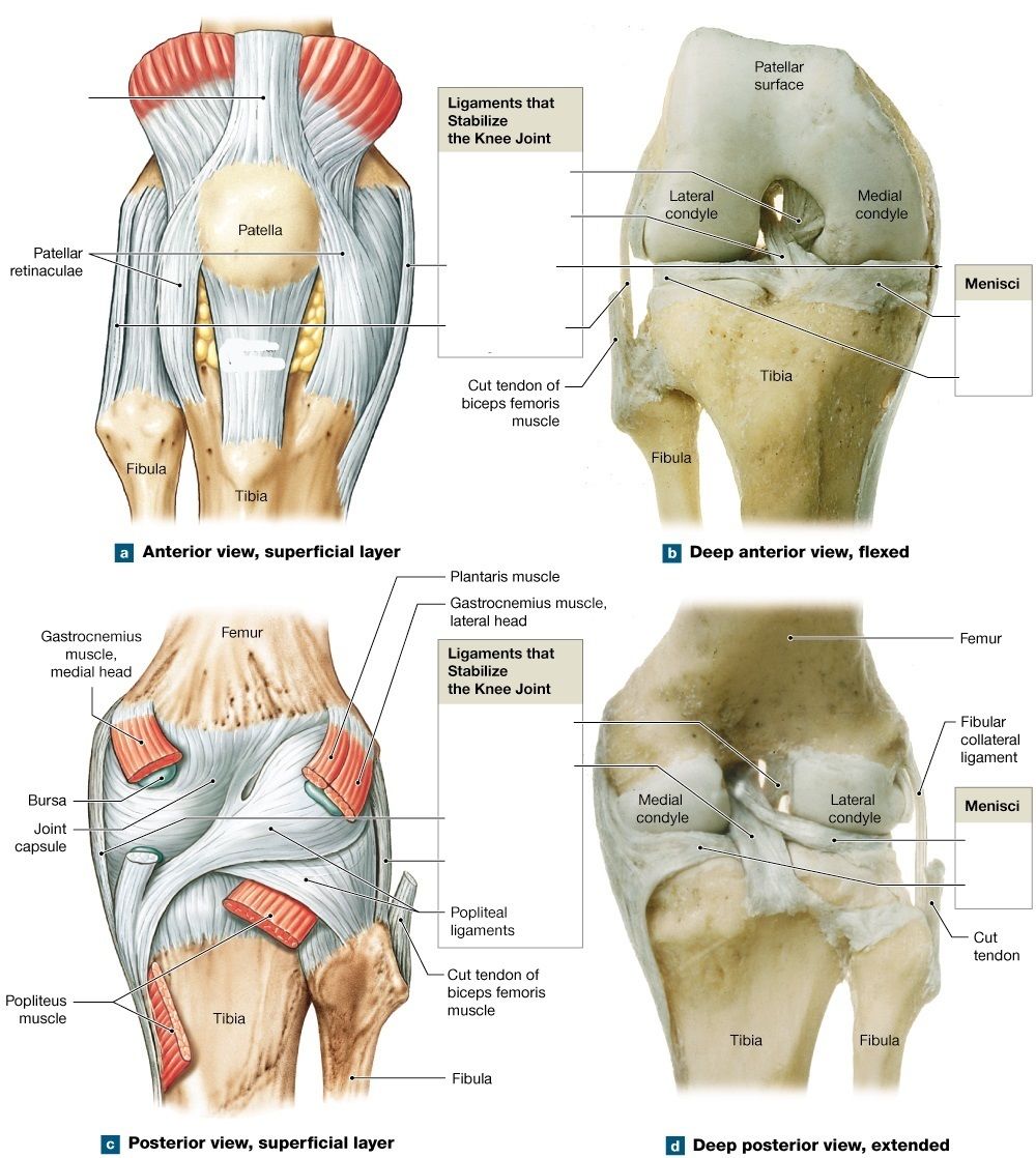
- Aquadriceps tendon
- Bpatellar ligament
- Cposterior cruciate ligament
- Danterior cruciate ligament
- Etibial collateral ligament
- Ffibular collateral ligament
- Gmedial meniscus
- Hlateral meniscus
- Ilateral meniscus
- Jmedial meniscus
- Kanterior cruciate ligament
- Lposterior cruciate ligament
- Mtibial collateral ligament
- Nfibular collateral ligament
45Label the picture
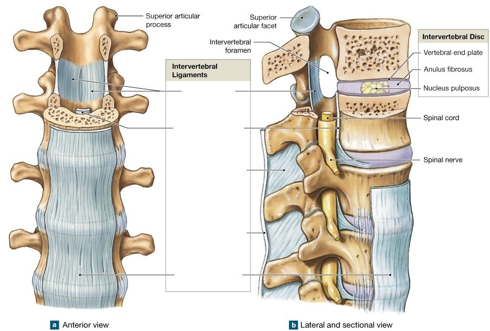
- Aligamentum flavum
- Bposterior longitudinal ligament
- Cinterspinous ligaments
- Dsupraspinous ligament
- Eanterior longitudinal ligament
46Label the picture
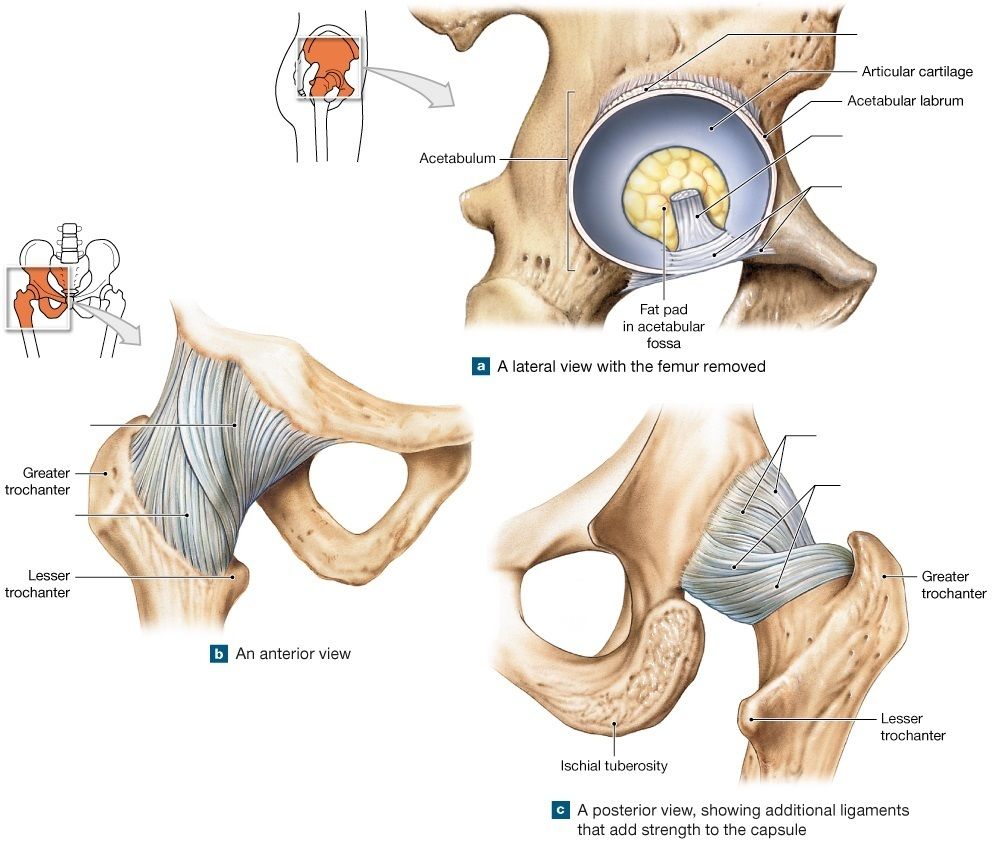
- Aligamentum teres
- Btransverse acetabular ligament
- Cpubofemoral ligament
- Diliofemoral ligament
- Eischiofemoral ligament
47Label the picture
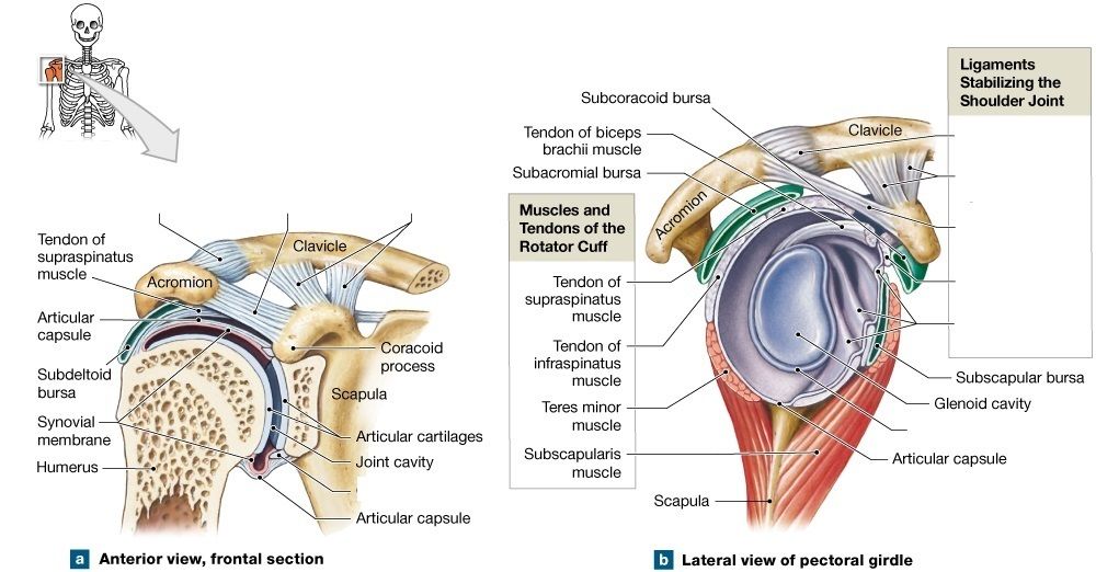
- Aacromioclavicular ligament
- Bcoraco-acromial ligament
- Ccoracoclavicular ligaments
- Dacromioclavicular ligament
- Ecoracoclavicular ligaments
- Fcoraco-acromial ligaments
- Gcoracohumeral ligament
- Hglenohumeral ligaments
48Label the picture
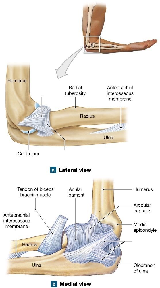
- Aanular ligament
- Bulnar collateral ligament
- Cradial collateral ligament
Información
- Descripción
- Anatomy and Physiology Test sobre A&P Lab Practical 2, creado por Tamara Podnosova el 25/09/2018.
- Autor
- Tamara Podnosova
- Idioma
- English
- Visitas
- 1.804
- Etiquetas
- Valoración
- Aún sin valoraciones Valora este test