
Test
Microbiology Lab Practical Staining
Este test tiene tipos de pregunta que se abren en el reproductor de siempre.
Preguntas del test (7)
1[blank_start]Simple[blank_end] staining uses [blank_start]basic[blank_end] [blank_start]dyes[blank_end] such as crystal violet, [blank_start]methylene[blank_end] blue, safrinin, [blank_start]malachite[blank_end] green, and basic fuchsin. These stains can show us if the cell is a [blank_start]prokaryote[blank_end] or [blank_start]eukaryote[blank_end]. They also show morphology such as [blank_start]shape[blank_end] and [blank_start]arrangement[blank_end].
- ASimple
- Bbasic
- Cdyes
- Dmethylene
- Emalachite
- Fprokaryote
- Geukaryote
- Hshape
- Iarrangement
2[blank_start]Differential[blank_end] staining uses two or more stains. Examples include the [blank_start]Gram[blank_end] stain and [blank_start]acid-fast[blank_end] stain. The latter is used for cells that have [blank_start]mycolic[blank_end] [blank_start]acid[blank_end] in their cell wall.
- ADifferential
- BGram
- Cacid-fast
- Dmycolic
- Eacid
3Step 1: [blank_start]Primary[blank_end] stain = crystal violet. All cells are [blank_start]purple[blank_end]. Step 2: Mordant = [blank_start]iodine[blank_end]. All cells are [blank_start]purple[blank_end]. Step 3: Decolorizer = [blank_start]alcohol[blank_end]. Gram negative cells are [blank_start]colorless[blank_end]. Gram positive cells are [blank_start]purple[blank_end]. Step 4: counterstain = [blank_start]safrinin[blank_end]. Gram negative cells are [blank_start]pink[blank_end]. Gram positive cells are [blank_start]purple[blank_end]. Acid fast cells are [blank_start]colorless[blank_end].
- APrimary
- Bpurple
- Ciodine
- Dpurple
- Ealcohol
- Fcolorless
- Gpurple
- Hsafrinin
- Ipink
- Jpurple
- Kcolorless
4The [blank_start]decolorizer[blank_end] creates holes in the [blank_start]lipopolysaccharides[blank_end] of Gram [blank_start]negative[blank_end] cell wall and washes out the [blank_start]iodine[blank_end] and [blank_start]crystal[blank_end] [blank_start]violet[blank_end]. The thick layer of [blank_start]peptidoglycan[blank_end] in Gram [blank_start]positive[blank_end] cell wall retains [blank_start]crystal[blank_end] [blank_start]violet[blank_end].
- Adecolorizer
- Blipopolysaccharides
- Cnegative
- Diodine
- Ecrystal
- Fviolet
- Gpeptidoglycan
- Hpositive
- Icrystal
- Jviolet
5Label:
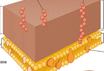
- Apeptidoglycan
- Bteichoic acid
- Clipoteichoic acid
- Dgram positive
6Label:
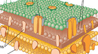
- Alipopolysaccharides
- Bporin protein
- Cphospholipids
- Dpeptidoglycan
- EGram negative
7Label:
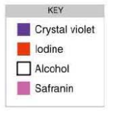
- Aprimary dye
- Bmordant
- Cdecolorizer
- Dcounterstain
Información
- Descripción
- Test sobre Microbiology Lab Practical Staining, creado por Meagan Mueller el 31/10/2016.
- Autor
- Meagan Mueller
- Idioma
- English
- Visitas
- 0
- Historial
- Creado el 31 oct 2016 · actualizado el 1 nov 2016
- Valoración
- Aún sin valoraciones Valora este test