
Sin título
Diapositivas del conjunto (35)
-
-
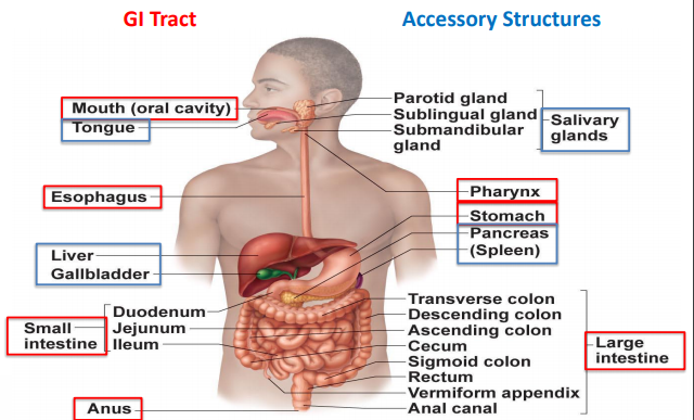
- Thick stratified squamous epithelium lining of the mouth
- Consists of the
- - Tounge - Composed of Skeletal Muscle. When MASTICATION is occurring the tongue is gripping the food, moving and mixing the food around the mouth with saliva to form a bolus. During swallowing the tongue collects the bolus, pushes it to the back of the mouth and down the pharynx. Produces speech
- - Pharynx - the common pathway to the respiratory system and digestive tract. Connects mouth to the oesophagus and trachea.
- - Oesophagus - Is a stratified squamous epithelium. Skeletal muscle in the beginning and smooth muscle at the bottom into the stomach. PERISTALSIS pushes bolus from the pharynx down to the stomach
-
Saliva Gland (Found in the mouth)
- A small intrinsic gland found under the mucous membrane of the mouth, lips, cheeks and tongue.
- - Parotid - present on either side of the mouth and in front of both ears
- - Submandibular or Wharton duct or submaxillary duct, is one of the salivary excretory ducts. drains saliva from each bilateral submandibular gland and sublingual gland to the sublingual caruncle at the base of the tongue
- - Sublingual - situated or applied under the tongue.
- Functions
- - Creates mucous - aids in swallowing
- - Begins Carbohydrate digestion (AMYLASE)
- - Cleans teeth
- - Inhibits bacteria by the enzyme creation of the enzyme that kills bacteria and immunoglobulin and it inhibits bacterial growth.
- - Creates bolus
- - Neutralise acidic foods (Phosphate and bicarbonate ions)
- - Dissolves food from taste receptors.
-
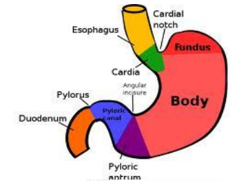
- - Located in the upper left-hand corner of the abdominal cavity
- - Elastic allowing variation in stomach diameter and stomach volume to allow high amounts of food.
- - Bolus enter by the cardiac sphincter and leaves by the pyloric sphincter
- - Regions of the cardia, fundus, body, and pylorus
- Stomach Lining
- - Mucosa
- - Submucosa
- - Muscularis Externa
- - Serosa
- Stomach Secretions
- - Hydrochloric Acid
- - Pepsinogen
- - Lipase
- - Rennin
- - Intrinsic Factor
- - Mucus
- - Hormones (Gastrin, Histamine, Serotonin and Somatostatin)
-
- Hydrochloric Acid
- Activates pepsin & lingual lipase Breaks up connective tissues & plant cell walls – Liquefies food to form Chyme Converts ingested ferric ions (Fe+3 ) to ferrous ions (Fe+2 ) Destroys ingested bacteria & pathogens
- Pepsinogen
- Inactive form of pepsin Requires HCl for activation Splits proteins into polypeptides & peptides Pepsin has an optimal pH range of 1.5 – 3.5
- Lipase
- Enzyme starts the breakdown of fats (lipids) into –Fatty acids –Glycerol
- Rennin
- Secreted by the gastric gland of children, not in adults Curdles milk by coagulating its proteins (Casein) Rennin ↓ with age ↓ dairy digestion
- Intrinsic Factor
- Essential for absorption of B12 by the small intestine
- Mucus
- Lines the stomach mucosa / protects against erosion, acids & enzymes Protects against gastric ulcers
- Hormones
- E.g.: Gastrin & histamine regulate HCl secretion E.g.: Serotonin & somatostatin regulate gastric motility
-
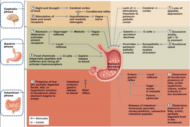
Stimulatory and Inhibitory Events
-
- Mucosa
- The innermost layer of the GI epithelium Surrounds the lumen Epithelium type varies depending on the organ function it lines – Stomach mucosa Secretion – Intestine Absorption
- Functions
- Secret mucus/digestive enzymes/hormones – Absorb the end products of digestion to blood – Protect against infectious disease
- Submucosa
- Areolar connective tissue layer Highly Vascular: Contains blood vessels Contains most of GI Nerves: – Regulating the movement of stomach/intestine/blood vessels vasoconstriction Attaches the mucosa layer to the muscular layer
- Muscular
- Mainly smooth muscle except – Mouth + pharynx + upper region of the oesophagus [Skeletal muscle Voluntary swallowing] – Anus (External anal sphincter) [Skeletal muscle Defecation] Consists (generally) of 2 layers of muscles: – Inner circular layer – Outer longitudinal layer Contraction of muscular moves food along the digestive tract.
- Serosa/Adventitia
- The outer most tissue layer If present: –It attaches the organ to its surrounding structures –Secretes serous fluid [Reduces friction as it moves over other organs in the abdominal cavity]
-
- Peristalsis
- Propulsion of food along GI tract by circular muscle layers • Strongest oesophagus & stomach • Weakest small intestine
- Segmentation
- Alternating ring-like, evenly spaced/localised contractions in the small intestine • Mixes chyme, bile acids & intestinal secretions
- Mass Peristalsis
- • Progressive, strong & vigorous peristaltic waves • Start in the transverse colon propel colon contents into rectum/anus
-
- Regions
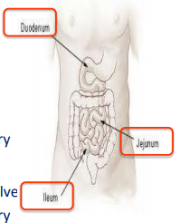
- Duodenum
- Mostly retroperitoneal, First 25 cm direct from the stomach – Curves around the head of the pancreas – Receiving bile and pancreatic juice
- Jejunum
- Next ~2.5m between Duodenum and Ileum – Covered with serosa and suspended by mesentery
- Ileum
- Last ~3.6m joins large intestine at ileocecal valve – Covered with serosa and suspended by mesentery
-
- Features
- Goblet Cells - Mucus-secreting cells
- Enteroendocrine cells - Hormone-secreting cells
- Paneth's cells - Secretes lysozyme (antibacterial enzyme)
- Peyer's patches - Aggregated lymphoid follicles at end of the small intestine
- Pits (crypt of Lieberkuhn) found between villi – Cells lining these pits secrete a watery mixture called intestinal juice
- Structural Modifications for absorption
- Circular folds
- - Permanent folds (~1 cm deep) that force chyme to slowly spiral through the lumen, allowing more time for nutrient absorption
- Villi
- - Fingerlike projections of mucosa (~1 mm high) with a core that contains a dense capillary bed and lymphatic capillary called a lacteal for absorption
- Microvilli
- - Cytoplasmic extensions of mucosal cell that give fuzzy appearance called the brush border that contains membrane-bound enzymes brush border enzymes, used for final carbohydrate and protein digestion
-
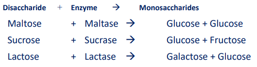
- Digestion of carbohydrates >>>>>>
- Digestion of protein peptides Pancreatic protease Single amino acids
- Fat digestion - secretions from pancreas and liver
- Salivary amylase turns Starch Oligosaccharides
- Brush border enzymes
- Dextrinase & Glucoamylase – act upon oligosaccharides with more than 3 simple sugars Maltase, Sucrase & Lactase
-
Absorption of Carbohydrate - SI
- Monosaccharides (glucose, fructose & galactose) are not small enough to diffuse directly through the plasma membrane into the cell TWO transport mechanisms that exist especially for glucose: Facilitated Diffusion Sodium-Glucose Co-transporter (SGLT-1) The movement of glucose is coupled to the gradient of Na+ Na+ flow down their gradient into the cell, providing the energy to transport glucose into the cell against its gradient Enter capillaries of the villus & carried to hepatic portal circulation
-
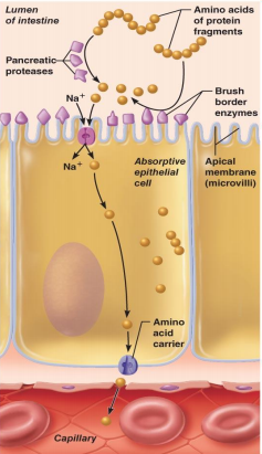
- 1.) Proteins and protein fragments are digested to amino acids by pancreatic proteases (typsin, chymotrypsin, and carboxy - peptidase), and by brush border enzymes (carboxypeptidase, aminopeptidase, and dipeptidase) of muscosal cells,
- 2) The amino acids are then absorbed by active transport into the absorptive cells, and move to their opposite side.
- 3.) The amino acids leave the villus epithelial cell by facilitated diffusion and the capillaries via the intercellular slefts.
-
- dual-action, with features of both Exocrine & Endocrine glands Exocrine gland: Secretes digestive enzymes through ducts Endocrine gland: Ductless; produce hormones that are released into the bloodstream Pancreatic juice consists of: – H2O – Sodium bicarbonate –Neutralises stomach’s acidic chyme – Lipase –Breakdown lipids into fatty acids & glycerol
- – Amylase CHO (polysaccharides, disaccharide maltose) – Trypsin, chymotrypsin & carboxypeptidase Proteins (polypeptides) broken down into short-chain amino acids – RNA & DNA digestive enzymes Nucleases
-
- Located under the diaphragm in the upper right quadrant • Consists of 4 lobes, which are made up of thousands of lobules • These lobules are the functional units of the liver • The liver can lose 3/4 of its cells (hepatocytes) before it stops functioning- the only organ in the body that can regenerate itself • The associated gallbladder sits in a pocket on the ventral surface
- Produces & secretes Bile – Bile emulsifies fats – Breaks fats down into smaller globules so that enzymes can digest them more efficiently
- digestive function is the production of bile Bile: fat emulsifier
- Metabolises
- - Carbohydrates
- - Proteins
- -Fats
- Stores
- - Carbohydrates
- - Fats
- -Vitamins and Minerals
-
- - •oxygenated blood flows in via the hepatic artery
- - •venous blood flows in via the hepatic portal vein carrying nutrients and toxic materials from the intestine, blood cells and their breakdown products from the spleen, and insulin and glucagon from the pancreas
- Sinusoids are vascular channels allowing exchange between blood & hepatocytes •Blood exits each lobule by small veins which combine to carry blood AWAY from the liver via the hepatic vein
-
- Bile produced in hepatocytes drains into canaliculi → exits the lobules via bile ducts → exits the liver via the hepatic duct → enters the duodenum via the common bile duct
-
- Carbohydrates
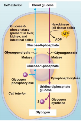
- - When glucose is ingested, insulin stimulates Glycogenesis (synthesis of glycogen) When glucose is in short supply, glycogen is broken down and released into the blood to be transported to cells The process of splitting glycogen into glucose is called Glycogenolysis
- When glycogen stores are depleted, the body can catabolise proteins and fats for energy OR The glycerol part of triglycerides, lactic acid & certain amino acids can be converted into glucose in a process known as Gluconeogenesis
-
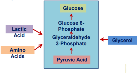
- Glucose
- Pyruvic Acid
- Amino Acids
- Lactic Acid -
- Glycerol
-
Liver Metabolism - Fats and Proteins
- Fats
- Triglycerides are routinely catabolised for energy… broken down into Fatty Acids and Glycerol If an immediate energy source is not required, these fatty acid and glycerol molecules are recombined back into triglycerides in a process known as Lipogenesis They can also undergo anabolic reactions to produce other lipids such as Lipoproteins, Phospholipids, and Cholesterol The breakdown of stored fats into fatty acid and glycerol is known as Lipolysis
- Proteins
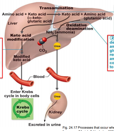
- Proteins are broken down into amino acids (AAs)… Amino acids are made up of two parts a carbon group and an amine group Transamination is where some AAs are converted into other AAs by transferring an amine group Hepatocytes convert some AAs to fatty acids (lipogenesis), ketone bodies (ketogenesis) or glucose (gluconeogenesis) some are oxidised in the Krebs Cycle Oxidative Deamination in the liver is the removal of an amine group (NH3 ) from glutamic acid Ammonia
-
- 1.) During transamination, an amine group is switched from an amino acid to a keto acid
- 2.) In oxidative deamination, the amine group of glutamic acid is removed as ammonia and combined with CO2 to form urea
- 3.) During keto acid modification the keto acids formed during transamination are altered so they can easily enter the Krebs cycle pathways
-
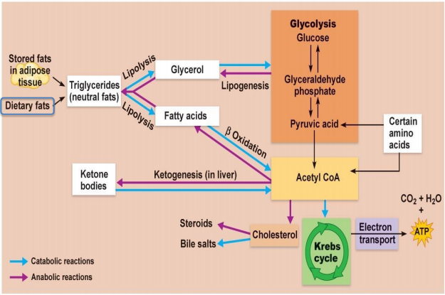
- Protein anabolism is the formation of peptide bonds between Amino Acids There are 20 Amino Acids 10 are Essential must be consumed in our diet as our body cannot synthesise them in adequate amounts As plants synthesise all AAs, humans can obtain all the essential ones by eating plants or animals that consume plants Non-essential AAs can be synthesised by body cells via transamination
-
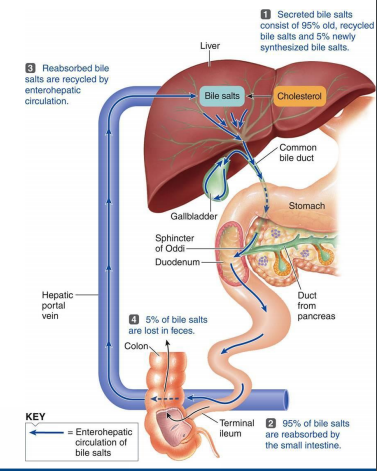
- The liver synthesises Bile Salts… • These are used in the small intestine for emulsification and absorption of lipids, cholesterol, phospholipids and lipoproteins
- 1.) Secreted bile salts consist of 95% old, recycled bile salts and 5% newly synthesized bile slats
- 2.) 5% of bile salts are lost in faeces
- 3.) Reabsorbed salts are recycled by enterohepatic circulation
- Bile is an emulsifying agent physical breakdown of lipids that exposes a greater surface area to lipases Bile is a yellow-green, alkaline fluid containing minerals, bile salts, cholesterol, triglycerides, bile pigments (bilirubin, biliverdin), phospholipids (lecithin) and electrolytes (Na, Cl, HCO3 ) Abnormal increase in bile salts in blood results in extraction by sweat glands, intense itching of the skin (pruritis) and interferes with absorption of water and salt
-
- Synthesis
- The liver synthesises Bile Salts • These are used in the small intestine for emulsification & absorption of lipids, cholesterol, phospholipids & lipoproteins • Bile salts are recycled in the body via Enterohepatic Circulation
- Excretion
- Bilirubin is derived from the heme of old erythrocytes It is absorbed by the liver from the blood and secreted into Bile It is metabolised in the small intestine by bacteria and then eliminated in faeces
- Processing
- The liver detoxifies certain substances and excretes drugs into Bile It can chemically alter and/or excrete thyroid or steroid hormones
- Storage
- In addition to glycogen & triglycerides, the liver stores Vitamins (A, B12, D, E & K) & Minerals (Iron & Copper), and releases them when needed elsewhere in the body
- Phagocytosis
- The Kupffer cells of the liver phagocytise aged red & white blood cells and some bacteria
- Activation of vitamin D
- In conjunction with skin & kidneys, the liver participates in synthesising the active form of vitamin D
-
- Most common liver diseases include: • Hepatitis A, B, C, E • Alcohol damage • Cirrhosis • Drug damage? • Symptoms can include: • Pale stools • Dark urine • Jaundice • Fatigue
-
- - Stores and concentrates bile
-
- of The Pancreas and Liver
- 1.) Chyme entering duodenum (Small Intestine) causes the release of cholecystokinin (CCK) and secretion from duodenal enteroendocrine cells.
- 2.) CCK (red dots) and secretin (yellow dots) enter the bloodstream.
- 3.) CCK induces secretion of enzyme-rich pancreatic juice. Secretin causes secretion of Hydrochloric basic rich pancreatic juice
- 4.) Bile salts and a lesser extent, secretin transported via bloodstream stimulate the liver to produce bile more rapidly
- 5.) CCK (via the bloodstream) causes the gallbladder to contract and hepatopancreatic sphincter to relax; bile enters duodenum.
- 6.) During cephalic and gastric phases, vagal nerve stimulation causes weak contractions of the gallbladder
-
- Begins as Caecum & appendix in the lower right corner of abdomen / ileocaecal valve Lined by simple columnar epithelium with mucus secreting goblet cells
- Main functions: • Reabsorb H2O & electrolytes from indigestible food materials • Absorbs Vitamin K & B from enteric bacteria (relating to the intestine) • Eliminate undigested residues
-
Regions of the Large Intestine
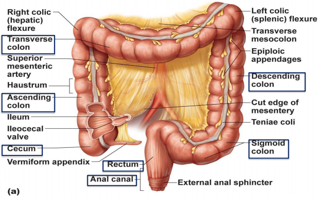
- Caecum: sac-like / appendix attached to its bottom – Vermiform Appendix: contains lymphoid tissue / immune defense Ascending colon: travels up the right side of the body to the level of right Kidney Transverse colon: travels across the abdomen; right to left toward spleen Descending colon: Runs down left side of abdomen / enters pelvis Sigmoid colon: S-shaped portion leading down pelvis Rectum: is straight portion / runs down through pelvis in front of sacrum & ending at anal canal Anus: 3cm long / 2cm wide. Has voluntary external sphincter & involuntary internal sphincter
-
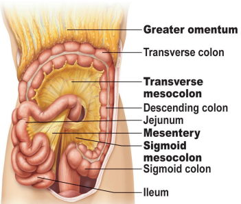
- Mesentery of small intestines holds many blood vessels Mesocolon anchors the colon to the back body wall
-
Digestion and Absorption - Carbohydrates
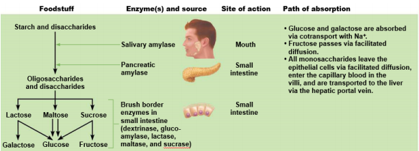
- - Glucose and galactose are absorbed via cotransport with Na+
- - Fructose passes via facilitated diffusion
- - All monosaccharides leave the epithelial cells via facilitated diffusion, enter the capillary blood in the villi, and are transported to the liver via the hepatic portal vein
-
Digestion and Absorption - Proteins
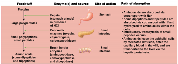
- - Amino acids are absorbed via cotransport with Na+
- - Some dipeptides and tripeptides are absorbed via cotransport with H+ and hydrolyzed to amino acids within the cells
- - Infrequently, transcytosis of small peptides occurs
- - Amino acids leave the epithelial cells by facilitated diffusion, enter the capillary blood in the villi, and are transported to the liver via the hepatic portal vein.
-
Digestion and Absorption - Fats
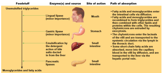
- - Fatty acids and monoglycerides enter the intestinal cells via diffusion. Fatty acids and monoglycerides are recombined to form triglycerides and then combined with other lipids and proteins within the cells. The resulting chylomicrons are extruded by exocytosis
- - The chylomicrons enter the lacteals of the villi and are transported to the systemic circulation via the lymph in the thoracic duct
- - Some short-chain fatty acids are absorbed, move into the capillary blood in the villi by diffusion, and are transported to the liver via the hepatic portal vein.
-
Digestion and Absorption - Nucleic Acids
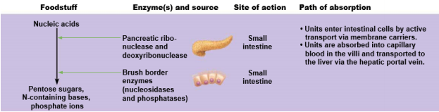
- - Units enter the intestinal cells by active transport via membrane carriers.
- - Units are absorbed into the capillary blood in the villi and transported to the liver via the hepatic portal vein.
-
- Mass peristaltic waves push faecal material from the sigmoid colon into the rectum Rectum involuntary contractions push the faeces downward Filling of the rectum stimulates stretch receptors Defecation reflex stimulates rectum longitudinal muscle contractions Relaxation of internal anal sphincter Voluntary relaxation of external sphincter (adults & older children)
- 1.) Distension, or stretch, of the rectal walls due to movement of feces into the rectum stimulates stretch receptors there. The receptors transmit signals along afferent fibres to spinal cord neurons.
- 2.) A spinal reflex is initiated in which parasympathetic motor (efferent) fibres stimulate contraction of the rectal walls and relaxation of the internal anal sphincter
- 3.) If it is convenient to defecate, voluntary motor neurons are inhibited, allowing the external anal sphincter to relax so that feces may pass.
Información
- Descripción
- Diapositivas sobre Sin título, creado por Natasha Hilton el 13/03/2018.
- Creado por
- Natasha Hilton
- Visitas
- 1
- Valoración