
Radiology with Hafferman

Diapositivas del conjunto (14)
-
- --KVP-- Kilo Voltage Peak
- Penetration power
- Increased to go through bone
- --MAS-- Milliampere second
- The contrast
- Increased for definition
- Lungs- Higher KVP, lower mas
- Abdomen- Lower (slightly) KVP, higher mas
-
- ~DV positioning
- -Rostral (nose) to 1st or 2nd cervical (neck) vertebra
- -Center directly over skull
- ~Lateral poisoning
- -Rostral (nose) to 1st or 2nd cervical (neck) vertebra
- -Lower jaws should overlap
- -Center directly over skull

- Pie de imagen: DV of skull and lateral of skull
-

- ~Lateral positioning
- -Use wedges (thin part under spine to straighten)
- -Base of skull to thoracic inlet
- -Centered where collar would be
- ~Ventraldorsal poisoning
- -Use wedges (?)
- -Base of skull to thoracic inlet
- Pie de imagen: Lateral view and VD view
-

- Pie de imagen: Lateral shoulder view
- ~Lateral poisoning
- -Each shoulder taken separately
- -Centered directly over joint (shoulder)
- -Roll shoulder outward
- -Humorous will align with trachea
-

- Pie de imagen: Elbow DV and lateral view
- ~DV positioning
- -Both front limbs for comparison
- -Centered directly over joint (elbow)
- -Shoulder to paw
- ~Lateral poisoning
- -90° angel at elbow
- -20° angel at carpus
- -Shoulder to paw
-

- Pie de imagen: Lateral view and DV view
- ~Lateral positioning
- -Carpus straight or slightly flexed
- -Both for comparison
- -Directly over joint (carpus)
- ~DV poisoning
- -Directly over joint (carpus)
- -Both legs for comparison
-
- Pie de imagen: VD chest and lateral chest
- Image colors:
- -When the image is white: KVP too low
- -When the image is dark: KVP too high
- ~Lateral positioning large breed
- -Thoracic inlet to three fingers length from the sternum (xiphoid)
- -Center of image in line with elbow
- -One line diaphragm in bottom of image
- ~Lateral positioning small breed
- -Thoracic inlet to two fingers length from the sternum (xiphoid)
- -Center image in line with elbow
- -One line diaphragm in bottom of image
-
- Image colors:
- -When the image is white: Mas decrease
- -When the image is dark: Mas increase
- ~Positioning large breed
- -Three fingers length from sternum (xiphoid) to point of greater hip (greater tubercle)
- -Center of image cranial to prepuce
- -One line diaphragm top of image
- ~Positioning small breed
- -Two figures length from sternum (xiphoid) to greater hip (greater tubercle)
- -Center of image cranial to prepuce
- -One line diaphragm top of image

- Pie de imagen: VD abdominal and Lateral abdominal
-
Spinal (thoracic and Lumbar) Rads

- ~Thoracic positioning
- -From above shoulder blades to last rib (TL junction)
- -Center directly above spine
- -Spine is the only thing in view
- ~Lumbar poisoning
- -1 finger above last rib to base of tail
- -Center directly over spine
- -Spine is the only thing in view
- Pie de imagen: Lateral Thoracic (above) and lateral Lumber (below)
-
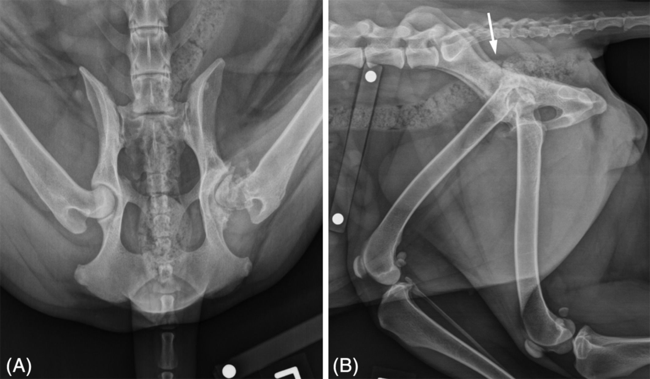
- ~Frog Leg
- -Naturally laying on back with legs splayed
- -More comfortable for P
- -Centered directly over hip joint
- ~Running Man
- -Lateral hip
- -Top leg loosely pulled forward (to offset legs/hip)
- -Bottom leg loosely pulled back (to offset legs/hip)
- -Centered on pint of hip (greater tubercle)
- Pie de imagen: Frog Leg (natural on back, VD) and Running Man (lateral)
-

- Butt shot is positioned to see the urethra and taken in lateral
- ~Positioning
- -Legs loosely pulled forward
- -Three fingers back from sternum (xiphoid) to three fingers past the point of hip (greater tubercle)
- Pie de imagen: Butt shot
-

- Taken as a lateral and VD/DV
- ~Positioning
- -Center directly over joint
- -Knee bent to 90° angel
- -Can be taken as VD or DV, DV will likely be more comfortable for -Good VD/DV will show fabella on wither ether side of the femur
- -Knee cap (patella) will be in center
- Pie de imagen: Lateral stifle and DV stifle
-

- ~DV positioning
- -Both front limbs for comparison
- -Centered directly over joint (paw)
- ~Lateral poisoning
- -Both front limb for comparison
- -Centered directly above joint (paw)
- -Flexion downward with spoon on top of paw to each toe is splayed laterally
- Pie de imagen: DV rad and lateral
-

- Pie de imagen: Lateral tail
- ~Lateral positioning
- -Point of hip (greater tubercle) to tip of tail (may need multiple views depending on length of tail
- -Center directly over tail bone
Información
- Descripción
- Radiology positioning
- Creado por
- Bree Eikenberry
- Visitas
- 57
- Valoración
Más sobre Radiographic Positioning