
Fichas
Radiographic Image Interpretation Flashcards
Fichas del conjunto (125)
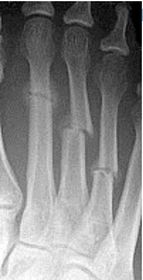
- 1Describe the alignment of metatarsal 2There is no
angulation nor displacement of the
distal segment relative to the
proximal segment. - 2Describe the alignment of metatarsal 3The distal
segment is displaced laterally
(50% apposition) and angulated
medially.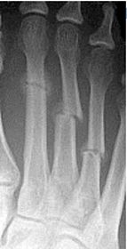
- 3Describe the alignment of metatarsal 4The distal
segment is displaced laterally
(75% apposition) but there is no
apparent angulation.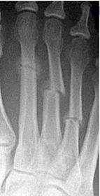
- 4Describe the tubulation of the metatarsalsMetatarsals are overtubulated - decreased girth.
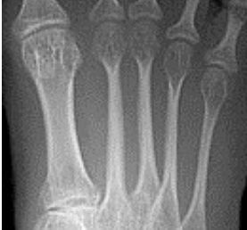
- 5Describe the tubulation of the metatarsalsMetatarsals are undertubulated - increased girth.
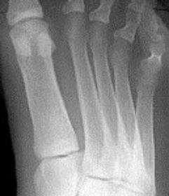
- 6Describe the normal variation of the sesamoid bonesTibial sesamoid is bipartite
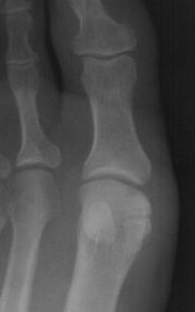
- 7What is this?Bipartite medial cuneiform
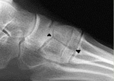
- 8What is this?Bipartite navicular
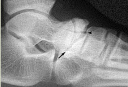
- 9What is this? Where is it typically found?Os interphalangeus.
Typically found on the inferior aspect of the hallux IPJ. It is rare to see this ossicle at the
IPJs of the lesser toes.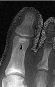
- 10What is this?Accessory sesamoids.
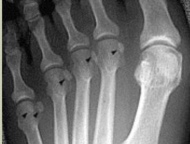
- 11What is this?Os Intermetatarseum
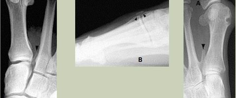
- 12What is this?Tibialis Anterior Tendon Sesamoid
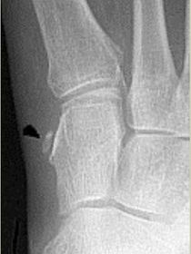
- 13What is this?Os Vesalianium
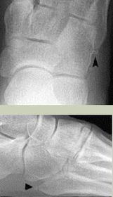
- 14What is this? What tendon is it found in?Os Peroneum.
Sesamoid bone found in peroneus longus tendon.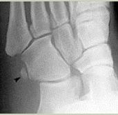
- 15What is this?Os Infranaviculare
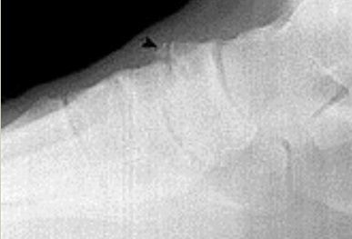
- 16What is this?Os Supranaviculare
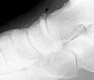
- 17What is this?Os Supratalare
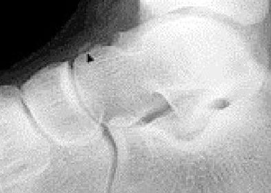
- 18What is this? What type is it?Accessory navicular (Os tibiale externum)
Type 1: Sesamoid in the tendon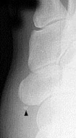
- 19What is this? What type is it?Accessory navicular (Os tibiale externum)
Type 2: Articulating accessory ossification centre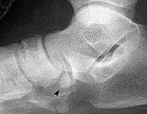
- 20What is this? What type is it?Accessory navicular (Os tibiale externum)
Type 3: Fused accessory ossification centre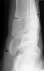
- 21What is this? What is it called when fused to the talus?Os trigonum
Called trigonal (Steida's) process when fused to the talus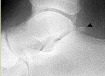
- 22What is this?Os calcaneus secundarius
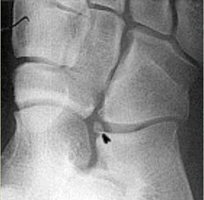
- 23What is this?Os supracalcaneum
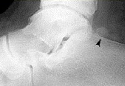
- 24What is this?Os subtibiale
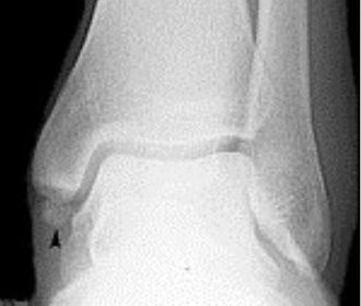
- 25What foot type does this calcaneal inclination angle indicate?Pes cavo-varus
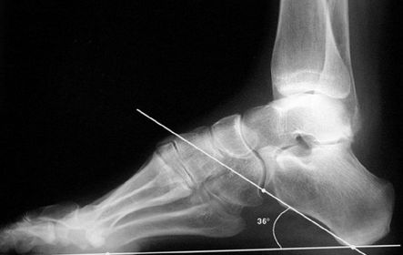
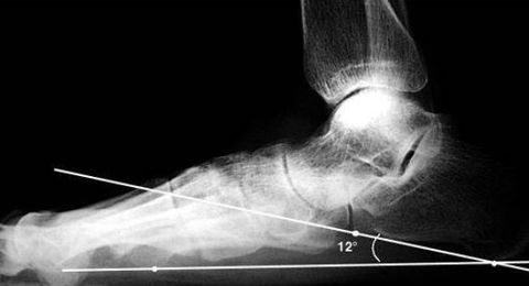
- 26What foot type does this calcaneal inclination angle indicate?Pes plano-valgus
- 27What is this angle called? What foot type does it indicate?Talar - 1st MT or Meary's Angle
Pes plano-valgus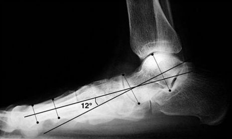
- 28What is this angle called? What foot type does it indicateTalar - 1st MT or Meary's Angle
Pes cavo-varus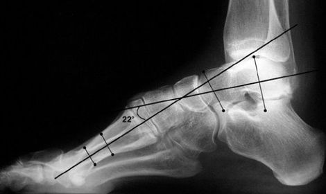
- 29What is this angle called? What is normal? What does an increased angle indicate?Talo-calcaneal angle.
25-45 degrees.
Increased angle = hindfoot valgus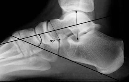
- 30What is this angle called? Is this normal or abnormal?AP - Talo-calcaneal angle
This is normal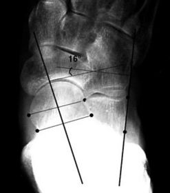
- 31What is this angle called? Is this normal or abnormal?AP-Talo-calcaneal angle
This is abnormal. The angle is reduced indicating pes cavo-varus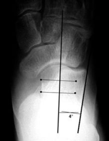
- 32What is this angle called? Is this normal or abnormal?Talo-navicular coverage.
This is normal.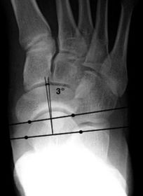
- 33What is this angle called? Is this normal or abnormal?Talo-navicular coverage.
This is abnormal as the talus is laterally deviated with medial overhang of navicular indicating pes cavo-varus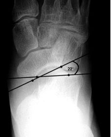
- 34What is this called? What does an anterior break indicate? What does a posterior break indicate?Cyma Line/Midtarsal joint.
Anterior break = pronated foot type
Posterior break = supinated foot type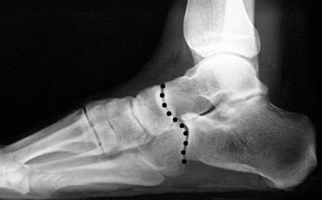
- 35What is this? What foot type is this visible in?Sinus tarsi/bullet hole sign.
Pes cavo-varus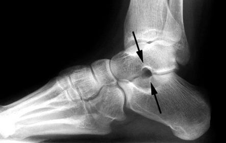
- 36What is this? What foot type is this visible in?Double talar dome sign.
Pes cavo-varus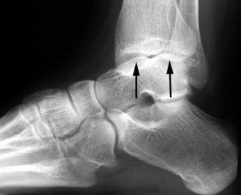
- 37What angle is this? Is this normal or abnormal?1st intermetatarsal angle
Abnormal. Greater than 9 degrees is abnormal. Usually due to metatarsus primus varus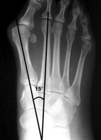
- 38What deformity is this?Metatarsus Adductus
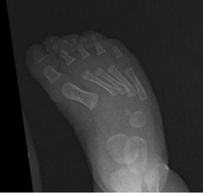
- 39What deformity is this?Metatarsus Adductovarus
(ladder like arrangement of the metatarsals in lateral view)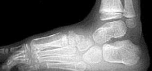
- 40What deformity is this?Skew foot
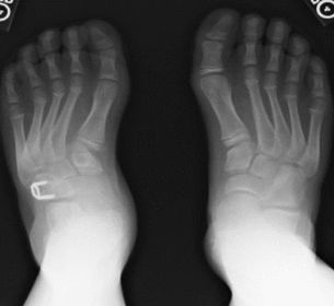
- 41What disease is this?Osteoarthritis
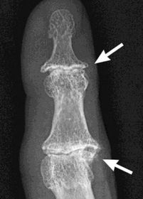
- 42What disease is this?Rheumatoid arthritis
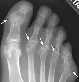
- 43What disease is this? What does the arrow indicate?Rheumatoid arthritis.
Arrow = Bone erosion secondary to inflammation of retrocalcaneal bursa.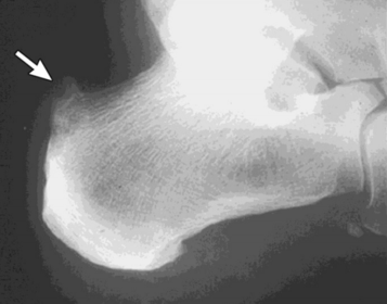
- 44What disease is this?Gout
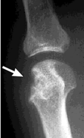
- 45What disease is this? What do the arrows indicate?Gout
Arrows = 'punched out' erosions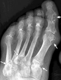
- 46What disease is this? What indicates this?Tophaceous gout.
Soft tissue swelling surrounding the index
finger PIPJ, with associated erosion and bone resorption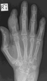
- 47What disease is this?Tophaceous gout.
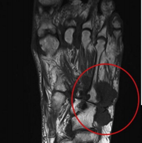
- 48What disease is this? What is shown by A, B and C?Psoriatic arthritis.
A = destructive changes
B = Pencil in cup deformity
C = IPJ fusion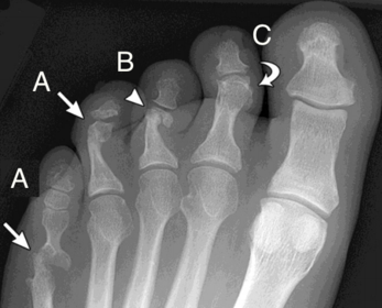
- 49What disease is this?Psoriatic arthritis
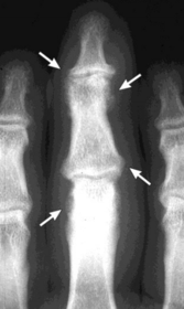
- 50What disease is this? What does the arrow show?Non-articular psoriatic arthritis
Arrow = 'fluffy spur'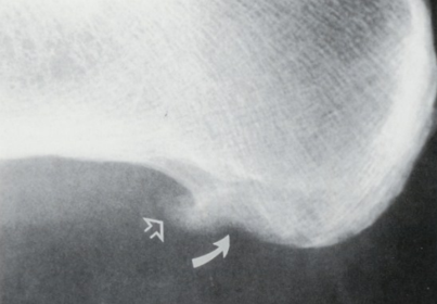
- 51What is this?Calcaneonavicular coalition
(AKA calcaneonavicular bar or anteater's nose)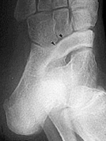
- 52What is this?Calcaneonavicular coalition
(AKA calcaneonavicular bar or anteater's nose)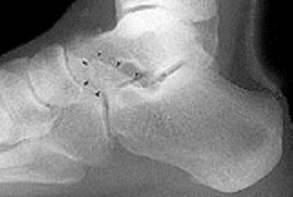
- 53What is this?Calcaneonavicular coalition
(AKA calcaneonavicular bar or anteater's nose)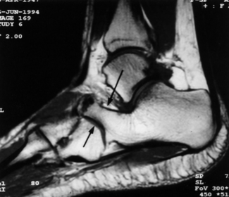
- 54What is this? What is shown by A and B?STJ coalition
A = talar neck spurring
B = Halo or 'C' sign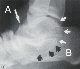
- 55What is this?Posterior talocalcaneal coalition
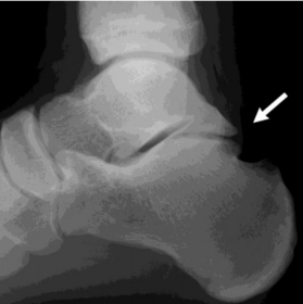
- 56What is shown by A and B?A = Talonavicular coalition
B = Calcaneocuboid coalition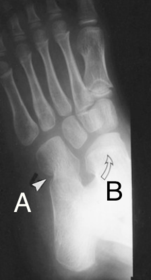
- 57What is this?Cuboid-navicular coalition
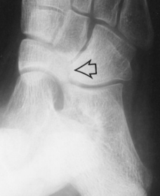
- 58What type of fracture is shown at the 2nd metatarsal?Transverse fracture
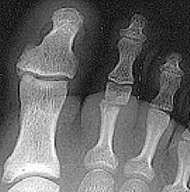
- 59What type of fracture is this?Oblique fracture
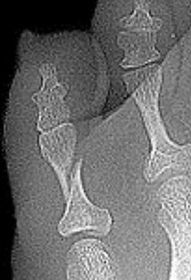
- 60What type of fracture is this?Spiral fracture
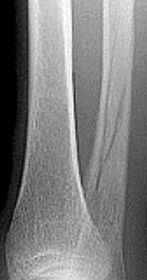
- 61What type of fracture is this?Comminuted fracture
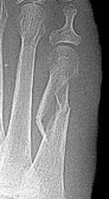
- 62What type of fracture is this?Osteochondral fracture
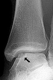
- 63What type of fracture is this?Impaction fraction
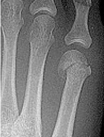
- 64What type of fracture is this?Intra-articular fracture
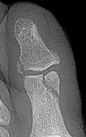
- 65What type of fracture is this?Avulsion fracture
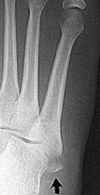
- 66What do the arrows show?Stress fracture
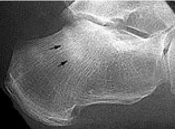
- 67Describe what has happened at the shaft of the 3rd metExuberant periosteal new bone production. May have resulted if a stress fracture was not treated and the patient continued weight bearing activities.
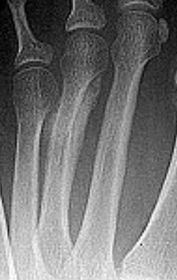
- 68What type of fracture is this?Greenstick fracture (common in paediatric patients)
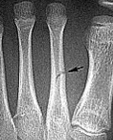
- 69Describe what the arrows are showingTorus fracture of the 1st met.
"Buckling" of the proximal one third
diaphysis is seen both
medially and laterally. Common in paediatric patients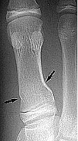
- 70What is the Salter Harris classification used for? What is the mnemonic for the 5 types?Used to classify fractures involving the growth plate.
S - slipped = type I
A - above = type II
L - lower = type III
T - through = type IV
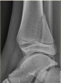
R - ruined/rammed = type V - 71What type of fracture is this?Salter-Harris Type II
- 72What type of fracture is this?Salter-Harris Type III
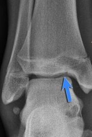
- 73What type of fracture is this? How does it occur?Tillaux fracture - a fracture of the anterolateral tibial epiphysis that is commonly seen in adolescents.
The fragment is avulsed due to the
strong ATF ligament in an external rotation injury of the foot.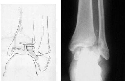
- 74What type of fracture is this?Jones fracture - fracture of the diaphysis of the 5th metatarsal. More distal than an
avulsion fracture.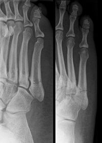
- 75What classification system is this? Describe the 3 classificationsWeber Ankle Fracture Classification
A = Fracture below syndesmosis
B = Fracture begins at joint level & extends proximally in an oblique fashion
C = Fracture above the joint line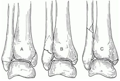
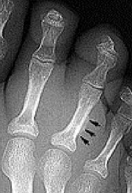
- 76What do the arrows indicate?Infectious periostitis
- 77What disease is this? What signs indicate this?Osteomyelitis.
Osteolysis (fragmentation), sequestra (dead/necrotic bone fragments floating
in pus), rarefacation (loss of bone density).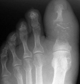
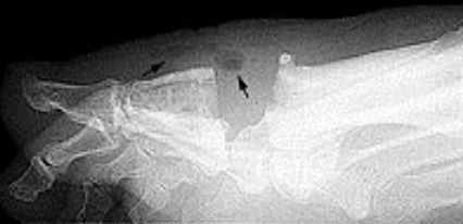
- 78What does the arrow show?Soft tissue emphysema
- 79What disease is this? What do A, B & C show?Osteomyelitis.
A = Soft tissue swelling
B = Periosteal reaction
C = Bony destruction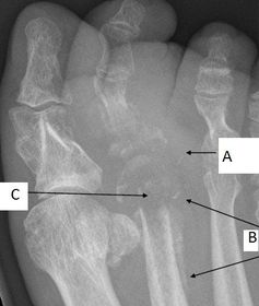
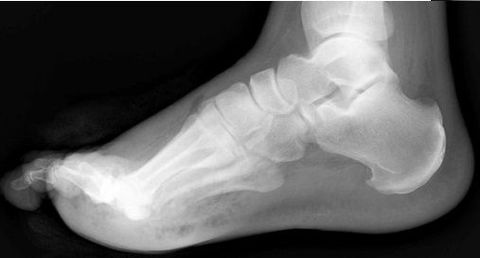
- 80What disease is this?Gas gangrene
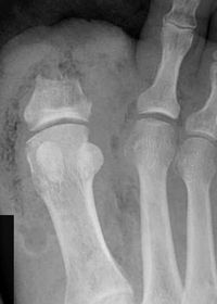
- 81What disease is this?Gas gangrene
- 82What disease is this? What causes it?Brodie's abscess - chronic bone abscess caused by organisms of low virulence. Patient most likely did not go through an acute phase (just chronic osteo)
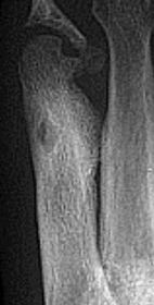
- 83What disease is this?Monckeberg’s arteriosclerosis
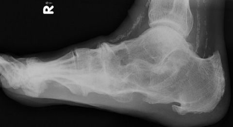
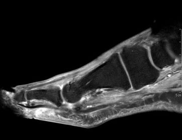
- 84What does this indicate?Increased signal and enhancement of the soft tissue plantar to the proximal phalanx suggestive of subcutaneous infection without abscess
- 85What does this indicate?Increased signal and soft tissue distension dorsally representing subcutaneous abscess
communicating with associated with plantar
ulcerative defect.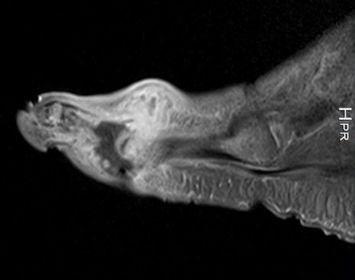
- 86What disease is this?Atrophic
neuroarthropathy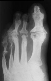
- 87What disease is this?Atrophic forefoot neuropathy
(diabetic osteolysis)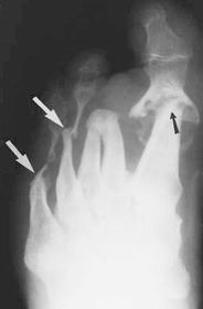
- 88What disease is this?Charcot Neuroarthropathy
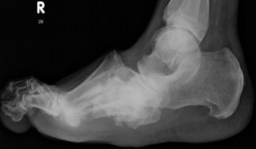
- 89This patient has taken a short course of prednisone. What does the image show?Osteonecosis of the talus
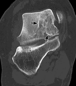
- 90What does this image show?Avascular necrosis of the talus, secondary to a talar neck fracture
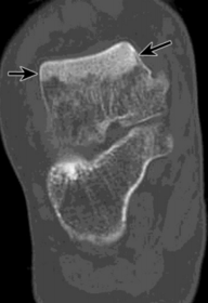
- 91What do these images show? What is this disease called? How does this differ from Meuller Wiess Syndrome?Osteochondrosis of the navicular.
Kohler's disease.
Kohler's disease only appears in children, Meuller Wiess Syndrome only appears in adults and is a spontaneus osteonecrosis of the navicular, not a true osteochondrosis.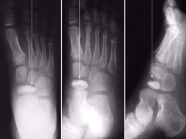
- 92Describe what is occurring at the head of the 3rd metFreiberg’s infarction (avascular necrosis of the metatarsal head)
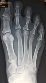
- 93What disease is this?Sever's disease
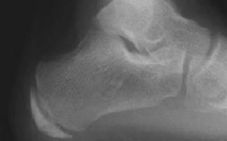
- 94What is this? What else could it be?Iselin’s disease
Traction apophysis, Os peronei, avulsion fraction, lis francs dislocation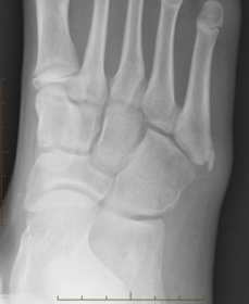
- 95Describe the patterns of destruction in A, B, C. Which is more malignant?A = geographic destruction
B = moth eaten destruction
C = permeative destruction
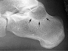
Malignancy = C>B>A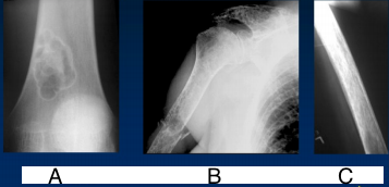
- 96What type of lesion is this?Geographic - slow growing lesion
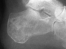
- 97What type of lesion is this?Moth eaten and permeative (mixed increased and decreased density throughout the entire calcaneus)
- 98Describe this lesionA lytic lesion of the proximal phalanx that
appears quite aggressive due to bone loss; however, the well-defined margins of the bone suggests a soft tissue tumor causing pressure atrophy against adjacent bone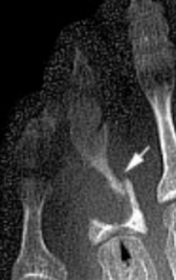
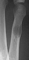
- 99What type of lesion is this? What type of periosteal reaction is this?Geographic. Continuous shell
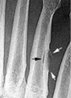
- 100What type of periosteal reaction is this?Interrupted
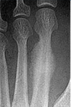
- 101What type of periosteal reaction is this?Complex
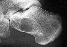
- 102What type of lesion is this?Unicameral bone cyst
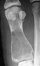
- 103What type of lesion is this?Giant cell tumour
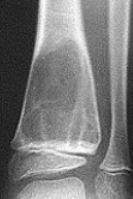
- 104What type of lesion is this?Aneurysmal bone cyst
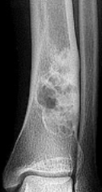
- 105What type of lesion is this?Chondromyxoid fibroma
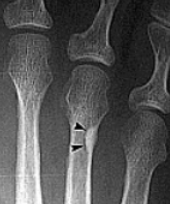
- 106What type of lesion is this?Osteogenic
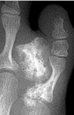
- 107What type of lesion is this?Chondrogenic
- 108What disease is this? What are the signs of this?Osteogenesis imperfecta
(“Brittle bone disease”)
Diffuse cancellous osteopenia (coarse
primary trabeculae), diminished tubular
bone girth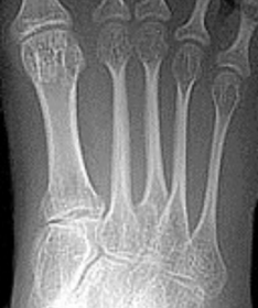
- 109What disease is this? What are signs of this?Osteopoikilosis
Defined homogeneous circular foci of increased density (bone islands), cortical-like densities in cancellous bone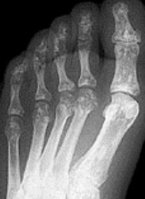
- 110What disease is this? What is it caused by? What indicates this?Osteopetrosis ("marble bone
disease")
Diffuse bone sclerosis due to osteoclasts
dysfunction.
"Bone within bone" appearance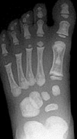
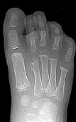
- 111What disease is this?Osteochondromatosis (multiple cartilaginous exostoses)
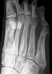
- 112What disease is this?Melorheostosis
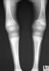
- 113What disease is this?Rickets
- 114What disease is this?Osteogenesis Imperfecta
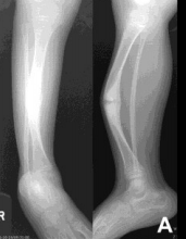
- 115What does this image show? What causes it?Regional bone loss
Disuse, immobilisation, paralysis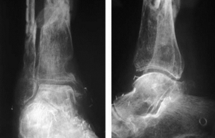
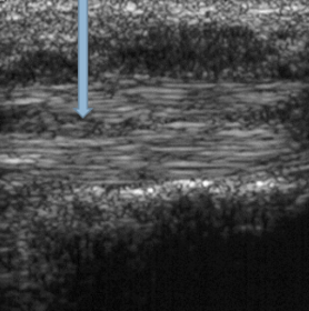
- 116In an ultrasound, what colour is bone, tendon and water (oedema)?Bone = white
Tendon = grey
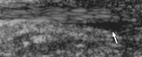
Water = black - 117What does the arrow indicate?A cleft (tear) in the tendon
- 118What does the red arrow show? What does the yellow arrow show?Red arrow = fluid
Yellow arrow = tendon tear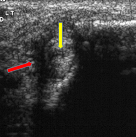
- 119What does the arrow indicate?Fluid
- 120Describe the tendon pathology as seen in an MRI of A-DA = normal tendon
B = Tenosynovitis
C = Tendonitis
D = Tendon Dysfunction (Functional Rupture)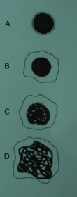
- 121What do the arrows indicate?Tenosynovitis
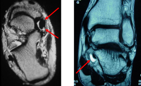
- 122What do the arrows indicate?Tendonitis
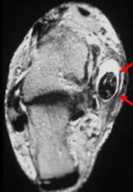
- 123What do the arrows indicate?Tendon Dysfunction – Functional Rupture
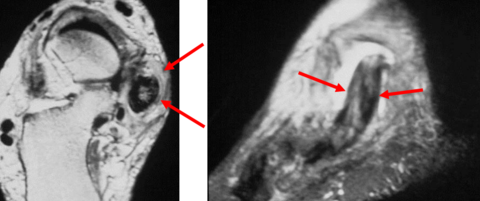
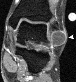
- 124What does this show?Ganglion in lateral ankle soft tissue
- 125What does the arrow show? What could this be?Teardrop-shaped hypointense lesion
in third interspace.
Could be morton's neuroma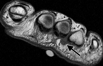
Información
- Descripción
- flashcards for radiographic image interpretation
- Creado por
- fliss29 · oct 2015
- Actualizado
- 1 feb 2018
- Visitas
- 709
- Etiquetas
- Valoración