11070674
Description
Flashcards by Agasana Viengmany, updated more than 1 year ago
|
|
Created by Agasana Viengmany
about 8 years ago
|
|
| Question | Answer |
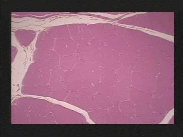
| Skeletal muscle Ep: epimysium Fa: Fasciculi Fi: individual muscle fiber BV: blood vessel | |
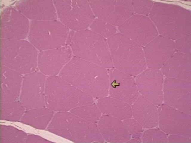
| Skeletal close view of a fasciculus which occupies almost the entire image; the thick white perimysium layer which surrounds the fasciculus; the individual muscle fibers, each of which is surrounded by a thin line (pink) of endomysium. | |
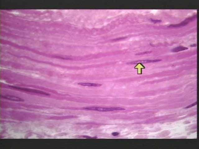
| Skeletal muscle arrow: endomysium | |
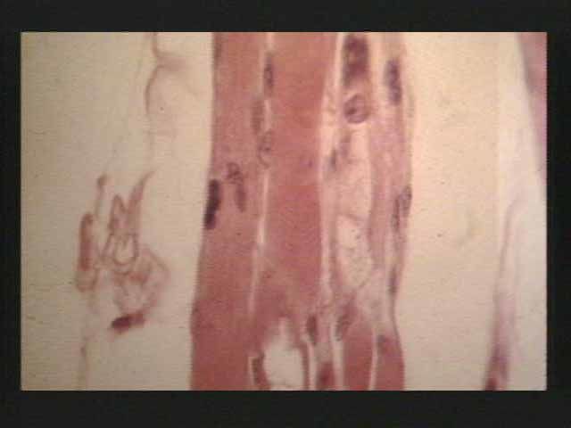
| Skeletal muscle shows dermatomyositis, inflammatory disease of muscle in which muscle fibers degenerate and die; untreated leads to progressive and sporadic weakening of muscles | |
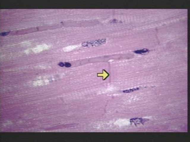
| Cardiac arrow points to an intercalated disc that serves as a bonding site between cardiac cells and a electronic junction that allows passage of the muscle membrane potential form one cardiocyte to another | |
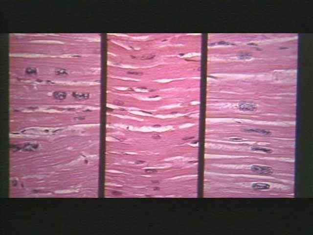
| Cardiac thin purple lines crossing the fibers are intercalated discs | |
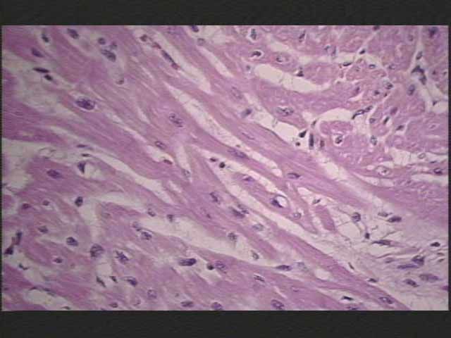
| Cardiac comparative example of normal cardiac tissue (center of the image) to hypertrophic tissue (left and right portion of image). Note that the hypertrophic cardiac fibers are much thicker than the normal cells. This is due to the proliferation of sarcoplasmic elements, such as myofilaments. | |
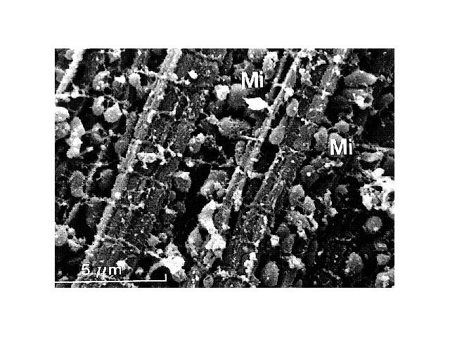
| Cardiac view of cytoplasm of cardiocyte running from top to bottom are rodlike myofibrils, crossed over by horizontal "t" tubulesthat arise from sarcoplasmic reticulum tubules (lighter colored tubes running beside and on top of the myofibrils). Note the numerous mitochondria ("Mi") located in between the myofibrils. | |
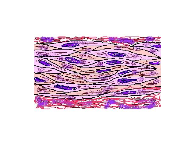
| Smooth muscle Smooth muscle fibers are long, spindle-shaped cells that contain a single nucleus and lack striations | |
| Smooth muscle flattened purple nuclei located in the middle of spindle-shaped cells. Although it is difficult to see the borders of these cells, you can see that they are tightly packed, which allows the transmission of excitatory muscle potentials directly from one cell to another. | |
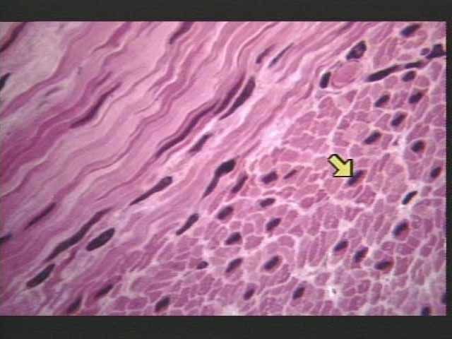
| Smooth muscle see a cross-sectional view of smooth muscle tissue (lower right) arrow points to a centrally-located nucleus in a smooth muscle cell. On the upper left of the photo you can see smooth muscle cells in a longitudinal view. | |
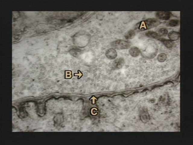
| Myoneural junction Structure a, b, c The nerve (axon terminal) is seen at the top of the slide; the muscle component at the bottom. Note the numerous synaptic vesicles ("B") in the nerve terminus, along with some darker-staining mitochondria ("A"). Toward the bottom of the slide identify the synaptic cleft ("C"--at the arrow) just below the basal lamina. Note also the highly infolded and thicker appearance of the muscle membrane (junctional folds) just below the synaptic cleft. (check pic on phone if confused) |
Want to create your own Flashcards for free with GoConqr? Learn more.