13533079
Description
Flashcards by Rasheed Assamidy, updated more than 1 year ago
|
|
Created by Rasheed Assamidy
almost 8 years ago
|
|
| Question | Answer |
| restoration | |
| secondary caries | |
| glandular odontogenic cyst | |
|
Image:
15 (binary/octet-stream)
|
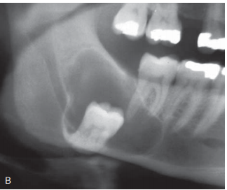
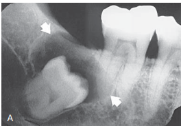
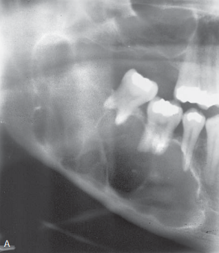
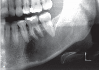
KOT DD. - dentigerous cyst but here it is not attach to CEJ - ameloblastoma, but it has a greater propensity to expand |
|
Image:
13 (binary/octet-stream)
|
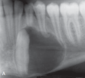
dentigerous cyst |
|
Image:
10 (binary/octet-stream)
|
condensing ostitis DD dense bone island |
|
Image:
6 (binary/octet-stream)
|
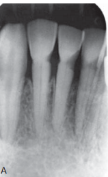
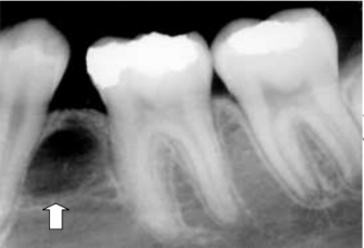
calculus |
|
Image:
4 (binary/octet-stream)
|
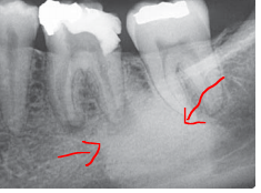
vertical bone loss of lingual plate |
|
Image:
12 (binary/octet-stream)
|
large radicular cyst |
|
Image:
9 (binary/octet-stream)
|
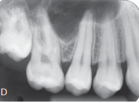
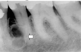
rerefying ostitis |
|
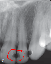
round radio opaque bodies are
Image:
7 (binary/octet-stream)
|
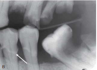
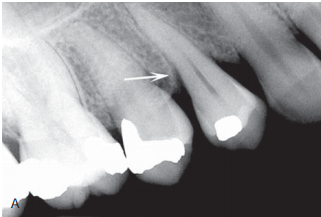
enamel pearl |
|
Image:
3 (binary/octet-stream)
|
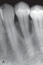
vertical bone loss |
|
Image:
8 (binary/octet-stream)
|
vertical bone loss + abrasion |
|
Image:
3 (binary/octet-stream)
|
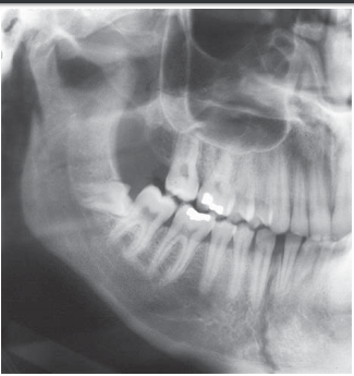
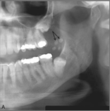
a fractured condylar head and displaced anteriorly |
|
Image:
12 (binary/octet-stream)
|
parasymphyseal region and a condylar neck fracture on the same side. |
| ameloblastoma hence root resorption DD KOT usually there is no root resorption or expansion | |
|
Image:
Pp (binary/octet-stream)
|
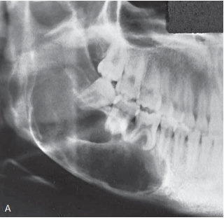
ameloblastoma , defined and course septa , root resorption and associated with expansion |
|
Image:
8 (binary/octet-stream)
|
ameloblastic fibroma The septa are infrequent and often very fine. DD - ameloblastoma ,the septa are more defined and coarse and in older age - Giant cell granulomas but usually anterior to the first molar in young patients - Odontogenic myxomas in older age |
|
Image:
1 (binary/octet-stream)
|
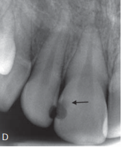
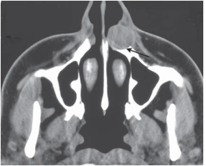
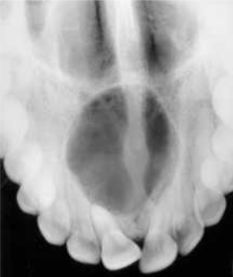
nasolabial cyst |
|
Image:
R P (binary/octet-stream)
|
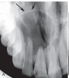
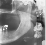
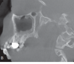
dome-shaped retention pseudocyst in the sinus |
| draping , pneumatization of maxillary sinus | |
|
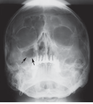
consider the ptrygoid plate fracture
Image:
L11 (binary/octet-stream)
|
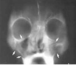
Le Fort II fracture |
|
Image:
Cln (binary/octet-stream)
|
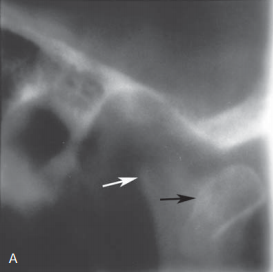
Calcified lymph nodes ''cauliflower shape'' DD Phlebolith |
|
Image:
Ndn (binary/octet-stream)
|
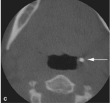
TONSILLAR CALCULI
CT scan to confirm
Image:
;; (binary/octet-stream)
|
|
Image:
لالا (binary/octet-stream)
|
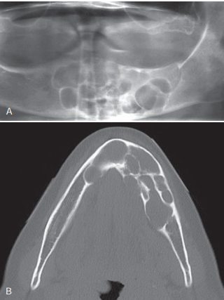
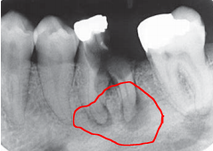
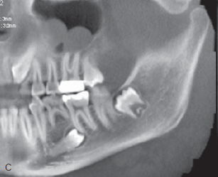
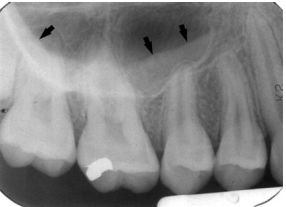
Buccal Bifurcation Cyst (paradental cyst) -Vital teeth roots tipped lingually due to expansion. |
|
Image:
Lpc (binary/octet-stream)
|
Lateral periodontal cyst: unilocular
- it can come multilocular (botryoid or “grape-like”) like this
Image:
Lld (binary/octet-stream)
|
|
Image:
Npc (binary/octet-stream)
|
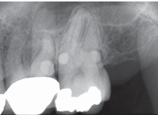
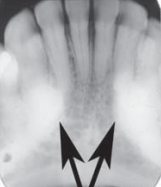
nasopalatine cyst the teeth are vital |
|
Image:
Sbc (binary/octet-stream)
|
simple bone cyst dd -KOTs usually have a more definite cortical boundary, and displace teeth -maintenance of some lamina dura and the lack of an invasive periphery and bone destruction we exclude malignancy |
|
Image:
Tori (binary/octet-stream)
|
mandibular tori |
|
Image:
Aot (binary/octet-stream)
|
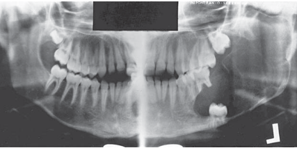
adenomatoid odontogenic tumor KOT exclude dentogerous cyst because the attachment is apical to CEJ. |
|
Image:
With4 (binary/octet-stream)
|
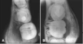
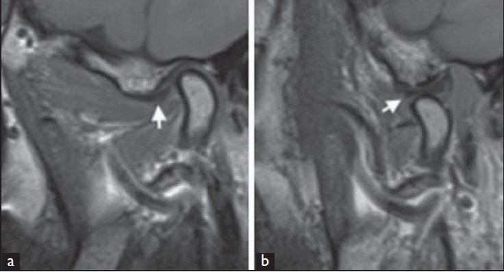
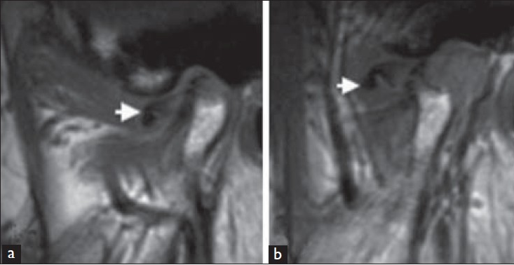
anterior disc displacement with reduction |
| anterior disc displacement without reduction | |
| antrolith DD Root fragment | |
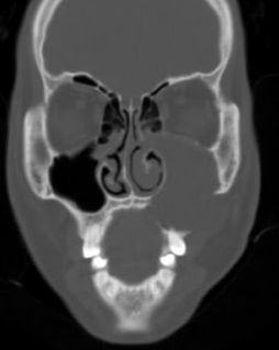
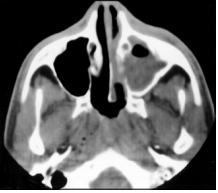
| mucocele in the left max. sinus Sinus margins are displaced outward and bone expands. teeth displaced | |
| sinusitis features :- - thickining of mucosa - sclerosis and thickening of the bony wall - if ostium get blocked this leads to fluid accumulation | |
|
Image:
''''' (binary/octet-stream)
|
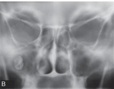
-fluid accumulation in the right sinus the arrow indicate air fluid level - complete radiopacification of the left maxillary sinus |
|
Image:
' (binary/octet-stream)
|
fluid accumulation in the left sinus |
|
Image:
Siniid (binary/octet-stream)
|
mucosal thickening |
Want to create your own Flashcards for free with GoConqr? Learn more.