34280231
Description
Flashcards by Haneen Kokash, updated more than 1 year ago
|
|
Created by Haneen Kokash
about 4 years ago
|
|
| Question | Answer |
|
Image:
Image (binary/octet-stream)
|
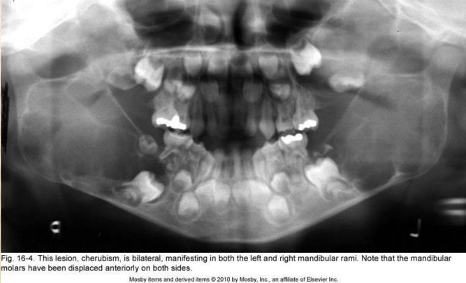
cherubism: bilateral origin in the midramus region the mandibular molars have been displaced anteriorly on both sides. it is central giant cell lesion |
|
Image:
Image (binary/octet-stream)
|
round lesion attached to cemento-enamel junction (CEJ) Corticated Border |
|
Image:
Image (binary/octet-stream)
|
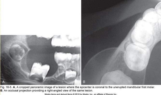
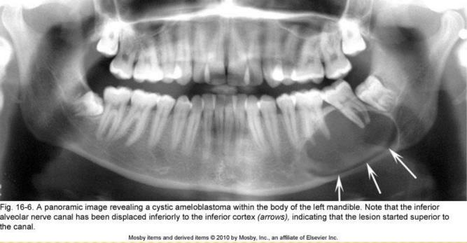
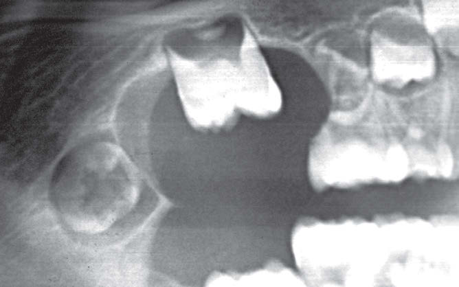
ameloblastoma a benign lesion periphery sup the alveolar canal canal shifted/displaced inf oval in shape teeth seperated or displacing the molars |
|
Image:
Image (binary/octet-stream)
|
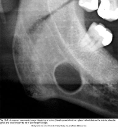
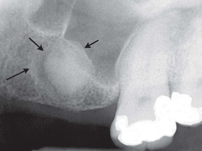
salivary gland defect or stafne bone cavity below the canal not odontogenic |
|
Image:
Image (binary/octet-stream)
|
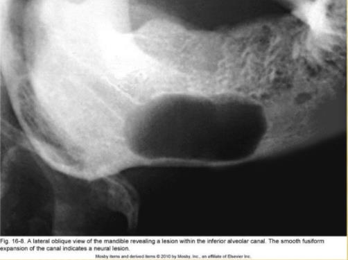
origin in inf canal it self could be neural(if symmetrical expansion) or vascular could be like hemangioma or neurofibroma |
|
Image:
Image (binary/octet-stream)
|
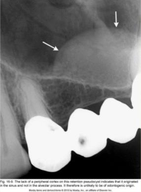
no cortical periphery so related to sinus like mucous retention psuodocyst Dome-shaped, smooth in appearance. |
|
Image:
Image (binary/octet-stream)
|
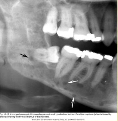
multiple malignant myeloma multiple,small punched-out lesions |
| The most common malignancy | squamous cell carcinoma |
| Punched-Out Border |
well defined
no bone
analogous to hole
surrounding has a normal appearance
ex: multiple myeloma
Image:
Image (binary/octet-stream)
|
| Corticated Border |
well defined
margin thin, uniform radiopaque line of reactive bone at the periphery of a lesion
commonly seen with cysts and benign slow-growing tumors
Image:
Image (binary/octet-stream)
|
| Sclerotic Margin |
wider zone of transition made up of a thick radiopaque border of reactive bone
not uniform
seen with periapical osseous dysplasia
indicate a very slow rate of growth or the potential for the lesion to stimulate the production of surrounding bone
Image:
Image (binary/octet-stream)
|
|
Image:
Image (binary/octet-stream)
|
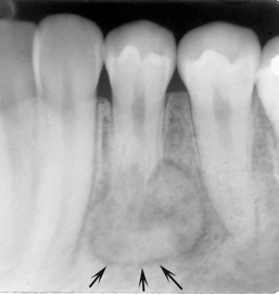
Sclerotic Margin periapical osseous dysplasia |
| Soft Tissue Capsule |
well defined
A radiopaque lesion
presence of a radiolucent line
This soft tissue capsule may be seen in conjunction with a corticated periphery
observed with odontomas and cementoblastomas
Image:
Image (binary/octet-stream)
|
|
Image:
Image (binary/octet-stream)
|
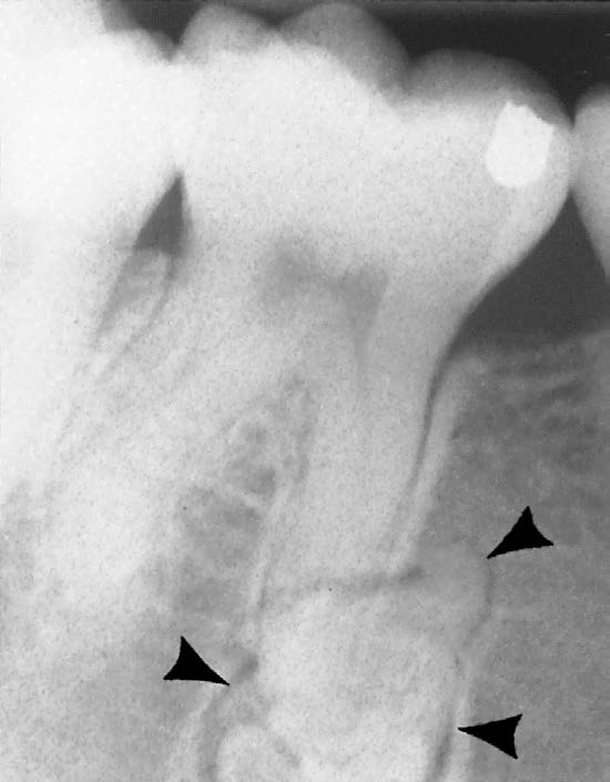
radiopaque mass associated with the root of the first bicuspid prominent radiolucent periphery (arrows) is characteristic of a soft tissue capsule of this benign cementoblastoma. |
|
so this is
Image:
Image (binary/octet-stream)
|
benign cementoblastoma. |
|
Image:
Image (binary/octet-stream)
|
well defined Thin, radiolucent periphery indicating a soft tissue capsule internal radiopaque structure odontoma |
|
so this is
Image:
Image (binary/octet-stream)
|
odontoma |
| Blending Border: |
ill defined border
gradual-wide zone of transition
Examples : sclerosing osteitis and fibrous dysplasia
Image:
Image (binary/octet-stream)
|
|
Image:
Image (binary/octet-stream)
|
sclerosing osteitis ill defined border - gradual |
| Invasive Border: |
ill defined border
of radiolucency with few or no trabeculae
wide zone of transition
associated → rapid growth and can be seen with malignant lesions.
Image:
Image (binary/octet-stream)
|
|
Image:
Image (binary/octet-stream)
|
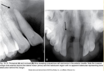
ill defined Invasive border squamous cell carcinoma(malignant) severe periodontitis, or due to LCH – langerhans cell histocytosis) |
|
Image:
Image (binary/octet-stream)
|
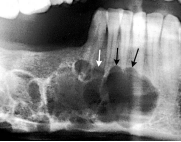
malignant neoplasm, in this case a lymphoma ill defined Invasive border |
|
Image:
Image (binary/octet-stream)
|
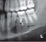
Scalloped shape keratocyst (benign) |
|
Image:
Image (binary/octet-stream)
|
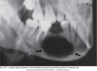
oval shaped residual cyst |
|
Image:
Image (binary/octet-stream)
|
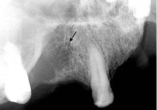
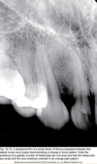
orange peel appearance ONLY associated with fibrous dysplasia. |
|
Image:
Image (binary/octet-stream)
|
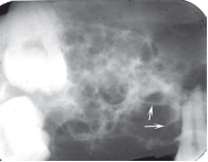
giant cell granulomas |
|
Image:
Image (binary/octet-stream)
|
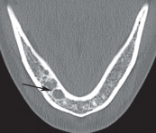
ameloblastoma - Periapical image small, soap bubble—like compartment |
|
Image:
Image (binary/octet-stream)
|
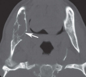
ameloblastoma - Axial CT |
|
Image:
Image (binary/octet-stream)
|
myxoma |
|
Image:
Image (binary/octet-stream)
|
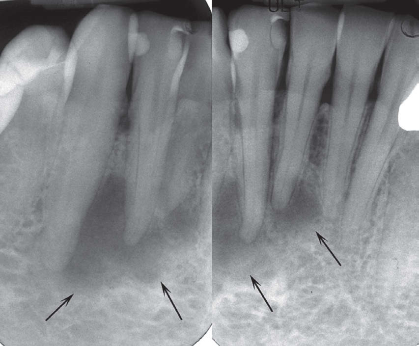
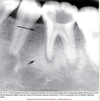
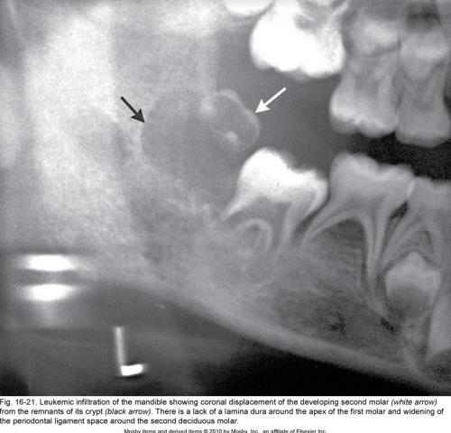
malignancy lesion widening of the PDL. |
|
Image:
Image (binary/octet-stream)
|
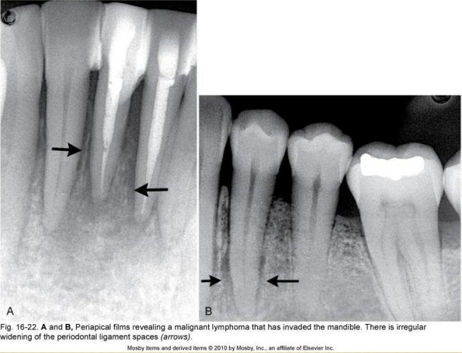
malignancy resulting in symmetrical expansions (in this case it is asymmetrical expansions). This is an example of a lymphoma |
|
Image:
Image (binary/octet-stream)
|
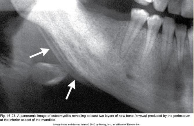
osteomyelitis. new periosteal bone indication of inflammation of bone |
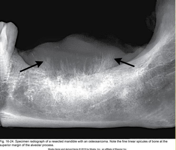
| sunray appearance or codman triangles indication of | osteomyelitis sunray appearance: 1. Malignancy (usually osteosarcoma, condrosarcoma). 2. Benign tumor like hemangioma |
|
Image:
Image (binary/octet-stream)
|
sunray appearance: 1. Malignancy (usually osteosarcoma, condrosarcoma). 2. Benign tumor like hemangioma |
Want to create your own Flashcards for free with GoConqr? Learn more.