1788475
| Question | Answer |
| Multifactorial Inheritance | Condition attributed to the additive effects of genetic and environmental factors |
| Hardy-Weinberg Equilibrium | The relative proportions of each genotype remains constant with frequencies of p^2, 2pq, and q^2 for that particular genotype. |
| Assortative Mating | Non-random mating Tendency to choose partners who share characteristics such as height, intelligence, or racial origin. |
| Consanguinity | Non-random mating. Marriage between blood relatives who have at least one common ancestor no more remote than a great-great-grandparent. |
| Coefficient of Relationship | Proportion of alleles shared by TWO individuals sharing common ancestry. COR = (1/2)^n n = # links via common ancester |
| Coefficient of Inbreeding | Proportion of homozygosity across all loci in ONE individual resulting from related ancestors. Equal to 1/2 of coefficient of relationship. COI = 1/2 x (1/2)^n n = # of links via common ancester |
| Founder Effect | Rare allele becomes very common if introduced into a small population, either due to chance or proliferative parent. |
| Centromere | Hold sister chromatids together. Site of kinetochore assembly during cell division. Several hundred kilo bases of repetitive DNA and proteins. 171 bp tandemly repeated alpha-satellite DNA clustered at all human centromeres. with sequence divergence allowing chr-specific FISH probe (except 13 and 21, 14 and 22 too similar for unique hybridization) |
| Metacentric Chromosome | Centromere is located centrally |
| Acrocentric Chromosome | The centromere is terminally located. Chr 13, 14, 15, 21, 22. Small p arm comprised of large, tandem arrays of rDNA genes (stalks). At end of stalks "satellites" with highly repetitive junk DNA. |
| Submetacentric Chromosome | The centromere is in an intermediate position. |
| Kinetochore | Site for attachment of the chromosome to the mitotic spindle. Only present during M phase. |
| Telomeres | Seal ends of chromosomes to protect from degradation and end-to-end fusions. Tandem repeats of TTAGGG sequence. Substrate for telomerase, shortens with age. True terminal deletions can generate new telomeres. TAR (telomere associated repeats) are 100-300 kb sutelomeric repeats. |
| Telomerase | Replaces the 5' end of the long strand, which would otherwise become progressively shorter. Active in germ cells, stem cells, and certain white blood cells. Activation implicated in cancer cell immortalization. |
| Interphase | Period between successive mitosis. Starts with G1 phase of cell growth. (Some cells arrest here, G0 phase) Followed by S phase of DNA synthesis. Completed by short G2 phase. |
| G1 Phase (Gap1) | Chromosomes become thin and extended. Cellular contents (not chromosomes) are duplicated. Variable length. In rapidly dividing cells, this lasts 10-12 hours. |
| S phase (synthesis) | DNA replication occurs. Chromatin of each chromosome is replicated leading to formation of two chromatids. 6-8 hours |
| Chromatin fibers | Tertiary coiling of Nucleosomes (DNA and histone proteins). |
| Euchromatin | Stains lightly (G-negative). Genes that are actively expressed. GC-rich. Rich in SINE and Alu repeats. Replicates early S phase. Housekeeping genes. |
| Heterochromatin | Stains darkly (G-positive). Made up largely of inactive, unexpressed, repetitive DNA. AT-rich. L1 (LINE) repeats. Replicates mid-late S phase. Genes are tissue specific. |
| G2 Phase (Gap2) | Chromosomes begin to condense in preparation for next mitotic division. Cell double checks the duplicated chromosomes for error making any needed repairs. 2-4 hours |
| Embryonic Cell Cycles | No cell growth. Very quick cell cycles. Only S and M phases. No Gap phases (no G1 or G2). |
| Mitosis (M phase) | Process of somatic cell division. Diploid cell yields 2 diploid cells. Pair of chromatids separate and disperse into daughter cells. |
| Prophase | Chromosomes condense. Mitotic spindle and centromeres form. Centromeres go to opposite poles of the cell as microtubules radiate from them. |
| Prometaphase | Nuclear membrane dissolves. Each chromosome becomes attached at its centromere (kinetochore) to a microtubule. |
| Metaphase | Chromosomes aligned at equatorial plane. Chromosome centriole attached to microtubule. Chromosomes maximally condensed resembling letter X in shape. |
| Anaphase | Centromere of each chromosome divide longitudinally. Two daughter chromatids separate to opposite poles. |
| Telophase | Chromosomes reach poles and begin to decondense. Nuclear envelopes begin to reform around chromosomes. Cytokinesis resulting in 2 new daughter cells. |
| Meiosis | Division for gametogenesis. One round of DNA synthesis followed by two rounds to cell division. A diploid cell leads to four haploid gametes. |
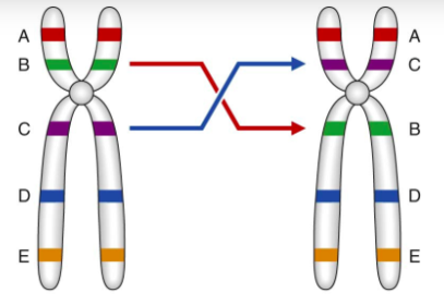
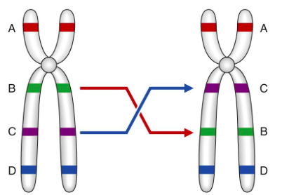
| Prophase I | Exchange of homologous segments between chromatids by crossing over or recombination (telomere>centromere; overall F>M, at telomere M>F) 5 stage subdivision: Leptotene, Zygotene, Pachytene, Diplotene, Diakinesis |
| Leptotene | Chromosomes begin to condense. Homologs align. |
| Zygotene | Homologous chromosomes align directly opposite each other (synapsis), held together by filamentous structures (synaptonemal complexes) |
| Pachytene | Each pair of homologous chromosomes (bivalent) are coiled. CROSSING OVER OCCURS, DNA exchanged between chromatids. |
| Diplotene | Homologous recombinant chromosomes separate, but remain attached where crossing over has occurred (chiasmata). |
| Diakinesis | Separation of homologous chromosome pairs. Chromosomes maximally condensed. |
| Oogenesis | Mitotic division in fetal development. Primary oocyte arrested in Meiosis I (Prophase I, diplotene = dictyotene). At ovulation complete Meiosis I to make a single secondary oocyte & polar body. . Meiosis II then commences, during which fertilization can occur. |
| Spermatogenesis | Begins at puberty. Requires 60-65 days. Enter Meiosis I to make secondary spermatocytes. Meiosis II forms 4 spermatids which develop into mature spermatozoa. Continuous though adult life. |
| Pseudoautosomal regions | Homologous segments of X and Y chromosomes. Pair during recombination during spermatogenesis with obligatory recombination in PAR1. |
| Colchicine | Prevents formation of mitotic spindle. Arrests cell division during metaphase. Used to prepare cells for karyotype. |
| Giemsa Banding | DNA-binding dye. Trypsin technique used for staining to denature protein content. Provides 400-500 bands per haploid set. Each band is 6000-8000 kb. Analytical sensitivity at 5 Mb. |
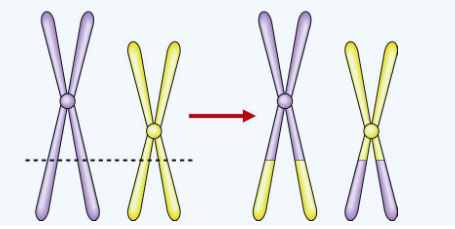
| Non Allelic Homologous Recombination (NAHR) | Unequal crossing-over between misaligned homologous chromosomes or sister chromatids. Causes recurrent rearrangements. |
| Non homologous end joining | Chromosome breakage followed by abnormal reconstitution. External: ionizing radiation, chemicals Internal: stressors, breakage syndromes |
| Reciprocal translocation. 1:500 individuals carries a balanced reciprocal translocation. t(11;22)(q23;q11) most common. | |
| Alternate Segregation. Normal or balanced haploid complement. | |
| Adjacent-1 segregation. Unbalanced haploid complement. Like centromeres segregate apart. | |
| Adjacent-2 segregation. Unbalanced haploid complement. Like centromeres segregate together. | |
| 3:1 Segregation | 3 chromosomes segregate to one gamete with only one chromosome in other gamete. More likely if one translocation product is much smaller than the other product. |
| Robertsonian translocation | Breakage of 2 afrocentric chromosomes at or close to their centromeres with subsequent fusion of their long arms. Most common 13q14q |
| Pericentric Inversion. During Meiosis I, inversion loop can lead to crossover, deleted & duplicated segments. Larger the size, smaller imbalance during crossover -> more likely to result in birth of abnormal infant. Smaller the size, large imbalance -> more likely to result in miscarriage. | |
| Paracentric inversion. Meiosis I crossover in the inverted segment, will result in recombinant chromosomes that are eccentric or dicentric. No viable offspring. | |
| Isochromosome | Loss of one arm with duplication of the other (centromere divided in the wrong direction). Most common is X chromosome long arm. |
| Ring Chromosomes | Break on each arm of a chromosome leaving 2 "sticky" ends that reunite as a ring. Two distal chromosomal fragments are lost. Unstable in mitosis. |
| Parental origin of 47,XXY | Paternal 45% Maternal 55% |
| Parental origin of 45,X | Paternal 80%. Maternal 20%. |
| Trisomy 16 | Most common autosomal aneuploidy in SAB. 100% cases from error in maternal meiosis I. |
| XIST (X-inactivated specific transcript) | Xq13.3 Expressed only from inactivated X Encodes an RNA product that coats the inactive X. Multiple genes escape silencing (~15%) |
| Sensitivity | Fraction of those affected that screen positive. Fraction of individuals with disease who have the susceptibility genotype. TP / [TP + FN] |
| Specificity | Fraction of unaffected that screen negative. Fraction without disease who do not have the susceptibility genotype. TN/[FP+TN] |
| Positive Predictive Value | Fraction of affected with positive screens. Proportion of individuals with susceptibility genotype who have or will develop a particular disease. TP / [TP+FP] |
| False negative rate | 1 - sensitivity (TP/[TP+FN]) |
| False positive rate | 1 - specificity (TN/[FP+TN]) |
| Probability Law of multiplication | Applies to independent events Defined by "both", "and", "all", or "none" Probabilities are multiplied |
| Probability Law of Addition | Applies to events that are mutually exclusive. Defined using "either" and "or" Probabilities are added |
| Carrier risk of AR disorder based on frequency | Carrier risk = square root of 4 x frequency of condition |
| Carrier risk for XLR "lethal" condition | Mother carrier risk is 2/3 Maternal grandmother risk is 1/3 |
| Bayesian Grid | Hypothesis: Is, Isn't Prior Probability: A, C Conditional Probability: B, D Joint Probability: AxB=E, CxD=F Posterior Probability: E/(E+F), F/(E+F) |
| Prior Probability | Probability of a hypothesis (i.e. disease) before additional information is available (i.e. test result) Basic Mendelian probabilities |
| Conditional Probability | If the particular hypothesis is true, probability of additional information. Reduced penetrance, variability. |
| Joint probability | Prior x conditional probability Probability of the particular hypothesis AND additional information |
| Posterior Probability | Probability of the particular hypothesis given additional information = Joint probability of the particular hypothesis divided by the sum of all joint probabilities. Fraction of total joint probability that is represented by scenario. |
| Law of Uniformity | When two homozygotes with different alleles are crossed, all of the offspring are identical and heterozygous. |
| Law of Segregation | Each person possesses 2 genes for a particular characteristic, only one of which can be transmitted at any one time. (rare exceptions - chromosome non-disjunction) |
| Law of Independent Assortment | Members of different gene pairs segregate to offspring independently of one another. (not always true, genes close together are 'linked') |
| Nucleosomes | DNA helix coiling around spherical histone beads |
| Fluorescent in-situ hybridization FISH | DNA probe labeled with fluorochrome. Hybridizes with patient's sample. Region where hybridization has occurred to be visualized using a fluorescence microscope. |
| Barr body | Inactive X chromosome, forms sex chromatin. Visualized during interphase in female somatic cells. Always late in replicating. |
| Heteromorphism | Pericentric inversion involving chromosome 9. Common structural variant or polymorphism. Not thought to be of any functional importance. |
| Large Pericentric Inversion | The duplicated and deleted segments will be relatively small so that survival to term and beyond becomes more likely. The larger the size the more likely an abnormal infant will be born. As opposed to small inversion that would more likely result in pregnancy loss. |
| Isochromosomes | Loss of one chromosome arm with duplication of the other. Centromere divided transversely rather than longitudinally. Most common is two long arms of X chromosome (15% of Turner syndrome) |
| Chimerism | Presence of two or more genetically distinct cell lines derived from more than one zygote. Dispermic chimeras - double fertilization. Two sperm fertilize two ova and the zygotes fuse to form one embryo. Blood chimeras - Exchange of cells via the placenta between non-identical twins in utero. |
| Lyonization | X-chromosome inactivation Occurs 15 to 16 days gestation. Either of the two X chromosomes can be inactivated, the same X chromosome is inactivated in all daughter cells. Abnormal X chromosome will be preferentially inactivated (actually, only those with normal X intact survive). |
| Autosomal Dominant | Vertical transmission. Father to son transmission confirms. Child has 50% chance of inheriting disorder or trait. |
| Pleiotropy | A single gene that may give rise to two or more apparently unrelated effects. Manifest in different systems of the body in a variety of ways. |
| Variable Expressivity | Clinical features of a disorder show striking variation from person to person even in the same family. |
| Reduced Penetrance | 'skipping a generation' Result of modification effects of other genes, integration of the gene with environmental disorders. No features despite inheriting gene is non-penetrance. |
| Co-Dominance | Two allelic traits that are both expressed in the heterozygous state. Example: blood group AB |
| Pseudodominance | A person homozygous for AR disorder has child with carrier of same AR disorder. Children have 1/2 chance of being affected. |
| Locus heterogenity | Disorder inherited in the same manner can be due to mutations in more than one gene. |
| Genocopies | Disorders with the same phenotype from different genetic loci |
| Phenocopy | Same phenotype of disorder from environmental causes |
| Y-linked inheritance Holandric inhirtance | Only males are affected. Hairy ears. H-Y histocompatibility antigen. Genes involved in spermatogenesis. |
| Digenic Inheritance | Disorder is shown to be due to the additive effects of heterozygous mutations at two different gene loci. Example in humans: Retinitis PIgmentosa can be caused by double heterozygosity for mutations in two unlinked genes (ROM1 and Peripherin) |
| Triallelic Inheritance | Example: Bardet-Biedl Syndrome that has multiple causative genetic loci. One form occurs when an individual is homozygous for mutations at one locus is also heterozygous for mutation at another Bardet-Biedl locus. |
| Anticipation | Onset of the disease occurs at an earlier age or with increased severity in the offspring than in the parents. Example: Myotonic dystrophy where CTG repeat expands further during maternal meiosis. Huntington disease where CAG repeat expands further during paternal meiosis. |
| Uniparental isodisomy | Two copies of the same homolog from one parent through an error in meiosis II |
| Uniparental heterodisomy | Two different homologs inherited from one parent due to an error in meiosis I |
| Homoplasmy | mitochondrial DNA from different mitochondria is identical |
| Heteroplasmy | If mutation occurs in mtDNA, there will be two populations of mtDNA. The proportion of mt with mutation in DNA varies between cells and tissues. |
| Estimating carrier frequency | Incidence for AR disorder in HWE is q^2 Square root = q. p+q=1. 1-q = p Carrier frequency = 2pq If p is close to one, rough estimate can be square root of disease incidence. For XLR disorder, frequency of affected males = q, affected females = q^2, and carrier females = 2pq |
| Estimation of Mutation Rates | Direct: counting new cases in defined number of births. 12 kid with AD condition in 100,000 births. 2 have affected parents. 10 de novo. 10 mutations/200,000 genes. Mutation rate of 1 per 20,000 gametes. Indirect: AD condition with reproductive fitness of zero (f=0), all cases new mutations. Incidence = I, mutation rate m, each child gets two alleles, either can mutate. I = 2m. If fitness greater than zero, genes lost through reproductive fitness must be balanced by new mutations. 2m=I(1-f). For AR, m=I(1-f). For XLR, m=[Incidence in males x (1-f)]/3 |
| Polymorphism | Occurence in the population of two or more genetically determined forms (alleles, sequence variants) in such that the rarest of them could not be maintained by mutation alone. By convention, at least two alleles, each with a frequency greater than 1%. |
| Polymorphic Information Content (PIC) | Value of particular polymorphic system is assessed by determining PIC. The higher the PIC the more likely the polymorphic marker will be of value in linkage analysis and gene tracking. |
| Odds Ratio | Radio of the odds of developing the disease in those with a specific marker to the odds of developing the disease without that specific marker. |
| Genome Wide Association | Compare variants across the entire genome. Hypothesis-free approach to determine which SNPs are involved in disease. P value threshold lower for statistical significance at 5 x 10^-8. Large sample sizes needed to achieve low P value. |
| Positive Predictive Value | Proportion of positive tests that are true positives. True Positives/(True Positives + False Positives) |
| Sensitivity | Proportion of cases that are detected. True Positives/(True Positives + False Negatives) |
| Specificity | Extent to which the tests detects only affected individuals. True Negative/(True negative + False Positive) |
| Syntenic | Genes that are on the same chromosome |
| Genetic Information Nondiscrimination Act (GINA) | 2008 Private employers with 15 or more employees are prohibited from deliberately seeking or using genetic information, including family history, to make an employment decision. Prohibits most group health insurers from denying insurance or adjusting group premiums based on the genetic information of members of the group. |
| Triploidy | 69,XXY>69,XXX (69,XYY very rare) No risk of recurrence Sx: >99% lost 1st trimester, 6-10% all SAB, 16-20% all chromosomally abnl SAB, dysplastic calvaria with large posterior fontanelle, 3/4 finger syndactyly, ASD, VSD, hydrocephalus, holoprosencephaly. Mat (digyny): small placenta, severe asymmetric IUGR with a large head Pat (diandry): hydropic large placenta, well grown to mod symmetric IUGR, nl or microcephalic head; MOST COMMON Eval: low maternal serum hCG |
| Timeline of Cytogenetics | 1923 - Y chr, 48 chr 1953 - hypotonic chr spread 1956 - 46 chr 1959 - Trisomy 21 down syndrome 1969-1970 - Banding techniques 1976-1980 - fragile sites 1990 - FISH 1992 - CGH (for tumors) 2001 - CMA |
| Epistasis | Gene interaction results in modification of phenotype |
| Transference | PATIENT redirects or projects feelings they have for another person into their relationship with their counselor. Ex: anger, mistrust, extreme dependence |
| Counter-Transference | COUNSELOR redirects feelings for another person into their relationship with their client. |
| Autosomal Recessive Inheritance | Carrier risk for unaffected full sibling of affected individual is 2/3 Carrier risk for full sibling of parent of affected individual is 1/2 Carrier risk for full sibling of parent of a CARRIER is 1/4. |
| X-linked Recessive inheritance | If males don't reproduce (early lethal), mutation rates are equal in males and females: carrier risk for mother is 2/3. Carrier risk for maternal grandmother and sisters is 1/3. Carrier risk for maternal aunt is 1/6. |
Want to create your own Flashcards for free with GoConqr? Learn more.