21406170
| Question | Answer |
|
Image:
Image (binary/octet-stream)
|
Simple epithelium -Single layer -Gaseous/fluid exchange |
|
Image:
Image (binary/octet-stream)
|
Emphysema -Fenestrations in alveolar wall -Enlarged airpsaces -May see inflammation -May see more macrophages |
|
Image:
Image (binary/octet-stream)
|
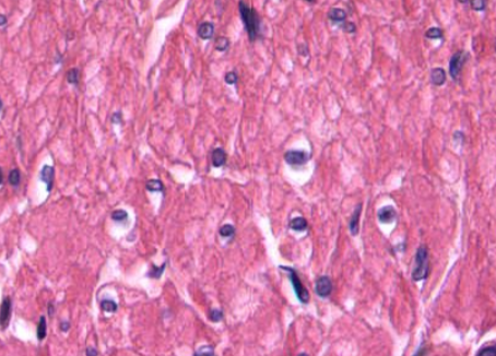
Medulla -Highly basophilic (catecholamines) -Capsule -Vascular -Venous channels -Ganglions present |
|
Image:
Image (binary/octet-stream)
|
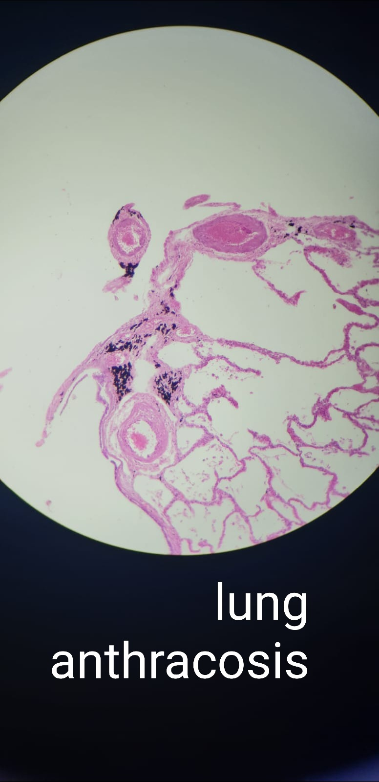
Lung anthracosis -Carbon accumulation -May see granulomas -Enlarged MPs - anthracotic -No necrosis -Multinucleated giant cells |
|
Image:
Image (binary/octet-stream)
|
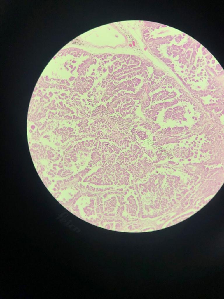
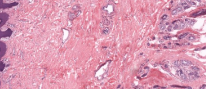
Bronchogenic Carcinoma -Thickened alveolar wall -Small alveolar space -Type II pneumocyte proliferation -FIbrosis |
|
Image:
Image (binary/octet-stream)
|
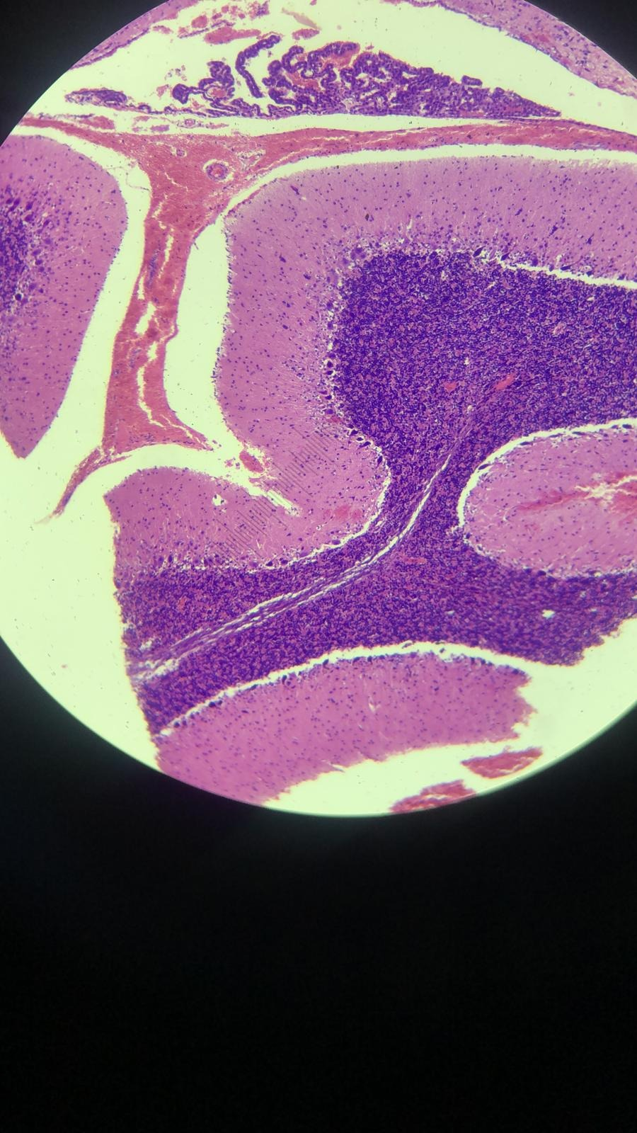
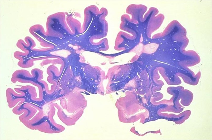
Cerebrum -White and grey matter -Vascular -Basophilic - catecholamines -Corpus callosum - middle space -Frontal lobe RHS -Amygdala lower LHS |
|
Image:
Image (binary/octet-stream)
|
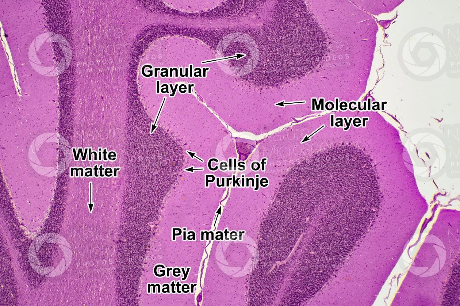
Cerebellum -Purkinje cells -White and grey matter -Highly basophilic - catecholamines -Granular layer -Pia mater -Nissl substance (purple) -Cauliflower appearance |
|
Image:
Image (binary/octet-stream)
|
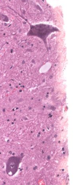
Spinal cord -Nissl substance (purple - basophilic RNA in RER) -Motor neurons -Axons |
|
Image:
Image (binary/octet-stream)
|
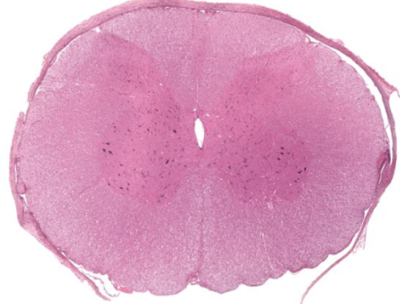
Spinal cord -Central canal - CSF -White and grey matter -Ventral and dorsal horns |
|
Image:
Image (binary/octet-stream)
|
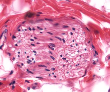
Peripheral nerve -Schwann cells |
|
Image:
Image (binary/octet-stream)
|
Peripheral nerve -Epineurium -Perineurium -Endoneurium |
|
Image:
Image (binary/octet-stream)
|
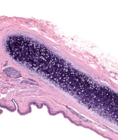
Corpus luteum -Fibrous scar visible -Thick wall - lutein cells -Central cavity - blood clot, replaced with connective tissue -Oocytes on periphery |
|
Image:
Image (binary/octet-stream)
|
Ovary -Largest - Graafian follicle -Antral follicle -Primary follicle (unseen) -Primordial follicle (unseen) -Capsule -Vascular |
|
Image:
Image (binary/octet-stream)
|
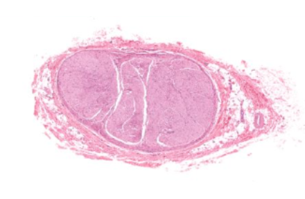
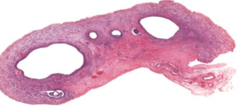
Testis (Epididymus) -Epididymus - single coiled duct -Muscular layer -Principal cells - tall coumnar, microvilli |
|
Image:
Image (binary/octet-stream)
|
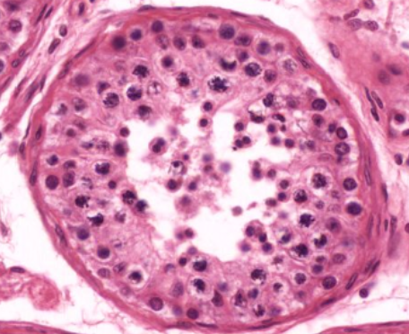
Testis (semniferous tubule) -Primary spermatocytes (rarely visible) -Spermatogonia (on BM) -Sertoli cells (large, prominent nucleus) |
|
Image:
Image (binary/octet-stream)
|
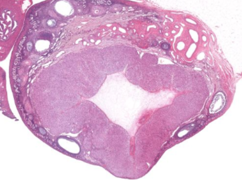
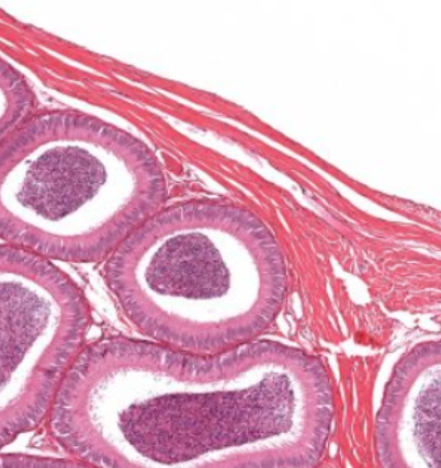
Testis -Thick tunica albuginea capusle -Connective tissue between lobules -Semniferous tubules - coiled |
|
Image:
Image (binary/octet-stream)
|
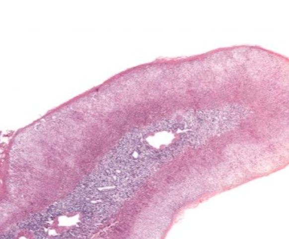
Spleen -Capsule -Trabeculae -White pulp (basophilic, lymphatic) -Red pulp (eosinophilic, RBCs) -Marginal zone (between red and white - interact) |
|
Image:
Image (binary/octet-stream)
|
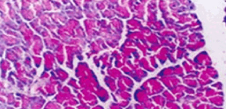
Pancreas (PAS) -Glycogen appears MAGENTA |
|
Image:
Image (binary/octet-stream)
|
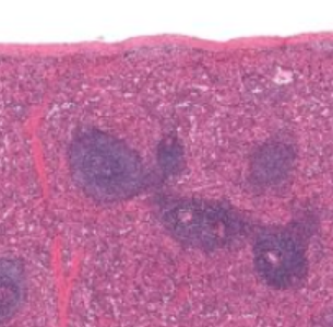
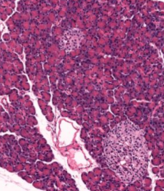
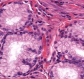
Pancreas -Islets of Langerhans (pale) -Parasympathetic ganglions (v similar appearance) -Exocrine cells (dark) -Capsule -Lymph nodes may be seen -Lobes/lobules |
|
Image:
Image (binary/octet-stream)
|
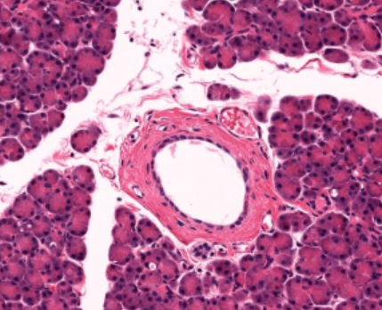
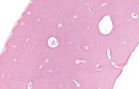
Pancreatic duct -Between lobules |
|
Image:
Image (binary/octet-stream)
|
Pancreas -Capsule -Lobes/lobules -Adipose tissue with lymph nodes |
|
Image:
Image (binary/octet-stream)
|
Stomach (PAS) -Mucins appear MAGENTA |
|
Image:
Image (binary/octet-stream)
|
Duodenum (glands) -Brunner's glands -Alkaline mucus in submucosa |
|
Image:
Image (binary/octet-stream)
|
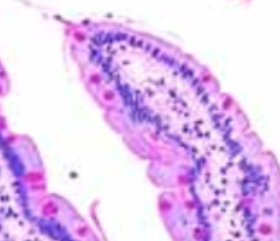
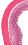
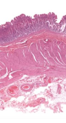
Duodenum -Thick SM - muscularis externa -Submucosa (glands only present in oesophagus/duodenum) -Mucosa - villi, brush border |
|
Image:
Image (binary/octet-stream)
|
Stomach -Mucosa - glands, gastric pits -Submucosa (connective tissue) -Muscularis externa -Adventitia (loose connective tissue) |
|
Image:
Image (binary/octet-stream)
|
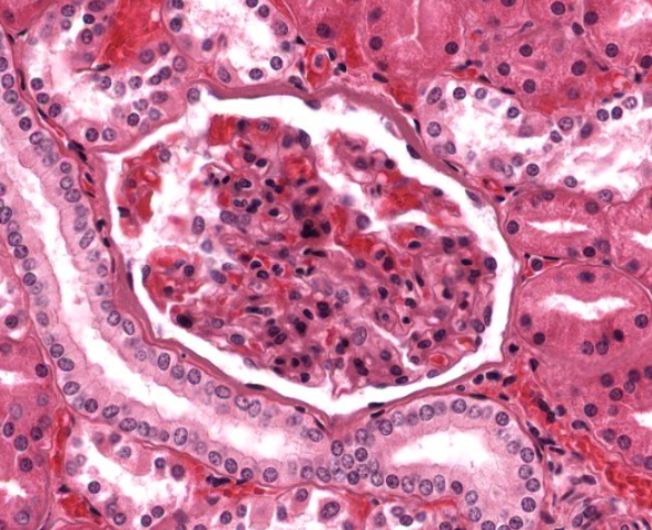
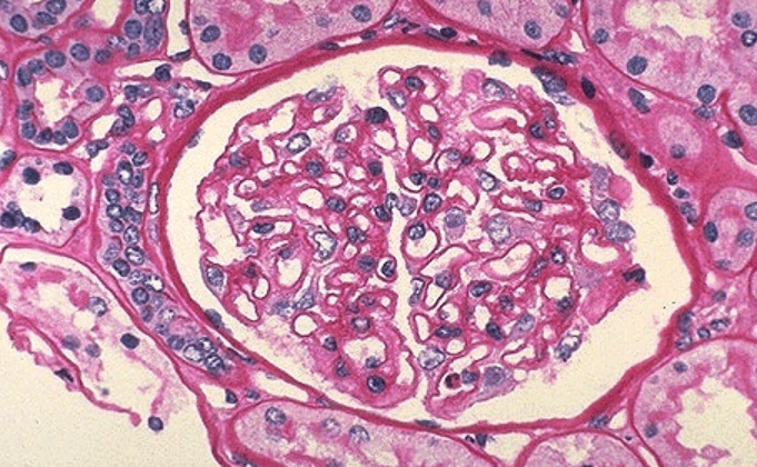
Kidney (PAS) (Renal corpuscle) -Basement membrane (red) -Vascular loops of glomerulus |
|
Image:
Image (binary/octet-stream)
|
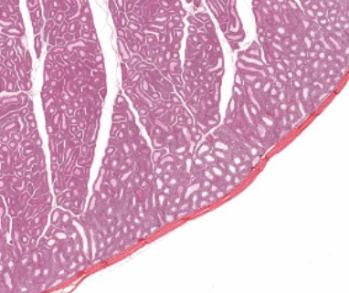
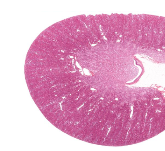
Kidney -Capsule -Cortex - ultrafiltration -Medulla - urine -Renal pelvis - to ureter -Hilium - exit |
|
Image:
Image (binary/octet-stream)
|
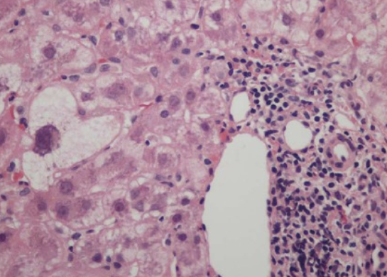
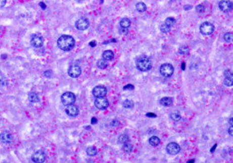
Hepatitis -Portal triad - inflammation, proliferation -"Ballooning" of hepatocytes -Necrosis (spotty) |
|
Image:
Image (binary/octet-stream)
|
Liver (PAS) -Glycogen appears MAGENTA |
|
Image:
Image (binary/octet-stream)
|
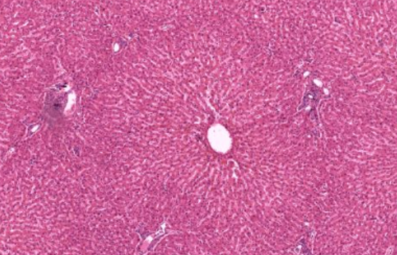
Liver -Hexagonal hepatocytes -Central vein -Portal triads -Sinusoids |
|
Image:
Image (binary/octet-stream)
|
Adrenal gland -Capsule -Cortex -Medulla -Medullar vessel - large vein |
|
Image:
Image (binary/octet-stream)
|
Trachea -Purple cartilage -Smooth muscle -Seromucous glands -Columnar epithelium, pseudo-strat |
|
Image:
Image (binary/octet-stream)
|
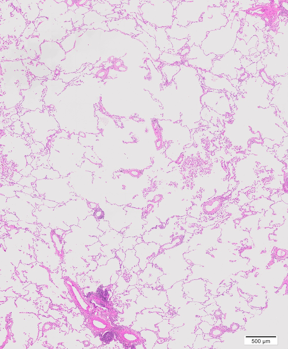
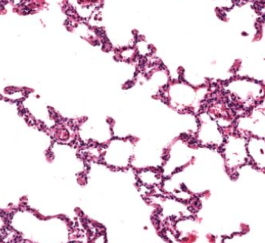
Lung (alveoli) -Type I and II pneumocytes -Air spaces |
|
Image:
Image (binary/octet-stream)
|
Larynx (MT) -Goblet cells -Pseudostratified ep -Laryngeal glands |
|
Image:
Image (binary/octet-stream)
|
Thyroid (MT) -Follicular cell - colloid (thyroglobulin and thyroxin) -Parafollicular cells -Simple cuboidal |
|
Image:
Image (binary/octet-stream)
|
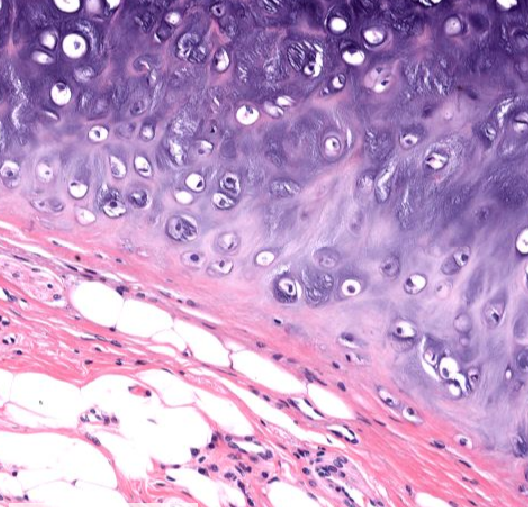
Cartilage -Purple - collagen (matrix) -Chondrocytes in matrix -Pink/purple - MSCs -Outer fibrous layer - fibroblasts -Pink - perichondrium - connective tissue |
|
Image:
Image (binary/octet-stream)
|
Skin (dermis) -Purple - papillary layer -Thick connective tissue layer -Glands |
|
Image:
Image (binary/octet-stream)
|
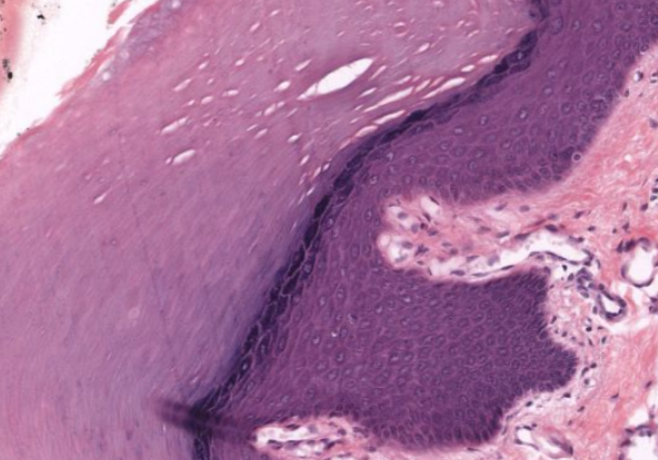
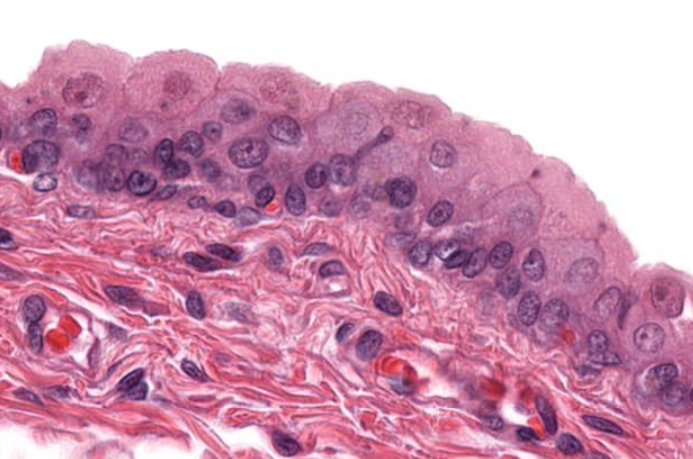
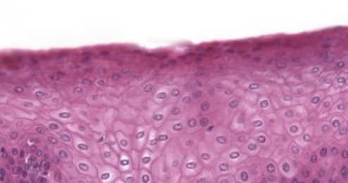
Epidermis (thick keratinised) -Dead cells on outside -Granular keratinocytes -Keratinocytes - purple layer (spinosum) -Dermis |
|
Image:
Image (binary/octet-stream)
|
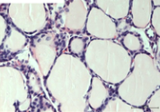
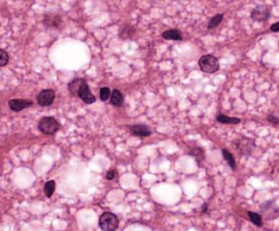
Adipose (brown) -Smaller -Droplets |
|
Image:
Image (binary/octet-stream)
|
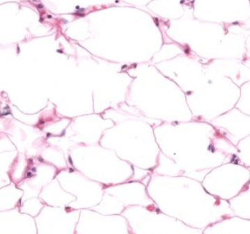
Adipose (white) -Large cells -Flattened nuclei -Appear empty - lipids removed during processing |
|
Image:
Image (binary/octet-stream)
|
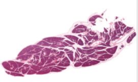
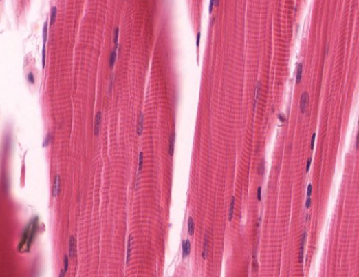
Skeletal muscle -Striated -Sarcomeres -Gaps between myofibrils -Satellite cells - dark, on surface |
|
Image:
Image (binary/octet-stream)
|
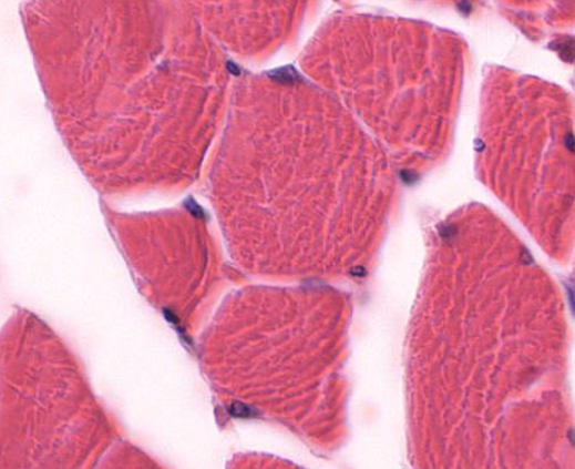
Skeletal muscle (longitudinal) -Polygon shape -Nuclei appear at periphery |
|
Image:
Image (binary/octet-stream)
|
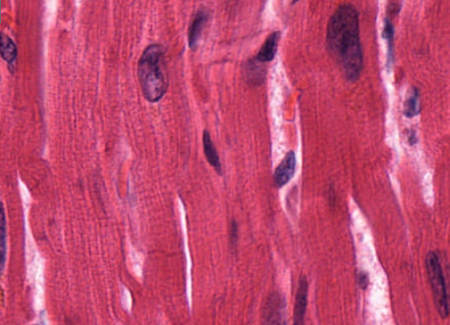
Cardiac muscle -Striations -Can be 2 nuclei -Branching -Intercalated discs |
|
Image:
Image (binary/octet-stream)
|
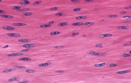
Smooth muscle -No striations -Spindle-shaped -Less gaps -Little detail |
|
Image:
Image (binary/octet-stream)
|
Smooth muscle (cross-section) -Central nuclei -Spindle-shaped cells |
|
Image:
Image (binary/octet-stream)
|
Compact bone -Osteons - unit -Large osteoblasts -Very small osteocytes -Haversian vessel - remnants of BV/nerve -Haversian canal - centre of an osteon -Haverian lamellae - concentric circles |
|
Image:
Image (binary/octet-stream)
|
Transitional epithelium (relaxed) -Apical - dome-shaped umbrella cells -Several layers |
|
Image:
Image (binary/octet-stream)
|
Transitional epithelium (stretched) -Umbrella cells flattened -Several layers |
|
Image:
Image (binary/octet-stream)
|
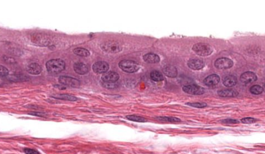
Stratified epithelium -Several layers e.g. oesophagus |
Want to create your own Flashcards for free with GoConqr? Learn more.