34628618
Description
Flashcards by Aquilina Laidlaw-allan, updated more than 1 year ago
|
|
Created by Aquilina Laidlaw-allan
about 4 years ago
|
|
| Question | Answer |
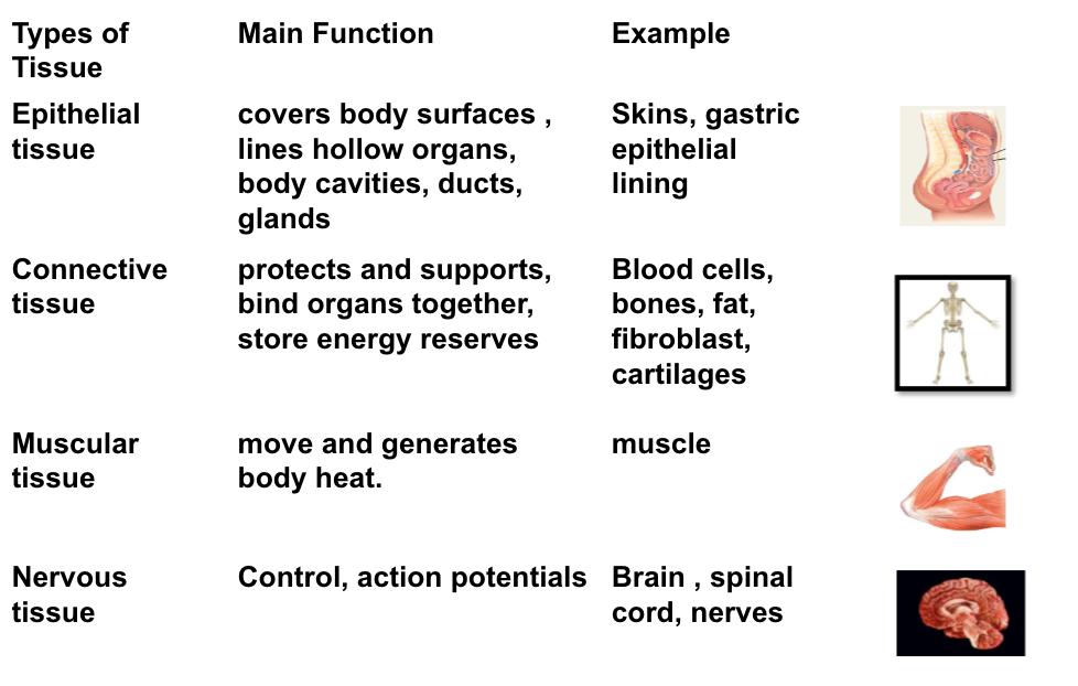
| Provide the functions and examples for each type of tissue: Epithelial tissue Connective tissue Muscular tissue Nervous tissue | |
| What are the general functions of the CNS? | Cognition Speech Conscious Coordination and planning Movement Motor function Sensory functions |
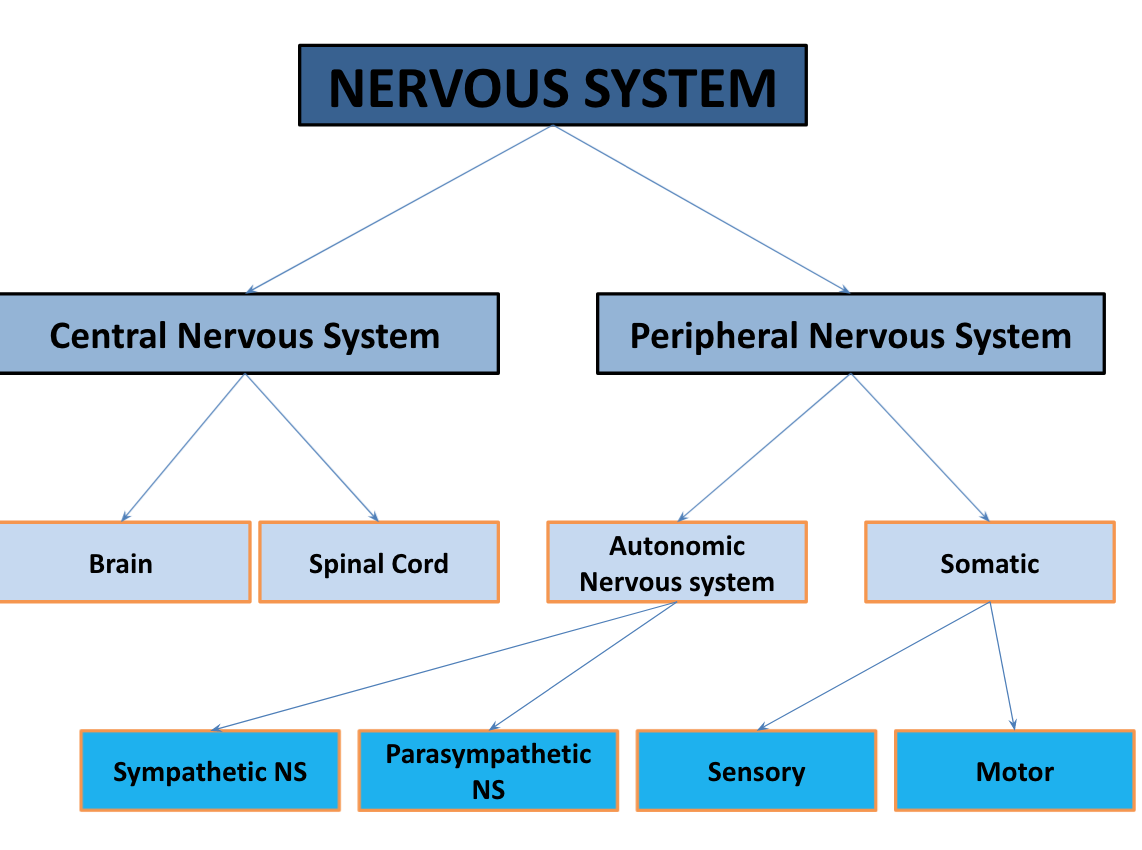
| Describe the sub-systems within the following: CNS PNS | |
| Describe right and left control of the brain. | The right brain controls the left half of the body. |
| Describe the anatomy of the meninges, white matter, and gray matter. | Meninges: cover the spinal cord White Matter: conduction tracts Gray Matter: - Dorsal (posterior) horns: Sensory tracts - Anterior (ventral) horns: Motor horns |
| Describe the anatomy of the ventral and dorsal roots. | Ventral root: the efferent motor root of a spinal nerve. At its distal end, the ventral root joins with the dorsal root to form a mixed spinal nerve.. Dorsal root: dorsal root ganglia – cell bodies outside the central nervous system, |
| What two elements is the nerve tissue composed of? | 1. The nerve cell or neuron 2. The neuroglia |
| What is the role of the synapses? | Facilitate nerve conduction |
| Describe the functional role of the neuron vs. the neuroglia. | Neuron: - Neuro transmission Neuroglia: - Neuroglia divide: Tumor (glioma grows) - Support neurons - Phagocytosis - Myelin synthesis - Blood brain barriers |
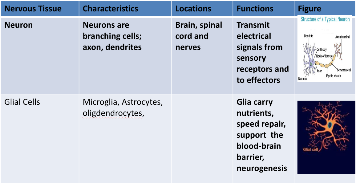
| Describe the characteristics, locations, functions of the neurons and glial cells. | |
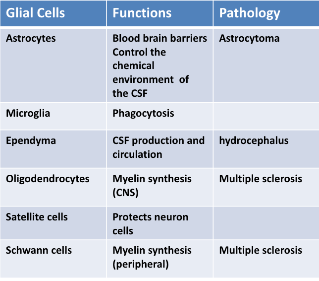
| Describe the types of glial cells, their function, and their pathology (if applicable). | |
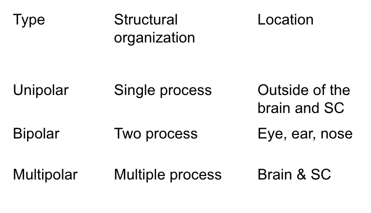
| Describe the structural organization and the location of the following structural classifications of neurons: Unipolar Bipolar Multipolar | |
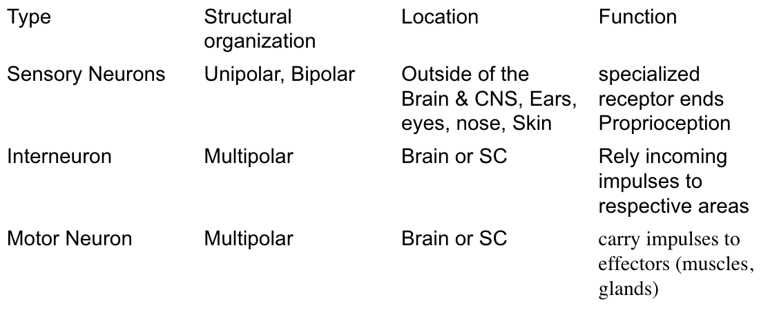
| Describe the structure, location, and function of the following functional classifications of neurons: Sensory neurons Interneurons Motor neurons | |
| Collection of neurons in CNS= ___ Collection of the neuron outside of the CNS= ___ | Collection of neurons in CNS= Nuclei Collection of the neuron outside of the CNS= Ganglia |
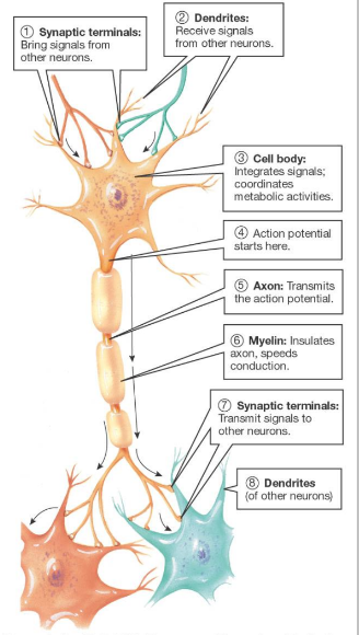
| Describe the following about the functional organization of neurons: Synaptic terminales Dendrites Cell body Axon Myelin Synaptic Terminale Dendrites | |
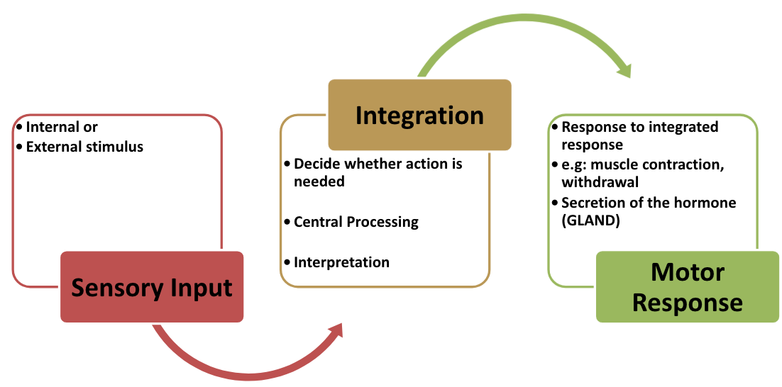
| Describe the process of: Sensory Input -> Integration -> Motor Response | |
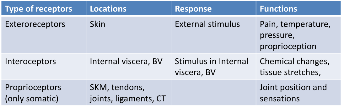
| Describe the location, response, and function of the following types of receptors: Exteroceptors Interoceptors Proprioceptors (only somatic) | |
| Describe the following terms of nerve (impulse) physiology: Reception Transmission Integration Response (effector) | Reception: Determine type of stimulus Transmission: Signal the intensity of a stimulus Integration: Integrate responses from many sources Response (effector): Initiate and direct operations |
| What is an action potential? What does it result in? | Electrical impulse generation resulting in actions such as: - Muscle contraction - Withdrawal |
| What occur with Na+ and K+ gates during an action potential? | Synchronized opening and closing of Na+ and K+ gates |
| Where is the AP transmitted? Via what? What is released during this process? | Transmitted directly to the next cell (via gap junctions) Release chemical (NT) |
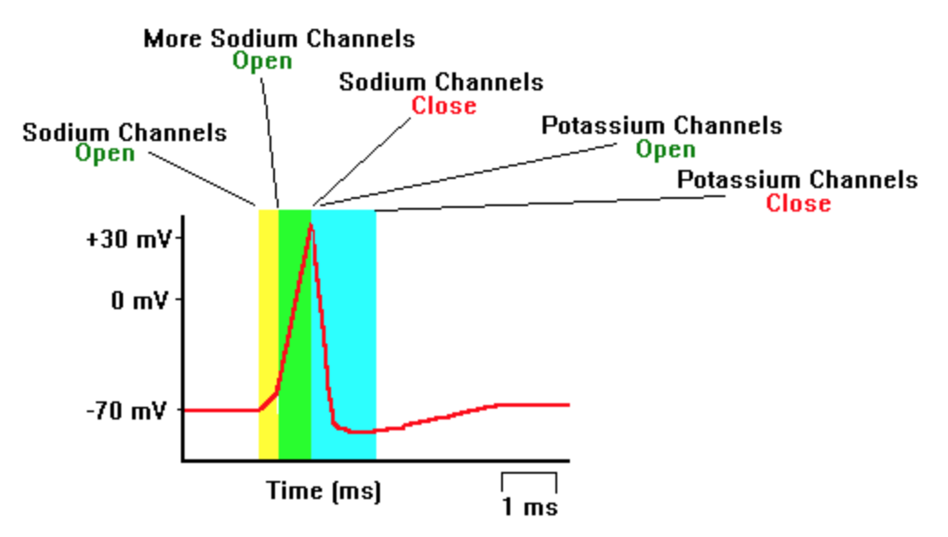
| Describe the following terms: Resting Membrane Potential Threshold Level Depolarization Repolarization Hyperpolarization | Resting Membrane Potential (-70mv): a relatively static membrane potential. Threshold Level (-55mv): critical level to which a membrane potential must be depolarized to initiate an action potential. Depolarization (+40mv): cell undergoes a shift in electric charge distribution, resulting in less negative charge inside the cell compared to the outside, therefore inner membrane = more positive (Na+ voltage-gated channels open). Repolarization (-70mv): change in membrane potential that returns it to a negative value just after the depolarization phase (Na+ channels close, K+ voltage-gated channels open). Hyperpolarization (-90mv): the membrane potential of a cell becomes more negative than it typically is. |
| Describe the opening and closing of sodium and potassium channels during the phases of an action potential. | |
| Describe transduction and translation in terms of generating an action potential. | Transduction: - stimulus induces change in sensor potential. Results in depolarization. - stronger the stimulus, the greater the amplitude of the sensor potential. Translation: - Once the sensor potential exceeds a certain threshold, it is transformed into an action potential, AP. |
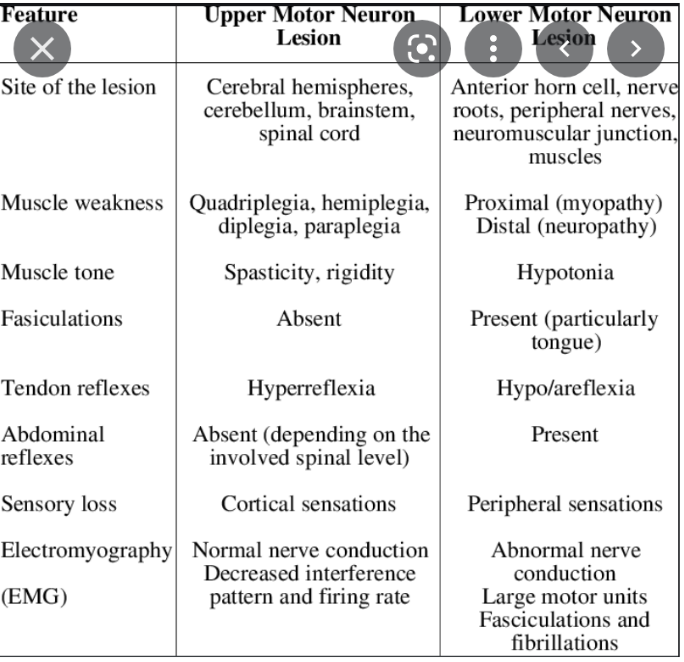
| Compare upper motor neurons (UMN) and their lesioning to lower motor neurons (LMN) and their lesioning. | The upper motor neurons originate in the cerebral cortex and travel down to the brain stem or spinal cord, while the lower motor neurons begin in the spinal cord and go on to innervate muscles and glands throughout the body. UMN are the neurons located higher than the anterior horn cells of the spinal cord (which includes brain AND spinal cord). Not every sensation has to travel up to the brain because the body works on the principle of compensation. For example: reflexive movement (if you touch something HOT and must jerk away immediately, it is too far for the reflex to travel…) LMN are the neurons located distal to the anterior horn cells of the spinal cord. Once you have a problem with the UMN or LMN it is an electrical problem. Whether the power plant is out or the wire on your street is out, electricity from your house is out either way. If electricity from main plant is out, effects many other areas. But, if it is only the wire to your house, it is just you that is affected. |
| Describe impulse propogation. | - An action potential is started in the dendrite - Impulses are transmitted across the synapse to another nerve - Neurotransmitter is released from a nerve’s axon terminal |
| Myelinated regions of axon are ______ ______. Electrical charge moves _____ ___ ____ rather than across the membrane. Action potentials occur only at _____ regions called: _____ __ _______ | Myelinated regions of axon are electrically insulated. Electrical charge moves along the axon rather than across the membrane. Action potentials occur only at unmyelinated regions: nodes of Ranvier. |
| Describe saltatory potentials. | - Saltatory = jumping. Makes electrical transmission RAPID. - Inward current movement at the rode of Ranvier (Axon) - Depolarizes new potential |
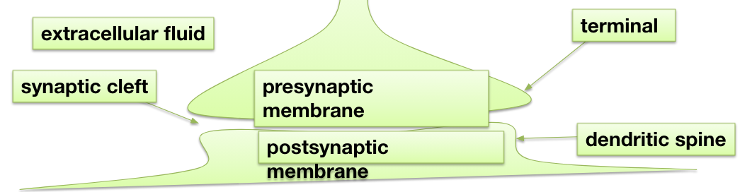
| Describe the structure and function of a synapse. | Structure: - Gap between: Two neurons + Neuron and muscle (NMJ) - Neurotransmitters mediated: Synaptic cleft and Synaptic vesicles Function: - Controls nerve impulse transmission - Neurotransmitter re-uptake |
| Describe Electrical Synapsis vs. Chemical Synapsis. | Electrical synapsis: In these synapses the membranes of the two cells actually touch, and they share proteins. This allows the action potential to pass directly from one membrane to the next. They are very fast, but are quite rare, found only in the heart and the eye. Chemical synapsis: In a chemical synapse, electrical activity in the presynaptic neuron is converted into the release of a chemical called a neurotransmitter that binds to receptors located in the plasma membrane of the postsynaptic cell. |
| An action potential causes ________ release from the ___________. Neurotransmitters _______ across the synaptic cleft. Neurotransmitters ____ to ______ within the postsynaptic membrane, altering the membrane potential. | An action potential causes neurotransmitter release from the presynaptic membrane. Neurotransmitters diffuse across the synaptic cleft. They bind to receptors within the postsynaptic membrane, altering the membrane potential. |
| Describe the process of neurotransmitters being released from the pre-synaptic membrane. | Synaptic vesicles, containing neurotransmitter, congregate at the presynaptic membrane. The action potential causes voltage-gated calcium (Ca2+) channels to open; Ca2+ ions flood in. Ca2+ causes vesicle membrane to fuse with presynaptic membrane. Vesicle contents empty into cleft: exocytosis. Neurotransmitter diffuses across synaptic cleft. |
| Describe Excitatory Synapses. | Excitatory synapses cause the post-synaptic cell to become less negative triggering an excitatory post-synaptic potential (EPSP) *Increases the likelihood of firing an action potential.* Opening of ion channels which leads to depolarization makes an action potential more likely, hence “excitatory PSPs”: EPSPs. Inside of post-synaptic cell becomes less negative. *Na+ channels (NB remember the action potential)* *Ca2+ . (Also activates structural intracellular changes -> learning.)* |
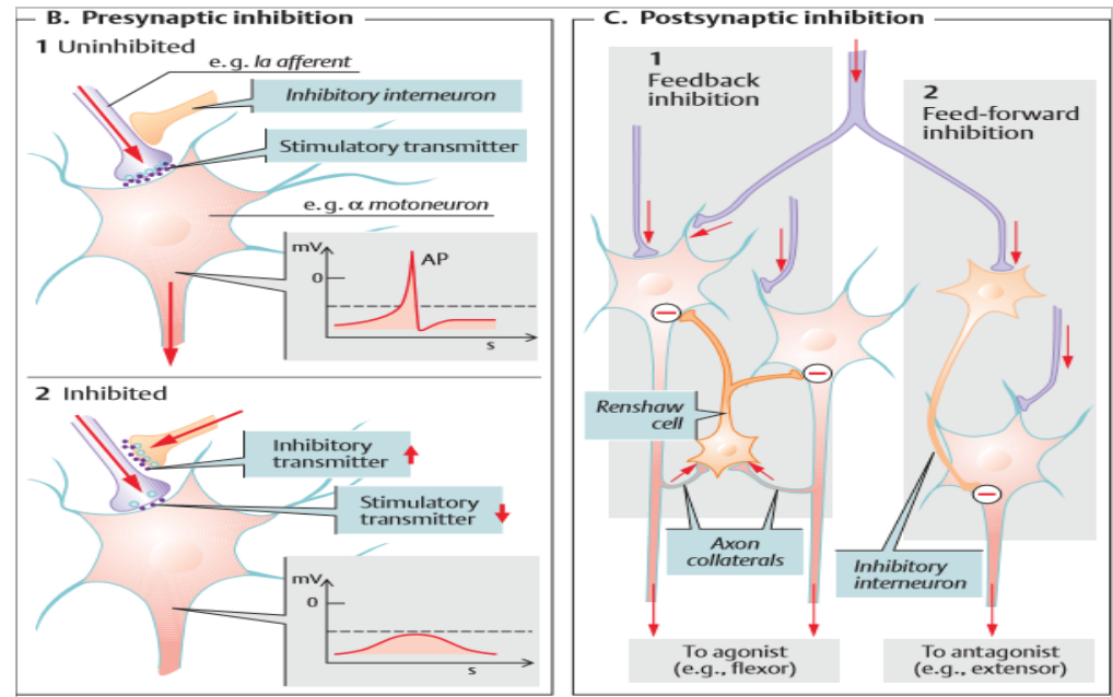
| Describe Inhibitory Synapses. | Inhibitory synapses cause the post-synaptic cell potential to become negative triggering an inhibitory post-synaptic potential *Decreases the likelihood of firing an action potential* Opening of ion channels which leads to hyperpolarization makes an action potential less likely, hence “inhibitory PSPs”: IPSPs. Inside of post-synaptic cell becomes more negative. *K+ (NB remember termination of the action potential)* *Cl- (if already depolarized)* |
| Describe pre-synaptic inhibition vs. post-synaptic inhibition. | |
| Describe a reflex arc. What structures does a reflex arc include? | Response to stimulus - Immediate withdrawal - Example: touching hot object - The pathway followed by nerve impulses that produce a reflex is a reflex arc. A reflex arc includes: a. sensory receptor b. sensory neuron c. integrating center (mono and polysynaptic) d. motor neuron e. effector: *somatic reflex – skeletal muscle, autonomic; *visceral reflex – smooth muscle, etc. |
| What are the key features of the CNS? | LOC/ sensorium Headache Altered sensation Numbness Tingling sensations Pain Altered JPS Unsteady gait, falls Urinary and bowel incontinence Speech Cognition |
| Describe Multiple Sclerosis (cause and symptoms). | - Pathology of Myelin - Demyelination Causes: - idiopathic - Inflammatory - Autoimmune Symptoms: - neuropathic pain - Vertigo - Aphasia - Unsteady gait and coordination - tremor |
| Describe the following in terms of MS Symptoms in order of frequency Lhermitte’s sign Uhthoff’s phenomenon SPMS Symptoms not commonly found in MS | symptoms in order of frequency: fatigue, depression, numbness, weakness, visual disturbance, bladder dysfunction, spasticity, impaired gait, cognitive disturbance, pain Lhermitte’s sign: flexion of neck causes electric shock sensation down back into limbs indicating cervical cord lesion Uhthoff’s phenomenon: worsening of symptoms (classically optic neuritis) in heat SPMS: classically weakness of legs in pyramidal distribution paired with cerebellar findings of arm (i.e. intention tremor) symptoms not commonly found in MS: visual field defects, aphasia, apraxia, progressive hemiparesis |
| Describe spastic paralysis vs. flaccid paralysis. | Spastic paralysis: - Damage to UMN (MS, cerebral palsy) - Hypertonic - Spastic/ Scissoring gait Flaccid paralysis: - Damage to LMN (anterior horn of the spinal canal) - Hypotonia - Fasciculation - Muscle atrophy - Loss of muscle function |
| Describe Amyotrophic Lateral Sclerosis (ALS/Lou Gehrig’s Disease) - Signs and Symptoms. | Progressive degeneration of motor neurons causing UMN and LMN symptoms Signs and Symptoms - limb motor symptoms: segmental and asymmetrical UMN and LMN symptoms of limbs - bulbar findings: dysarthria, dysphagia, tongue atrophy and fasciculations - pseudobulbar affect or emotional lability - sparing of ocular muscles and of sphincters |
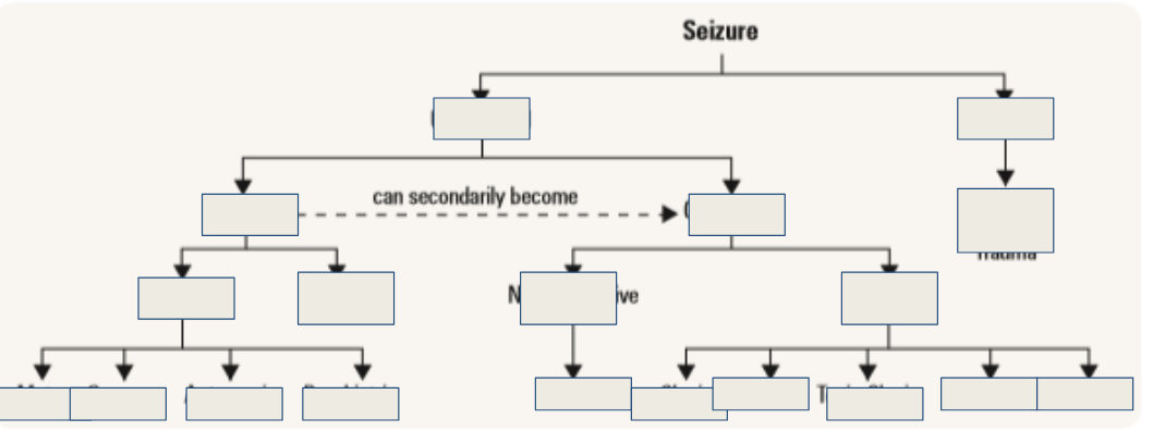
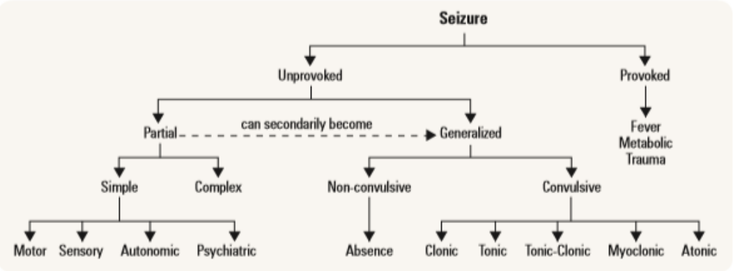
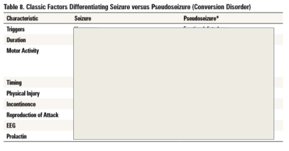
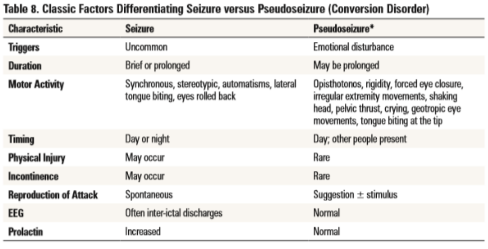
| Describe epilepsy. | - A seizure is the event - Epilepsy is the disease associated with spontaneously recurring seizures - A brief disruption in normal brain activity that interferes with brain function. - Type of seizure depends on which area of the brain is involved. - People may experience an alteration in behavior, consciousness, movement, perception and/or sensation. |
Want to create your own Flashcards for free with GoConqr? Learn more.