35605609
Description
Flashcards by Nicole Servetnik, updated more than 1 year ago
|
|
Created by Nicole Servetnik
about 2 years ago
|
|
| Question | Answer |
| this is the most abundant type of blood cell | red blood cell |
| these are biconcave cells that deliver oxygen to tissues, are very numerous | red blood cells |
| these cell fragments do not have a nucleus and help to form blood clots (small purple crescents between RBCs) | platelets (thrombocytes) |
| these cells are separated into granulocytes and agranulocytes | white blood cells |
| these white blood cells contain many dark staining granules in the cytoplasm and the nucleus typically takes on a multi-lobed appearance | granulocytes |
| these white blood cells contain very few dark staining granules in the cytoplasm | agranulocytes |
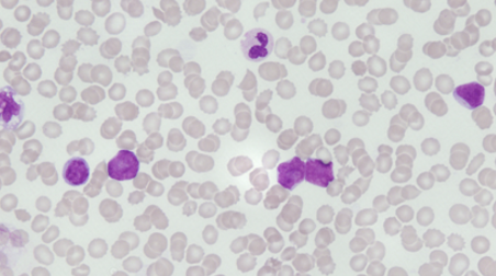
| these 3 white blood cell types are granulocytes | neutrophils, eosinophils, basophils |
| these 2 white blood cell types are agranulocytes | lymphocytes, monocytes |
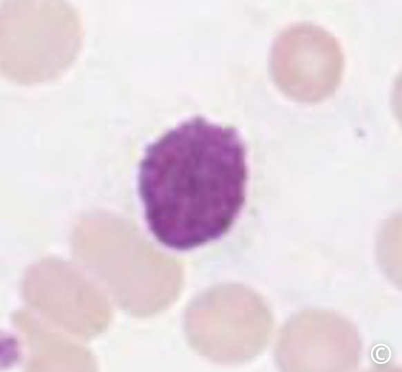
| this white blood cell releases chemicals that change blood pressure. look for dense staining of granules in cytoplasm that obscure nucleus. | basophil |
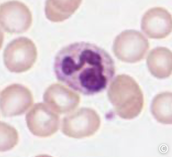
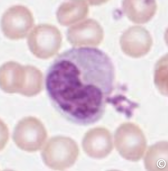
| this white blood cell responds to the site of an acute injury or infection and performs phagocytosis of debris. look for a nucleus of 3-4 dark staining lobes with many dark staining granules in the cytoplasm. | neutrophil |
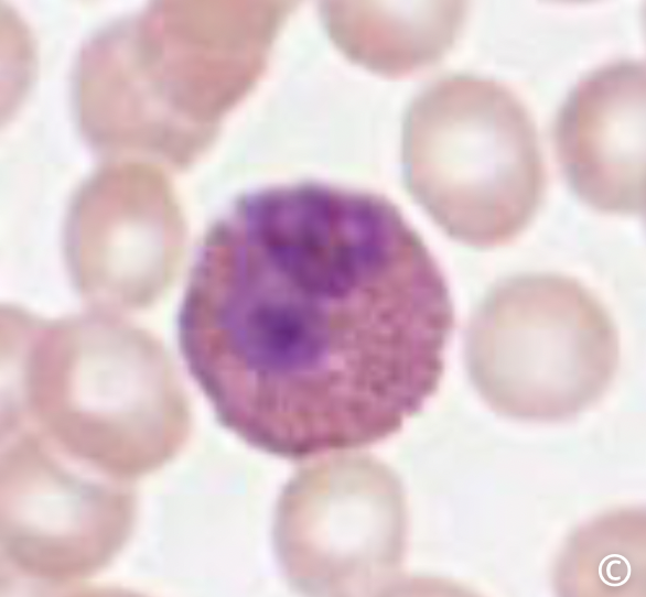
| this white blood cell responds to allergens and chronic inflammation. they release chemicals that change blood pressure. look for nuclei with two lobes and many dark staining granules in the cytoplasm. | eosinophil |
| what is the order of white blood cells in order of most to least abundant | neutrophils, lymphocytes, monocytes, eosinophils, basophils |
| what is this cell | neutrophil |
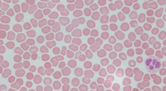
| what is this cell | eosinophil |
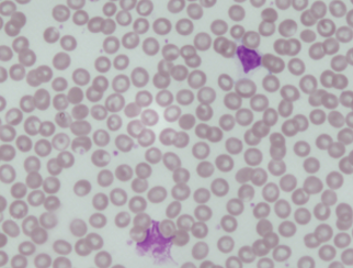
| what is this cell | basophil |
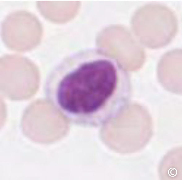
| what is this cell | lymphocyte |
| what is this cell | monocyte |
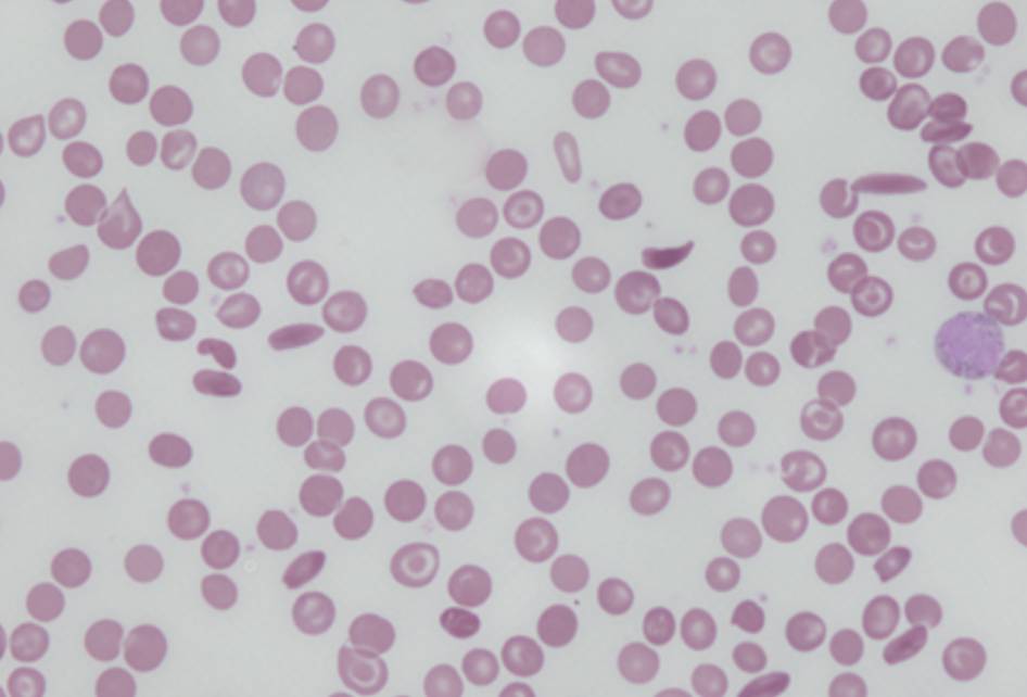
| what is this blood disease | sickle cell anemia |
| what is this blood disease | infectious mononucleosis |
| this is a viral blood disease caused by the Epstein-barr virus which results in an increased production of monocytes and lymphocytes (agranulocytes) | infectious mononucleosis |
| what is this blood disease | polycythemia |
| this is the abnormal increase in the number of erythrocytes which increases viscosity of blood. often a result of bone marrow cancer. | polycythemia |
| what is this blood disease | leukemia |
| this type of cancer involved an overproduction of leukocytes. cells and immature/abnormal in appearance and lack normal function. can be acute or chronic. | leukemia |
| this is the main component of RBCs and has four folded proteins called globins. the red pigment is called heme | hemoglobin |
| hemoglobin is important because each one of these molecules binds an oxygen | iron |
| typical range of hemoglobin for a male | 13.5-17.5 g/dL |
| type range of hemoglobin for a female | 12.0-15.5 g/dL |
| the Hb-O2 curve is this shape | sigmoidal |
| this is a diagnostic test to see the portion of RBC's in a blood sample | hematocrit |
| in a completed hematocrit, the upper clear yellow layer is ___, the Buffy coat is ___, and the lower dark red layer is ____. | plasma, leukocytes and platelets, RBCs |
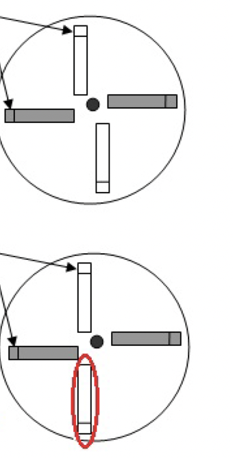
| what is the proper way to balance centrifuge tubes? top or bottom picture? | top |
| what is the calculation for hematocrit packed red blood cell volume? | (packed RBC/total volume)x100 |
| the hematocrit is (higher/lower) than normal if a patient has anemia | lower |
| the hematocrit is (higher/lower) than normal is a patient has polycythemia | higher |
| the hematocrit is (higher/lower) than normal if a patient has compensated blood loss | lower (but is not much lower than normal since plasma and RBCs are lost in equal proportions) |
| the hematocrit is (higher/lower) than normal is a patient is suffering with dehydration | higher |
| the hematocrit is (higher/lower) than normal is a patient is blood doping | higher |
| what happens to the hematocrit when you have leukemia | lower hematocrit, larger Buffy coat |
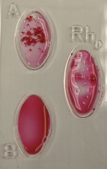
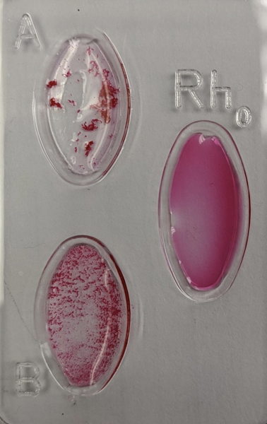
| this is determined by which antigen is present on a person's blood cells | blood typing |
| Blood type A has these antibodies and can receive blood from ___ and donate blood to ___. | anti-B antibodies. receive from A and O. donate to A and AB. |
| Blood type B has these antibodies and can receive blood from ___ and donate blood to ___. | anti-A antibodies. receive from B and O. give to B and AB. |
| blood type AB has these antibodies and can receive blood from ___ and donate blood to ___. | no antibodies. receive from A, B, AB, and O. give to AB. |
| blood type O has these antibodies and can receive blood from ___ and donate blood to ___. | anti-a and anti-b antibodies. receive from O. give to O, A, B, and AB. |
| what is this blood type? | A+ |
| what is this blood type? | AB- |
| when you plot the amount of oxyhemoglobin vs oxygen concentration you obtain a | oxygen-hemoglobin dissociation curve |
| hemoglobin exhibits this kind of binding | cooperative binding |
| what is p50? | half saturation pressure |
| this shift to the Hb-O2 curve means p50 is higher, oxygen affinity is lower, there is more CO2, less H+, higher DPG, or higher temperature | right shift |
| this shift to the Hb-O2 curve means p50 is lower, oxygen affinity is higher, there is less CO2, more H+, lower DPG, or colder temperature | left shift |
| this was used to monitor the proportion of oxyhemoglobin present in the sample | spectrophotometer |
| what wavelength do you use on the spectrophotometer for hemoglobin? | 660 nm |
| this is the wavelength where the substance shows max absorbance | lambda max |
| this plot shows the intensity of color related to the amount of substance present | standard curve |
| Calculation for saturation of hemoglobin | (A-B)/(A-C) X 100% |
| For the saturation of hemoglobin, what did A, B and C stand for in the saturation equation? | A - absorbance after deoxygenation B - absorbance after each step C - absorbance before deoxygenation |
| this is the pressure at which half of hemes are bound to oxygen | p50 |
| what is beers law and what does each component stand for | A = EbC A - absorbance C - concentration of absorbing molecule E - molar absorptivity (measurement of how strongly it absorbs light) b - cell path length |
| which has higher oxygen affinity? myoglobin or hemoglobin? | myoglobin |
| which has higher oxygen affinity? fetal hemoglobin or adult hemoglobin? | fetal hemoglobin |
| the positive electrode for ECG is placed on the | left arm |
| the negative electrode for ECG is placed on the | right arm |
| the earth electrode for ECG is placed on the | right leg |
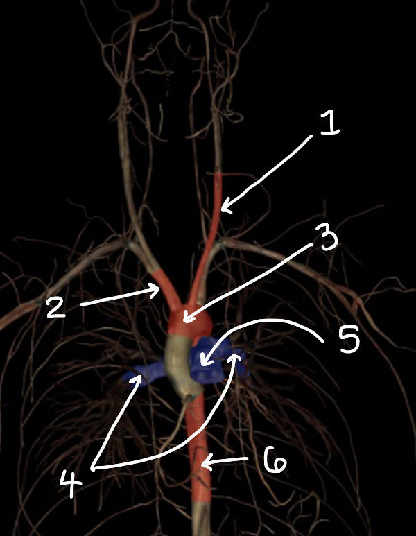
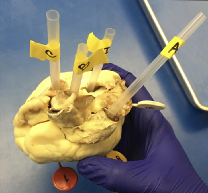
| what structure is labeled 1 | common carotid artery |
| what structure is labeled 2 | brachiocephalic trunk |
| what structure is labeled 3 | aortic arch |
| what structure is labeled 4 | pulmonary artery |
| what structure is labeled 5 | pulmonary trunk |
| what structure is labeled 6 | thoracic aorta |
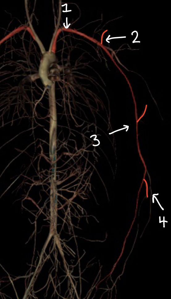
| what structure is labeled 1 | subclavian artery |
| what structure is labeled 2 | axillary artery |
| what structure is labeled 3 | brachial artery |
| what structure is labeled 4 | radial artery |
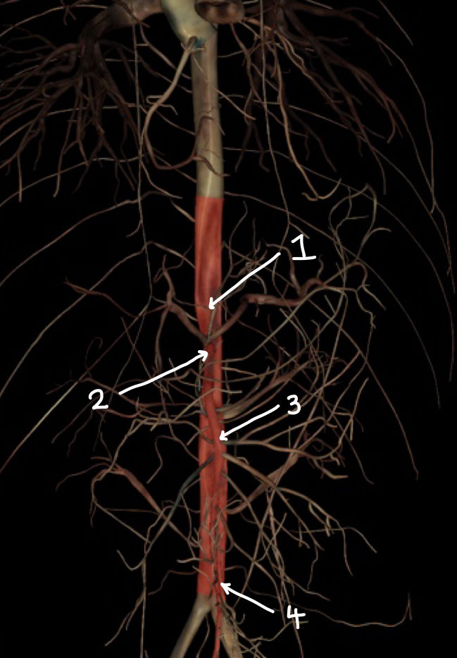
| what structure is labeled 1 | abdominal aorta |
| what structure is labeled 2 | celiac trunk |
| what structure is labeled 3 | superior mesenteric artery |
| what structure is labeled 4 | inferior mesenteric artery |
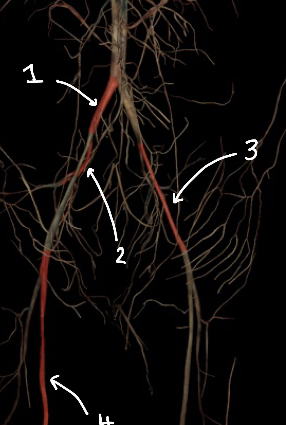
| what structure is labeled 1 | common iliac artery |
| what structure is labeled 2 | internal iliac artery |
| what structure is labeled 3 | external iliac artery |
| what structure is labeled 4 | femoral artery |
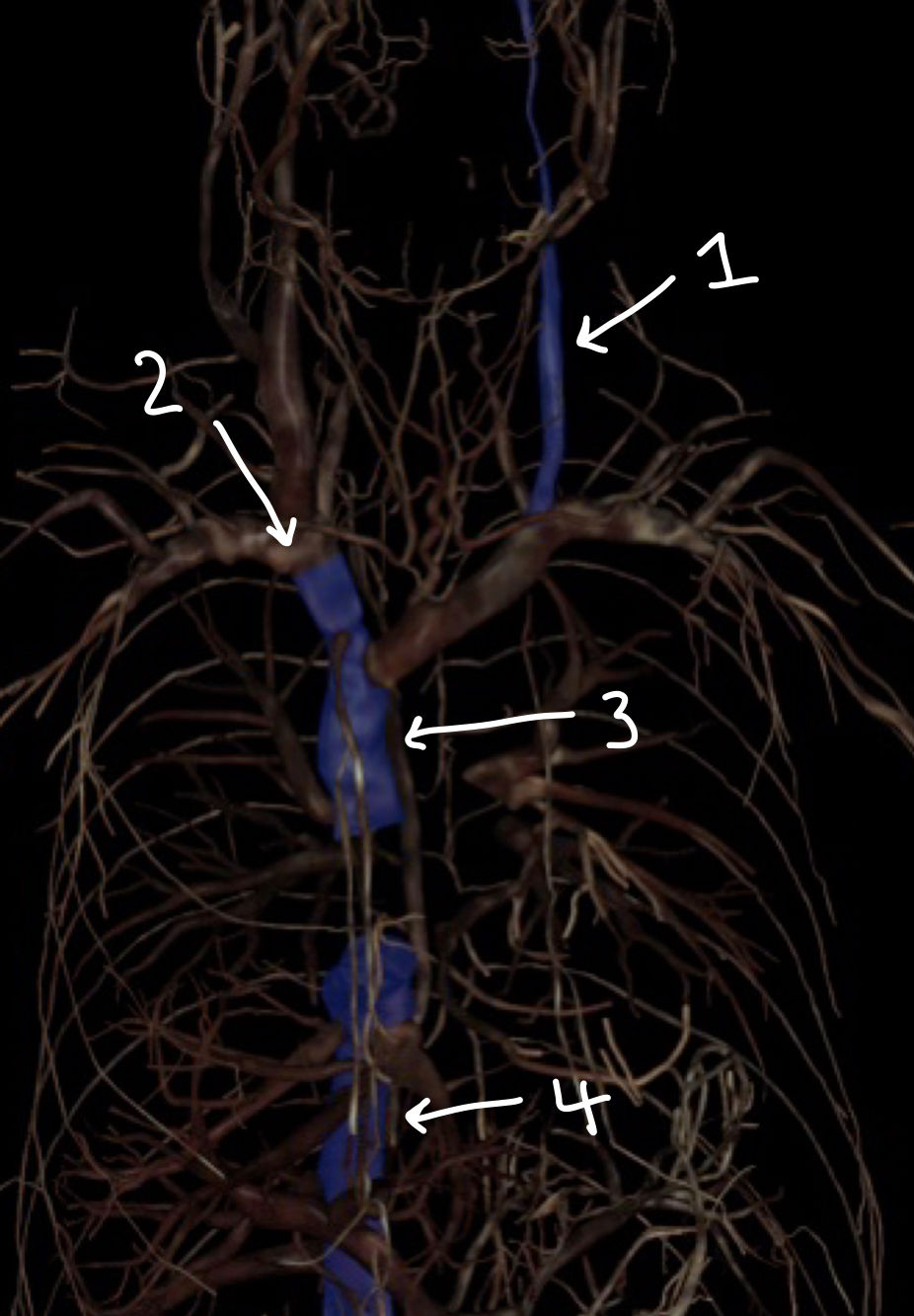
| what structure is labeled 1 | internal jugular vein |
| what structure is labeled 2 | brachiocephalic vein |
| what structure is labeled 3 | superior vena cava |
| what structure is labeled 4 | inferior vena cava |
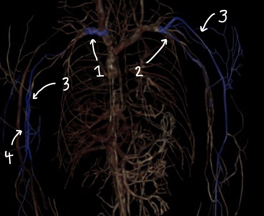
| what structure is labeled 1 | subclavian vein |
| what structure is labeled 2 | axillary vein |
| what structure is labeled 3 | cephalic vein |
| what structure is labeled 4 | basilic vein |
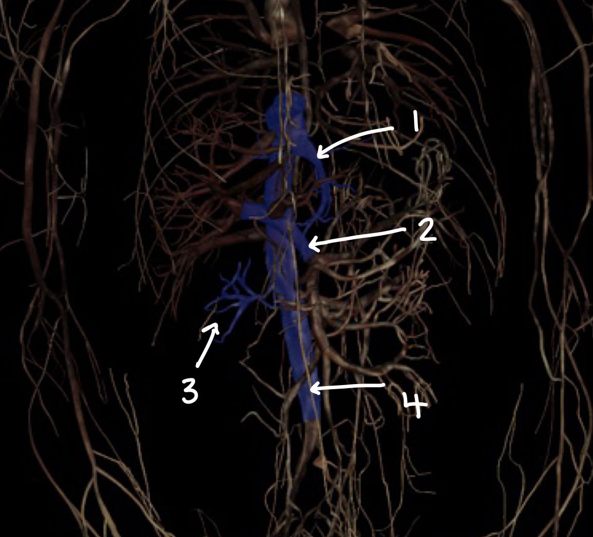
| what structure is labeled 1 | hepatic vein |
| what structure is labeled 2 | hepatic portal vein |
| what structure is labeled 3 | renal veins |
| what structure is labeled 4 | inferior vena cava |
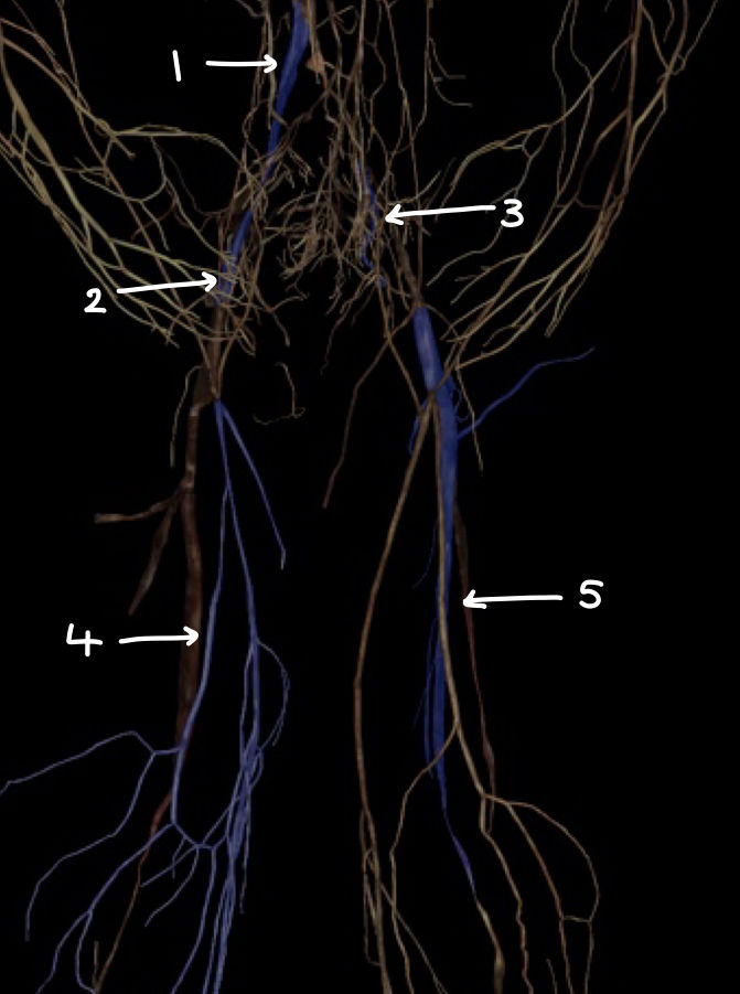
| what structure is labeled 1 | common iliac vein |
| what structure is labeled 2 | external iliac vein |
| what structure is labeled 3 | internal iliac vein |
| what structure is labeled 4 | great saphenous vein |
| what structure is labeled 5 | femoral vein |
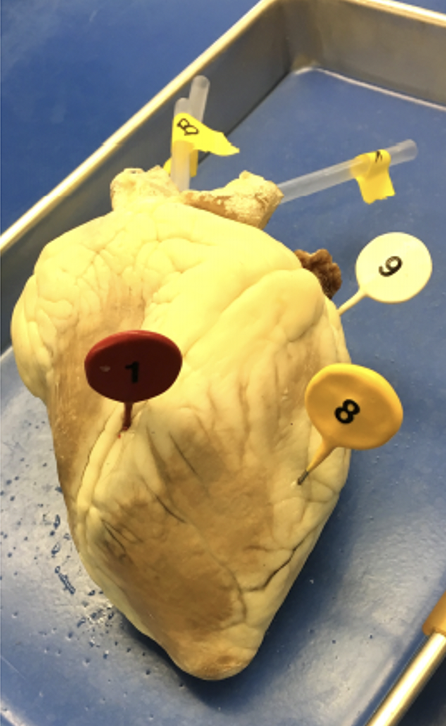
| what is A | pulmonary trunk |
| what is B | aorta |
| what is C | superior vena cava |
| what is D | pulmonary vein |
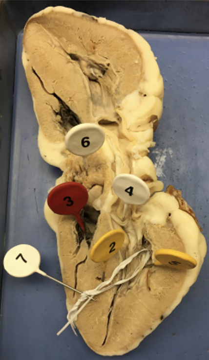
| what is 2 | interventricular septum |
| what is 3 | tricuspid valve |
| what is 4 | bicuspid valve |
| what is 5 | papillary muscle |
| what is 6 | pectinate muscle |
| what is 7 | chordae tendinae |
| what is 1 | interventricular sulcus |
| what is 8 | ventricle |
| what is 9 | atrium |
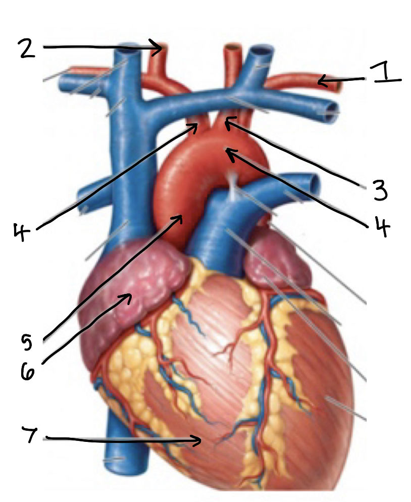
| what is 1 | subclavian artery |
| what is 2 | common carotid artery |
| what is 4 on the left side (anatomical position) | aortic arch |
| what is 4 on the right side (anatomical position) | brachiocephalic trunk |
| what is 5 | ascending aorta |
| what is 6 | atrium |
| what is 7 | ventricle |
| arteries carry blood (away from/to) the heart | away from |
| veins carry blood (away from/to) the heart | to |
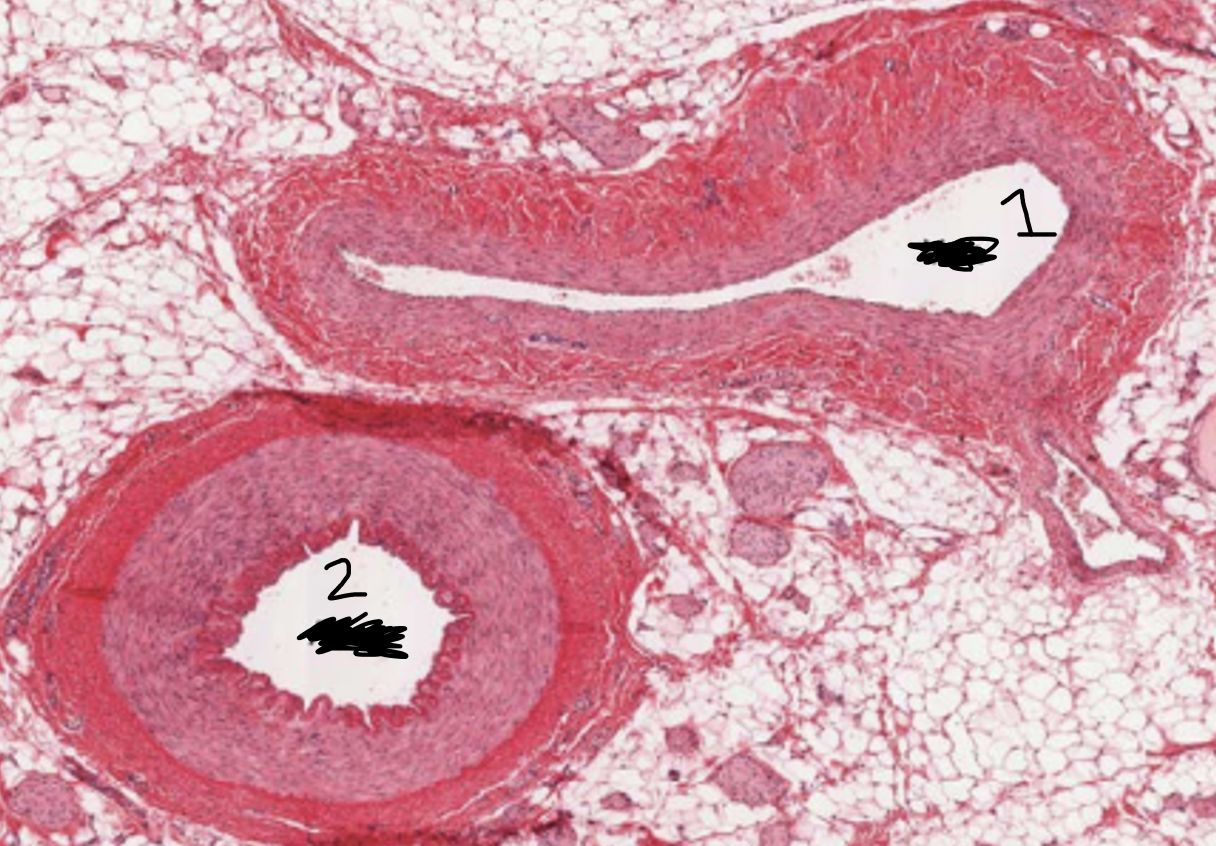
| the size of the lumen is bigger on an artery or a vein | vein |
| the thickness of the wall is larger on an artery or a vein | artery |
| what is 1 and 2 | 1 - vein 2 - artery |
| the lub sound (first heart sound) is created because these valves close | AV valves |
| the dub sound (second heart sound) is created because these valves close | SL valves |
| in this wave of the ecg, there is atrial depolarization | p wave |
| in this wave of the ecg, there is atrial repolarization and ventricular depolarization | qrs wave |
| in this wave of the ecg, there is ventricular repolarization | t wave |
| how is p wave amplitude and duration affected after exercise | amplitude goes up and duration goes up |
| how is qrs amplitude and duration affected after exercise | both stay pretty similar |
| how is the t wave amplitude and duration affected after exercise | amplitude increases and duration decreases |
| when does the first heart sound happen based on an ecg | 0.05 seconds after R |
| when does the second heart sound happen based on ecg | 0.05 seconds after T |
| what is the calibration force for the force transducer setup | 49 mN |
| the positive lead attaches to this part for the frog experiment | hook with heart |
| the negative lead attaches to this part for the frog experiment | right shoulder |
| the earth lead attaches to this part for the frog experiment | left thigh |
| how many atria and ventricles do frogs have compared to humans | frogs have 2 atria and 1 ventricle humans have 2 atria and 2 ventricles |
| is there oxygen separation in the frog heart | no |
| where are the pacemaker cells in a frog compared to human | frog - SV node human - SA node |
| a decrease in temperature results in this change in heart rate. this is because proteins and enzymes require higher temperatures to work efficiently. | decrease in heart rate |
| an increase in calcium concentrations leads to these changes in force and HR. the reasoning is that calcium facilitates the cross bridge formation between thick and thin filaments which increases contractility. | increase in force, little or no increase in HR |
| an increase is potassium concentration leads to this change in HR and force. this is because potassium depolarizes the membrane potential of pacemaker cells and decreases driving force. | increase in HR then decrease in HR no change in force |
| when adding isuprel to the frog heart, this is how HR and contractile force changed. This is because it activates beta1 adrenergic receptors which causes Gs stimulation, increases adenyl cyclase, increase cAMP, increase HCN channel activity, increase pKa, increase calcium concentration. | increase HR increase contractile force |
| when adding Ach to a frog heart, this happens to HR. This is because Ach activates muscarinic receptors and causes Gi stimulation which decreases adenyl cyclase, cAMP, HCN channel activity, decrease pKa, decrease calcium concentration. | decrease HR |
| When adding atropine then Ach to the frog heart, this is what happens to HR and force. This is because atropine is a plant alkaloid that blocks muscarinic receptors. | normal HR normal force |
| When mechanically stretching the frog heart, this is the expected observation. This is because of the frank-starling law | increase contractile force no decline phase due to pericardium |
| absolute refractory in a frog heart causes this channel to inactivate | Na |
| in frog heart experiment, which peak represents atrial contraction and ventricular contraction (big peak of little peak) | atrial contraction - little peak ventricular contraction - big peak |
| what are the primary lymphoid organs | thymus bone marrow |
| what are the secondary lymphoid organs | spleen, lymph nodes, tonsils, appendix |
| this lymphoid organ is bi-lobed, Capsule- connective tissue Cortex- T cells, dendritic cells, macrophages, epithelial cells Medulla- more mature T cells, macrophages, dendritic cells, Hassall’s corpuscles | thymus |
| this region is the darker region of thymus due to presence of lymphocytes, in comparison to this region, which has less dense concentration | cortex - dark medulla - light |
| this lymphoid organ filters blood and is the site of initiation of immune response | spleen |
| this pulp in the spleen serves as site of storage of RBCs and filters them | red pulp |
| this pulp in the spleen contains clusters of T cells, B cells and Macrophages | white pulp |
| what is the difference between indirect and direct ELISA? | indirect - antigen coated direct - antibody coated |
| this lymphocyte doesn't involve antibodies, matures in the thymus, and directly kills infected host cells | T lymphocyte |
| this lymphocyte secretes antibodies, matures in the thymus and bone marrow, and provides defense against pathogens | B lymphocyte |
Want to create your own Flashcards for free with GoConqr? Learn more.