4045767
Description
Flashcards by Riki M, updated more than 1 year ago
More
|
|
Created by eimearkelly3
about 10 years ago
|
|
|
|
Copied by Riki M
about 10 years ago
|
|
| Question | Answer |
| What are the two types of circulatory system? | Open and closed |
| Explain an open-circulatory system | The heart pumps blood into open-ended vessels. The blood leaves these vessels and flows all around the cells of the animal's body. The blood flows back to the heart, entering it through openings in the heart wall e.g. crabs, lobsters, insects, spiders, slugs, snails |
| Explain a closed circulatory system | Blood remains in a continuous system of blood vessels, i.e. blood is always enclosed in blood vessels Exchange of material is possible through the thin capillary walls e.g. earthworms, humans |
| Why is a closed circulatory system more efficient than an open one? | Allows the blood to be pumped around the body faster --> higher metabolic rate, faster exchange of material Allows the blood flow to different organs to be increased or decreased |
| What are the 3 components of the circulatory system? | > blood > blood vessels > heart |
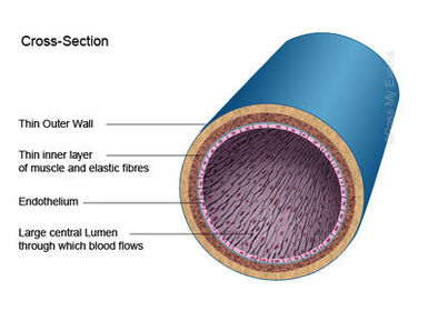
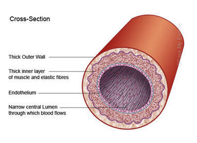
| What are the 3 types of blood vessels? | >Arteries >Veins >Capillaries |
| What is the role of the arteries? | Carry blood away from the heart, divide into smaller vessels called arterioles, oxygenated blood with the exception of the pulmonary artery |
| What is the role of the veins? | Carry blood to the heart, divide into smaller vessels called venules, deoxygenated blood with the exception of the pulmonary vein |
| What is the role of the capillaries? | Tiny vessels that link arteries and veins |
| Tough, inelastic protein in arteries and veins | Collagen (prevents walls from over-expansion) |
| What is the middle layer in arteries and veins? | Muscle and elastic fibre |
| What is the inner single layer of living cells surrounding the lumen? | Endothelium |
| Vein | |
| Artery | |
| Capillary walls are _____ and are made of ______ | permeable ; single layer of endothelium cells |
| What is blood pressure? | The force the blood exerts against the wall of a blood vessel |
| What is the function of valves? | To prevent the backflow of blood |
| systolic | contraction |
| diastolic | relaxation |
| average systolic pressure | 110 -140 mm Hg |
| average diastolic pressure | 75-80 mm Hg |
| Device used to measure blood pressure? | Sphygmomanometer |
| What is the location of the heart? | Between the two lungs (slightly to the left side of the chest) just above the diaphragm in the thoracic cavity. |
| Double membrane surrounding the heart | Pericardium |
| What type of pump is the heart? | A double pump |
| Which ventricle is thickest? | Left (pumps blood all around the body) |
| Wall that divides the heart | Septum |
| Four chambers of the heart | Two atria Two ventricles |
| Thickness of walls in the atria | Thin |
| Tough chords / heart strings | Tendons |
| Tendons are attached to the heart wall by projections called | papillary muscles |
| What is the valve on the right side of the heart? | Tricuspid valve |
| What is the valve on the left side of the heart? | Bicuspid valve |
| What are the valves that allow blood to flow into the aorta and pulmonary artery? | Semilunar valves |
| Deoxygenated blood enters the heart through the ? | Venae Cavae |
| Blood flows out of the heart to the lungs through the ? | pulmonary artery |
| Oxygenated blood enters the heart through the ? | pulmonary veins |
| The oxygenated blood flows out of the heart and around the body through the ? | aorta |
| Blockage of the coronary arteries can result in a ? | heart attack |
| What are the two circuits? | Pulmonary circuit Systemic circuit |
| Pulmonary circuit | the blood is pumped to the lungs to lose carbon dioxide and gain oxygen and is then returned to the heart |
| Systemic circuit | heart - body - heart |
| Heartbeat is controlled by the ? | SA node (Pacemaker) |
| What is the SA node and where is it located? | A small bundle of specialised tissue located close to the entry of the superior vena cava within the right atrium wall |
| The SA node sends out ? | regular electrical impulses that cause the right atrium to contract. |
| What is the role of the AV node? | It sets the rhythm of your heart contractions. |
| A record of the electrical activity of the heart | ECG (electrocardiogram) |
| filling phase | diastole (approx 0.4 secs) -relaxation (passive) |
| emptying phase | systole (approx 0.4 secs) 1. Atrial systole (0.1 secs) 2. Ventricular systole (0.3 secs) |
| Where is the AV node located and what does it do? | Is located between the right atrium and left ventricle. Receives the signal from the SA node and causes ventricle to contract. |
Want to create your own Flashcards for free with GoConqr? Learn more.