293593
Description
Flashcards by eimearkelly3, updated more than 1 year ago
|
|
Created by eimearkelly3
over 12 years ago
|
|
| Question | Answer |
| Two types of circulatory system: | Open and closed |
| Open-circulatory system | The heart pumps blood into open-ended vessels. The blood leaves these vessels and flows all around all of the cells of the animal's body. The blood flows back to the heart, entering it through openings in the heart wall e.g. crabs, lobsters, insects, spiders, slugs, snails |
| Closed circulatory system | Blood remains in a continuous system of blood vessels, i.e. blood is always enclosed in blood vessels Exchange of material is possible through the thin capillary walls e.g. earthworms, humans |
| Reasons for a closed system being more efficient: | Allows the blood to be pumped around the body faster --> higher metabolic rate, faster exchange of material Allows the blood flow to different organs to be increased or decreased |
| 3 components of the circulatory system | blood, blood vessels, heart |
| 3 types of blood vessels | Arteries Veins Capillaries |
| Arteries | Carry blood away from the heart, divide into smaller vessels called arterioles, oxygenated blood with the exception of the pulmonary artery |
| Veins | Carry blood to the heart, divide into smaller vessels called venules, deoxygenated blood with the exception of the pulmonary vein |
| Capillaries | Tiny vessels that link arteries and veins |
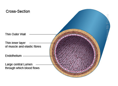
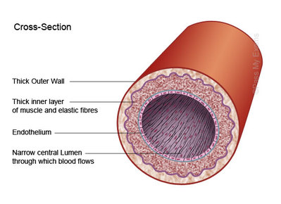
| Tough, inelastic protein in arteries and veins | Collagen (prevents walls from over-expansion) |
| Middle layer in arteries and veins | Muscle and elastic fibre |
| Inner single layer of living cells surrounding the lumen | Endothelium |
|
Image:
examtime (image/jpg)
|
Vein |
|
Image:
examtime (image/jpg)
|
Artery |
| Capillary walls are _____ and are made of ______ | permeable ; single layer of endothelium cells |
| Blood pressure | The force the blood exerts against the wall of a blood vessel |
| Function of valves | To prevent the backflow of blood |
| systolic | contraction |
| diastolic | relaxation |
| average systolic pressure | 110 -140 mm Hg |
| average diastolic pressure | 75-80 mm Hg |
| Device used to measure blood pressure | Sphygmomanometer |
| Location of the heart | Between the two lungs (slightly to the left side of the chest) just above the diaphragm in the thoracic cavity. |
| Muscle of the heart | Cardiac muscle |
| Double membrane surrounding the heart | Pericardium |
| Fluid in the pericardium | Pericardial fluid |
| Cardiac muscle is | Slow to fatigue |
| What type of pump is the heart? | A double pump |
| Which ventricle is thickest? | Left (pumps blood all around the body) |
| Wall that divides the heart | Septum |
| Four chambers of the heart | Two atria Two ventricles |
| Thickness of walls in the atria | Thin |
| Tough chords / heart strings | Tendons |
| Tendons are attached to the heart wall by projections called | papillary muscles |
| Valve on the right side of the heart | Tricuspid valve |
| Valve on the left side of the heart | Bicuspid valve |
| Valves that allow blood to flow into the aorta and pulmonary artery | Semilunar valves |
| Lord | Left oxygenated Right deoxygenated |
| Deoxygenated blood enters the heart through the | Venae Cavae |
| Blood flows out of the heart to the lungs through the | pulmonary artery |
| Oxygenated blood enters the heart through the | pulmonary veins |
| The oxygenated blood flows out of the heart and around the body through the | aorta |
| What is a portal system? | A blood pathway that begins and ends in capillaries |
| The cardiac muscle is supplied with blood from the | coronary/cardiac arteries branching from the aorta at the point where it leaves the heart, just beyond the semilunar valve |
| Blockage of the coronary arteries can result in a | heart attack |
| chest pains often preceding a heart attack | angina |
| two circuits | Pulmonary circuit Systemic circuit |
| Pulmonary circuit | the blood is pumped to the lungs to lose carbon dioxide and gain oxygen and is then returned to the heart |
| Systemic circuit | heart - body - heart |
| Heartbeat is controlled by the | Pacemaker |
| What is the pacemaker and where is it located? | A small bundle of specialised tissue located close to the entry of the superior vena cava within the right atrium wall |
| The pacemaker sends out | regular electrical impulses |
| AV node | atrio-ventricular node |
| function of the pacemaker | to control the rate of the heartbeat |
| A record of the electrical activity of the heart | ECG (electrocardiogram) |
| Nerves connecting the pacemaker to the brain (one reduces rate of heartbeat, the other increases) | Medulla oblongata |
| SA node / pacemaker | Sino-atrial node |
| noise made when the bicuspid and tricuspid valves close | lub |
| noise made when the semilunar valves close | dub |
| filling phase | diastole (approx 0.4 secs) -relaxation (passive) |
| emptying phase | systole (approx 0.4 secs) 1. Atrial systole (0.1 secs) 2. Ventricular systole (0.3 secs) |
| heart murmur | an abnormal sound associate eith the heartbeat, may indicate damage to one or more of the valves |
| Smoking | Causes heart disease (nicotine increases blood pressure and carbon monoxide interfere with the transport of oxygen in the body's cells - high levels cause the hardening of the arteries) |
| Diet | A build up of cholesterol from animal fats can lead to a blockage in the cardiac artery leading to a heart attack, eating less fatty meats and fatty dairy products can reduce this risk) |
| Exercise | increases heat rate which strengthens the cardiac muscle making it more efficient at pumping blood, this improve the oxygen supply to the cardiac muscle and reduces blood pressure |
Want to create your own Flashcards for free with GoConqr? Learn more.