4288720

| Question | Answer |
| 3.3: Organisms exchange substances with their environment | N/A |
| 3.3.1: Surface area to volume ratio | N/A |
| What is the relationship between size and surface area to volume ratio? | As size increases, surface area to volume ratio decreases. |
| What is the relationship between size and metabolic rate? | As size increases, metabolic rate decreases. |
| How does surface area to volume ratio affect exchange of substances with the environment? | The larger the surface area to volume ratio, the greater the rate of exchange as there is a larger surfaces over which exchange can occur. |
| What are some adaptations that can increase surface area to volume ratio? | - Thin bodies - Thin protrusions e.g. fins |
| 3.3.2 Gas exchange | N/A |
| What is Fick's Law? | Rate of diffusion ∝ (surface area x difference in concentration) / Distance |
| How do single-celled organisms exchange gases with their environment? | Gases diffuse across the cell membrane in and out of the cell. |
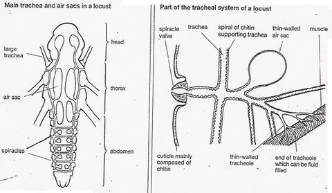
| How do insects exchange gases with their environment? | - Valves on the outside surface allow gases to diffuse in and out - Network of chitin-supported tubes called tracheae connect the body and transport gases - Smaller tubes called tracheoles branch off into muscle tissue and carry gases to respiring cells |
| How are insects adapted to increase the rate of diffusion of gases? | - Many have thin bodies - Contractions of abdominal muscles maintains constant flow of air and thus increases the concentration gradient - Short distance from outside to muscle tissues |
| What does the gas exchange network of an insect look like? | |
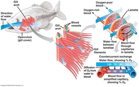
| How do fish exchange gases with their environment? | - Water flows in through mouth and out through gills - Oxygen contained in water diffuses into capillary network in gill lamella - Capillary network transports oxygen into bloodstream |
| How are fish adapted to increase the rate of diffusion of gases? | - Thin, flat bodies - Countercurrent flow of water and blood maintains concentration gradient - Large surface area of gills due to long, thin filaments ridged with thin lamella |
| What does the gas exchange system of a fish look like? | |
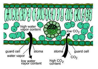
| How do plants exchange gases with their environment? | - Pores on the underside of leaves called stomata allow gases to diffuse in and out - Carbon dioxide diffuses through air gaps in spongy mesophyll layer to reach palisade mesophyll layer where it is used in diffusion |
| How are plants adapted to increase the rate of diffusion of gases? | - Thin, flat leaves - Air gaps in mesophyll layer reduce diffusion pathway |
| What does the gas exchange system of a plant look like? | |
| How do terrestrial insects reduce water loss through their gas exchange network? | - Many have small surface area to volume ratio e.g. ladybirds - Spiracles can close to reduce loss of water through diffusion - Body surface is coated with a waterproof layer to prevent diffusion of water - Body hairs trap a layer of moist air, reducing the concentration gradient |
| How do plants reduce water loss through their gas exchange network? | - Top surface of leaf is coated with a waxy cuticle that reduces water loss through diffusion - Guard cells on either side of stomata can close to reduce loss of water through diffusion - Many plants have needle-like leaves with reduced surface area to volume ratio |
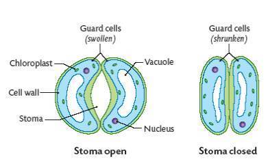
| What does the structure of a guard cell look like? | |
| How does the structure of a guard cell relate to its function? | - Large vacuole causes cell to become turgid when full of water and flaccid when without water, opening the stomatal pore when water is plentiful and closing it when water is scarce - Thicker interior cell wall creates a tight seal and reduces water loss through evaporation |
| What is the term for a plant specially adapted to reduce its water loss? | Xerophytic |
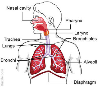
| How do mammals exchange gases with their environment? | - Gases enter through the mouth and nose and travel down the trachea (cartilage-ringed tube) - Tube splits into two bronchi, each leading into one of the two lungs - Bronchi branch-off into smaller bronchioles and further into tiny sacs cal |
| What organs make up the human gas exchange system? | - Trachea - Bronchi - Bronchioles - Alveoli - Lungs |
| What does the human gas exchange system look like? | |
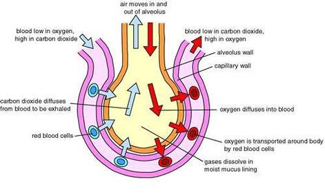
| What does the structure of an alveolus look like? | |
| How does the structure of an alveolus relate to its function? | - Sac able to accommodate large volume of air - Covered with network of capillaries, increasing the area over which diffusion can occur - Thin walls of both alveolus and capillaries reduce distance over which gases need to diffuse - Constant flow of blood maintains concentration gradient |
| What is ventilation? | The movement of air in and out of the lungs. |
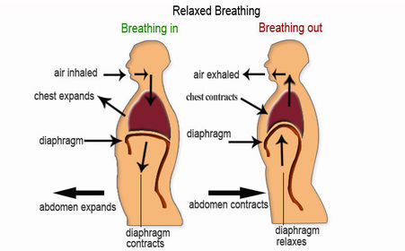
| How is air drawn into the lungs during inspiration? | - Diaphragm contracts and moves downwards, decreasing the pressure in the thorax - Outer intercostal muscles contract and inner intercostal muscles relax, pulling the rib cage up and outwards; increasing the volume of the thorax - Air is drawn into the lungs because of the pressure difference |
| How is air forced out of the lungs during expiration? | - Diaphragm relaxes and moves upwards, increasing the pressure in the thorax - Inner intercostal muscles contract and outer intercostal muscles relax, pulling the rib cage down and inwards; decreasing the volume of the thorax - Air is forced out of the lungs by the pressure difference |
| What does the structure of the thorax look like? | |
| What is pulmonary ventilation rate (PVR)? | The volume of air that can be breathed into a persons lungs in one minute. Pulmonary ventilation rate = tidal volume × breathing rate |
| How can lung disease affect ventilation? | - Alveoli are less or no longer elastic, reducing efficiency of ventilation - Airways become swollen, reducing the volume of air that can pass through - Increased mucus production can block airways and fill lungs, preventing air from passing |
| What are risk factors associated with lung disease? | - Smoking - Living in a polluted area - Working in conditions with a risk of inhaling chemicals |
| How can the impact of these risk factors be reduced? | - Quitting smoking - Reducing air pollution - Wearing of safety equipment e.g. gas mask |
| What is the difference between correlation and causation? | - Correlation is the mutual relationship or pattern between two factors - Causation means that one factor is the direct cause of the other |
| 3.3.3: Digestion and absorption | N/A |
| What is digestion? | The hydrolysis of large molecules into smaller ones for absorption across cell membranes. |
| How are carbohydrates digested? | - Amylase produced in the mouth hydrolyses the alternate glycocidic bonds of the starch molecule to produce maltose molecules - Maltose is hydrolysed into α glucose by maltase produced in the ileum - Sucrase hydrolyses the glycocidic bond in sucrose to produce α glucose and fructose - Lactase hydrolyses the glycocidic bond in lactose to produce α and fructose |
| How are lipids digested? | - Lipases hydrolyse esther bonds found in triglycerides to release monoglycerides and fatty acids - Lipids are firstly split up into smaller beads called micelles by Bile Salts produced in the liver during emulsification, increasing the surface area over which lipases can act |
| How are proteins digested? | - Endopeptidases hydrolyse the peptide bonds between amino acids in the central region of protein molecules, forming a series of peptide molecules - Exopeptidases hydrolyse the peptide bonds on the terminal amino acids of the peptide molecules formed by endopeptidase action. In this way they progressively release dipeptides and single amino acids - Dipeptidases hydrolyse the bond between the two amino acids of a dipeptide. Dipeptidases are membrane bound, in that they form part of a cell-surface membrane of the epithelial cells lining the ileum |
| What is co-transport? | Simultaneous transport of molecules across a membrane, through a transport protein |
| How is the ileum adapted to absorb products of digestion? | - Villi - Microvilli - Single cell thick walls |
| What are the steps involved in co-transport of substances into the ileum? | - Sodium ions in cells transported into blood and potassium is transported from the blood into cells via active transport through the antiport (sodium-potassium pump)(requiring ATP), lowering the concentration gradient of sodium inside - Glucose and sodium in the lumen enter cells through co-transport through the symport (requiring ATP), sodium moves along concentration gradient, glucose against it - Glucose enters the blood through facilitated diffusion through carrier proteins along the concentration gradient |
| 3.3.4.1 Mass transport in animals | N/A |
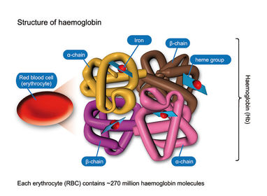
| What is haemoglobin? | - group of chemically similar molecules found in many different organisms - protein with a quaternary structure. |
| What does the structure of haemoglobin look like? | |
| What is 'loading'? | The process of oxygen 'associating' with each of the four heme groups in the quaternary structure. |
| What is 'unloading'? | The process of oxygen 'disassociating' with each of the four heme groups in the quaternary structure. |
| What is affinity? | The attraction between oxygen and the heme groups of haemoglobin molecules. |
| What is the oxygen dissociation curve? | A graph mapping the relationship between the partial pressure (concentration) of oxygen and the saturation of haemoglobin molecules with oxygen. |
| How does the structure of haemoglobin change after each successive oxygen molecule associates with its heme groups? | With every oxygen molecule associating with haemoglobin, the quaternary structure changes to better accommodate further oxygen molecules and thus increase affinity. |
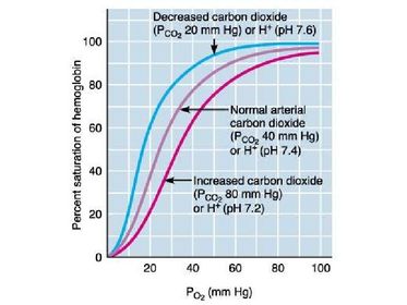
| What is the Bohr effect? | The effect of carbon dioxide on oxygen dissociation with haemoglobin, with increased carbon dioxide concentration increasing the rate at which oxygen 'unloads'. |
| What causes the Bohr effect? | Carbon dioxide is acidic, thus increased concentration will cause oxygen to dissociate from haemoglobin faster as the acidic condition cause bonds in the quaternary structure to break down, changing the shape of the molecule and reducing it |
| What effect does the Bohr effect have on the oxygen dissociation curve? | Increased carbon dioxide shifts the curve to the right, reduced carbon dioxide shifts it to the left. |
| What does the Bohr effect look like on an oxygen dissociation curve? | |
| What are some adaptations to the haemoglobin of animals? | - animals living in high altitudes have haemoglobin with a much greater affinity for oxygen so they can provide their cells with enough oxygen despite its scarcity in the air. |
| What is the pattern of blood circulation in mammals? | Right side of heart -> lungs -> left side of heart -> rest of body -> right side of heart |
| What is the name of the blood vessels supplying the heart muscle with oxygenated blood? | Coronary arteries |
| What are the four main blood vessels carrying blood to and from the heart? | - Vena cava - Pulmonary Artery - Pulmonary Vein - Aorta |
| What is the name of the blood vessel carrying oxygenated blood to the kidneys? | Renal artery |
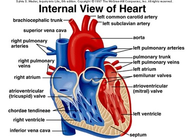
| What are the names of the four heart chambers? | - Right atrium - Right ventricle - Left atrium - Left ventricle |
| What are the names of the four main heart valves? | - Atrioventricular valves (separating atria and ventricles) - Semi-lunar valves (separating ventricles and main arteries) |
| What does the structure of the heart look like? | |
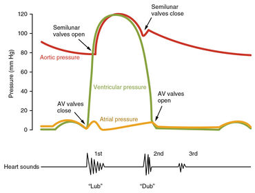
| What are the three stages of the cardiac cycle? | - Atrial systole - Ventricular systole - Diastole |
| What happens during the atrial systole phase? | - Atrial walls contract - Blood is forced through atrioventricular valves into ventricles - Blood pressure in ventricles increased |
| What happens during the ventricular systole phase? | - Atrial walls relax - Atrioventricular valve closes - Ventricular walls relax - Semi-lunar valves open - Blood forced through semi-lunar valves into pulmonary aorta/aorta - Pressure in ventricles decreased |
| What happens during the diastole phase? | - All valves close - All muscles relax - Blood flows into atria |
| What does a graph of the cardiac cycle mapping chamber pressure look like? | |
| What is the change to pressure of the heart's chambers during the cardiac cycle? | - Atrial pressure - spikes at atrial systole when blood forced out, slowly climbs back up at ventricular systole as blood flows back in, - Ventricular pressure - small spike at atrial systole when blood pumped in, large spike at ventricular systole when muscles contract |
| What is the change to volume of the heart's chambers during the cardiac cycle? | - Atrial volume - increases as blood flows in during diastole, decreases during atrial systole when muscles contract - Ventricular volume - increases during atrial systole when blood flows in, decreases during ventricular systole when muscles contract |
| Why is it important that pressure is maintained in the heart? | So that blood only flows in the forwards direction |
| What are the four main blood vessels in the body? | - Veins - Arteries - Arterioles - Capillaries |
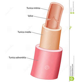
| What is the structure of a vein like? | - Thin muscular layer - Thin elastic layer - 1-cell-thick endothelium - Wide lumen - Valves at intervals |
| What does the structure of a vein look like? | |
| How does the structure of a vein relate to its function? | - Thin muscle and elastic layer as blood pressure low - Overall thin walls to allow skeletal muscle contractions to maintain flow against gravity - Wide lumen to allow maxiumum bloodflow - Valves prevent backflow of blood when pressure low |
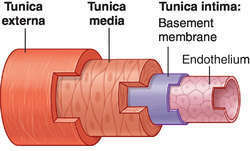
| What is the structure of an artery like? | - Thick muscular layer - Thick elastic layer - 1-cell-thick endothelium - Thin lumen |
| What does the structure of an artery look like? | |
| How does the structure of an artery relate to its function? | - Thick muscular layer to accommodate higher pressure blood and - Thick elastic layer to stretch and relax in time with heart contractions to maintain blood flow - Thin lumen due to high pressure |
| What is the structure of an arteriole like? | - Thicker, smooth, muscular wall than arteries - Thicker than arteries - One-cell thick endothelium - Thinner lumen than veins |
| What does the structure of an arteriole look like? | |
| How does the structure of an arteriole relate to its function? | - Thicker muscular wall to regulate blood flow to capillaries through muscular contraction and dilation - Thicker elastic wall due to lower pressure, helps to maintain flow by stretching at systole and springing back at diastole |
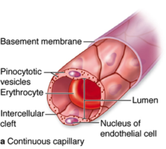
| What is the structure of a capillary like? | - No muscular layer - No elastic layer - 1-cell-thick endothelium - Thin lumen |
| What does the structure of a capillary look like? | |
| How are capillaries effective as exchange surfaces? | - Provide a large surface area for exchange - So narrow that red blood cells squeezed flat as they pass, bringing them even closer to cells needing oxygen - Numerous and highly branched, providing oxygen for all cells - Very short diffusion pathway due to thin walls |
| What is tissue fluid? | Fluid immediately surrounding cells in tissue - Allows substances to move between cells and the blood - comprised of water and dissolved substances such as glucose, amino acids, fatty acids, ions in solution, oxygen - takes waste such as CO2 & urea |
| How is tissue fluid formed? | Hydrostatic pressure in capillary (caused by heart pumping) must be greater than hydrostatic pressure outside the capillary, and osmotic pressure pushing inwards (as the blood has a higher water potential) in order for untrafiltration to take place - Ultrafiltration of substances by pressure out of capillaries forms tissue fluid |
| How does tissue fluid return to the capillaries? | Once tissue fluid has exchanged metabolic materials with the cells, it must return to the blood - loss of tissue fluid lowers hydrostatic pressure in capillaries, thus by the venule end of the capillary the hydrostatic pressure inside is lower than the hydrostatic pressure outside, tissue fluid also has lower water potential due to loss of water but maintainence of proteins - tissue fluid returns to the capillary by osmosis |
| What is the lymphatic system? | Not all tissue fluid returns to the capillaries, some is carried by the lymphatic system and drained back into the bloodstream via two ducts that join veins close to the heart - contents moved hydrostatic pressure of tissue fluid & contraction of body muscles - valves in lymph vessels ensure that tissue fluid only travels towards the heart |
| What is cardiac output? | Means of calculating the pumping power of the heart Cardiac output = stroke volume × heart rate |
| What are some risk factors associated with heart disease? | - Smoking - High cholesterol (can clog arteries) - Lack of exercise - Obesity |
| 3.3.4.2 Mass transport in plants | N/A |
| What is the xylem? | Hollow, thick walled tubes made up of xylem vessels (dead cells), which no cell walls on top or bottom - transports water from the soil to leaves in the process of transpiration |
| What is transpiration? | Process of movement of water from roots to leaves of flowering plants, where it evaporates through the stomata of the leaf |
| How does water move across the cells of a leaf? | Mesophyll cells lose water by evaporation through their cell walls to the empty spaces by stomata - loss of water lowers water potential, so water moves from neighbouring cells via osmosis - water moves via osmosis along the Symplastic pathway (through cytoplasm of cells connected by plasmodesmata) or Apoplastic pathway (through connected cell walls) - movement of water by osmosis perpetuates the water potential gradient, causing water from the xylem to replace lost water in the leaf cells |
| How does water enter the xylem? | Water enters root hair cells via osmosis, and moves along root cortex cells via the Symplastic pathway or Apoplastic pathway until it reaches the xylem vessels, which it enters via plasmodesmata |
| How does water move up the xylem? | As water evaporates from mesophyll cells into air gaps by stomata and thus out of the leaf, more molecules of water are drawn up the xylem by cohesion - transpiration pull - water column flows |
| What is cohesion-tension theory? | Water molecules form hydrogen bonds between molecules as they are polar - cohesion between molecules allows them to flow - water forms a continuous flow up the xylem into the mesophyll cells in the leaves, drawn by transpiration pull |
| What evidence is there for cohesion-tension theory? | - When the xylem is cut no water flows out, thus the transport is not by pressure or pumping - When the xylem is cut and air enters it, water can no longer travel to reach the leaves |
| What is phloem? | Transport tissue, made up of long, thin, mostly hollow cells called sieve tube elements (living specialised cells) arranged end to end. End walls are perforated to form sieve plates. - Associated with companion cells which run along one side - transports sugars produced during photosynthesis (and inorganic ions) from sources to sinks (cells which use or store sugar, respiring) by translocation |
| What is translocation? | Process of transportation of organic molecules and mineral ions are transported from one part of the plant to another - explained by the mass flow theory |
| How does sucrose enter the phloem? | Sucrose is manufactured in cells with chloroplasts (sources), - sucrose diffuses via facilitated diffusion into companion cells - hydrogen ions are actively transported from companion cells into the spaces between cell walls - hydrogen ions diffuse through carrier proteins into sieve tube elements - sucrose molecules move via co-transport along with hydrogen ions (in co-transport proteins |
| What is mass flow? | Bulk movement of substances through a given area in a specific time - passive process, but establishment of a hydrostatic gradient requires active transport of sugars requiring ATP |
| How does sucrose mass flow through the phloem? | - sieve tubes have a lower water potential due to sucrose being transported in - xylem has much higher water potential, so water moves from the xylem into the phloem by osmosis, creating high hydrostatic pressure in the source end of the phloem - at sink cells, sucrose is used during respiration or stored, creating a low sucrose content - sucrose is actively transported into sinks from sieve tubes, lowering their water potential - water moves into sink cells by osmosis, lowering the hydrostatic pressure in the sink end of the phloem - mass flow of sucrose solution down the hydrostatic gradient to the sink cell end |
| What evidence is there supporting the mass flow hypothesis? | - when phloem is cut, a solution of organic molecules flows out (thus phloem is where organic molecules travel) - concentration of sucrose is higher in leaves than in roots - metabolic poisons/lack of oxygen inhibit translocation of sucrose in the phloem |
| What evidence is there opposing the mass flow hypothesis? | - function of sieve plates is unclear - not all solutes move at the same speed - sucrose moves at the same speed to all regions, not faster to regions with lower sucrose concentration |
| How can radioactive isotopes be used to test the mass-flow hypothesis? | - Radioactive isotopes present in atmosphere in which plant grows, traced as they move throughout plant using autodradiography. As isotopes incorporated into sugars made by photosynthesis, they follow the path of sugars in plant. Parts of plant cause x ray film to blacken when placed on it, and are found to correspond to location of phloem tissue |
| How can ringing be used to test the mass-flow hypothesis? | - ring of bark removed from tree, also removing outer layers of tissue including phloem - visible bulge develops above ring, as sugars cannot travel below it - below the ring, the plant withers as it is denied sugars - this suggests that the phloem transports sugars in the tree |
Want to create your own Flashcards for free with GoConqr? Learn more.