476032
Description
Flashcards by gina_evans0312, updated more than 1 year ago
|
|
Created by gina_evans0312
almost 12 years ago
|
|
| Question | Answer |
|
Image:
Right_Lung (image/jpg)
|
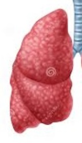
Right Lung- Identifiable by the three grooves |
|
Image:
Left_Lung (image/jpg)
|
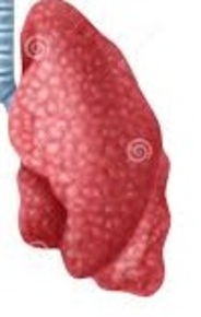
Left Lung- two lobes |
|
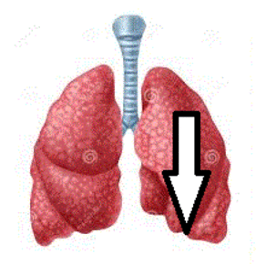
Image:
Cardiac_Notch (image/jpg)
|
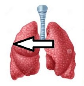
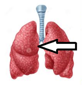
Cardiac Notch |
| Costal Lung Surface | |
|
Image:
Medial_L (image/png)
|
Medial Lung Surface |
| Diaphragmatic Lung Surface | |
| Bronchopulmonary Segment | Branches between segmental brachii and alveoli |
| No of Bronchopulmonary Segments | 10 |
| Bronchopulmonary Segments Supplier | Each have own artery |
| Bronchopulmonary Segments Separation | Done by connective tissue |
| X-Rays of Lungs are done | Back to Front |
| Fluid on X-Ray | Appears whitish |
| Air on X-Ray | Dark & Obscures faint rib lines |
| Pneumonia | Lung tissue infection- fluid accumulation & inflammation |
| In any case of lung pathology | You lose sharp angle of lung edge |
Want to create your own Flashcards for free with GoConqr? Learn more.