5816368
Description
Flashcards by poiznpixel.studi, updated more than 1 year ago
|
|
Created by poiznpixel.studi
almost 8 years ago
|
|
| Question | Answer |
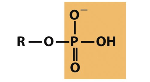
| Phosphoryl Group | |
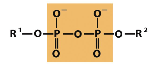
| Phosphoanhydride | |
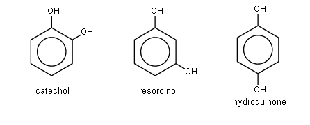
| Catechol, Resorcinol, Hydroquinone | |
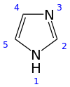
| Imidazole | |
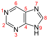
| Purine | |
| When pKa = pH then what does this tell us about the solution? | The solution will have an equal concentration of conjugate base and of the original acid. |
| Henderson–Hasselbalch equation | |
| What is [OH-] in terms of pH | 10^-(14-pH) |
| What is [H+] in terms of pH | 10^(-pH) |
| Buffers | http://chemcollective.org/activities/tutorials/buffers/buffers3 |
| What do we know about the solution when the solution is neutralized? | ??? |
| 4 weak forces relevant to this course | Vanderwaals Hydrophobic Effect Electrostatic Forces Hydrogen Bonding |
| Which AA isomer is more common | L |
| What is a typical pKa for a carboxylic acid | 2 |
| What is a typical pKa for a terminal {\alpha} amino acid | 9 |
| You need to prepare an acetate buffer of pH 5.62 from a 0.646 M acetic acid solution and a 2.85 M KOH solution. If you have 525 mL of the acetic acid solution, how many milliliters of the KOH solution do you need to add to make a buffer of pH 5.62? The pKa of acetic acid is 4.76. | see the sapling |
| Describe Myoglobin | Highly alpha helical |
| Proximal histidine | On the F helix (myoglobin) and is coordinated to the Fe(II) in the heme |
| Compare Fe2+ and Fe3+ | 2+: More stable and can bind to the proximal histidine but is too large to fit into the ring 3+: when the O2 binds the Iron becomes 3+ which is smaller and pulls the proximal helix closer initiating the binding event |
| Super Oxide Structure | Forms when Iron binds to the O2 atom. Has potential to leave and become a ROS. Bound by the distal histodine |
| Hemoglobin | 2 alphaBeta dimers Tetramer Allostericly regulated - binding generates conformation change which affects funcion |
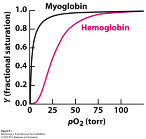
| Oxygen Binding Curves | Fractional saturation vs pO2 |
| Fractional saturation | fraction of possible sites bound to substrate in a POPULATION |
| R state of Hemoglobin | Relaxed state Less ion pair inter actions Compressed Quaternary Change Higher O2 binding affinity Equilibrium towards R favors O2 release |
| T State of Hemoglobin | Tense State More ion pair interactions More stable in absence of O2 Lower O2 binding affinity Equilibrium towards T favors O2 release Bicarbonate binding site stabilized |
| Positve Hemoglobin Effectors | O2 |
| O2 binding event chain | O2 binds Iron ion moves into heme plane Iron pulls the proximal His and the F helix C-terminus of F helix lies at the interface between the αβ dimers Shift in the interface contacts moves the distal His E7 and Val E11 out of the oxygen’s path to the Fe ion on another subunit Makes it easier for O2 to bind to the other subunit! |
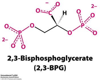
| Negative Hemoglobin Effectors | 2,3-bisphosphoglycerate (2,3-BPG) Protons CO2 all listed heterotrophic shift curve to the right and facilitates RELEASE |
| 2,3 bisphosphoglycerate | Only binds to T state and stabilizes the T state. Binds to highly positive (2lys, 4his) residues at the center of the T state |
| Why is carbon monoxide poisonous | hemoglobin prefers carbon monoxide about 200% more times to O2 |
| Bohr Effect | Metabolizing tissues generate large amounts of H+ and CO2 so: "The O2 affinity of Hb decreases as pH decreases." |
| What happens as the terminal His is protonated | It forms a salt bridge to the negative aspartate and the C-terminus is attracted to the Lysine 40 |
| Summarize Hemoglobin activity in the Lungs | pH is ”high" (pH ~7.4). ([H+] is low.) [CO2] is low because it's being gotten rid of (exhaled). [O2] is high. Ligand concentration conditions all favor R state. Result: O2 binds tightly. (That's what you want, to BIND O2, maximal "loading” in the lungs.) |
| Summarize Hemoglobin activity in tissues | pH is “low” (pH ~7.2) ([H+] is high) because catabolism (breakdown of nutrients) produces protons (acid, especially lactic acid in active muscle tissue). [CO2] is high because CO2 = end product of oxidation of C atoms in catabolism of nutrients. [O2] is low. Ligand concentration conditions all favor T state. Result: O2 binds weakly. (That's what you want, to DISSOCIATE O2, maximal "unloading".) |
| Structural Features of Sickle-cell Anemia | Hydrophobic "patch" allows the tetramers to stick together. This forms a fibrous crystal and precipitates eliminating it's ability to cary O2. The patch is more available in the non oxygenated state (T) (15deg rotation) Stabilizing R state is treatment option |
Want to create your own Flashcards for free with GoConqr? Learn more.