6152709
Description
Flashcards by Ashutosh Kumar, updated more than 1 year ago
|
|
Created by Ashutosh Kumar
over 9 years ago
|
|
| Question | Answer |
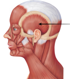
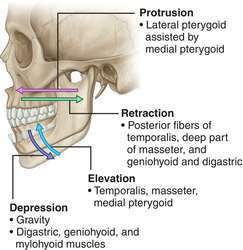
| Name Origin: Insertion: Function: Innervation: | Temporalis Origin: Floor of the temporal fossa and deep surface of the temporal fascia. Insertion: Coronoid process and anterior border of the ramus of the mandible. Function: Raises the mandible and posterior fibres can retract/retrude the mandible. Innervation: Deep temporal branches of the mandibular (V3). |
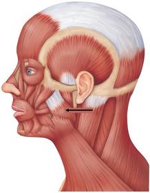
| Name Origin: Insertion: Function: Innervation: | Masseter Origin: Zygomatic arch. Insertion: Lateral surface of the ramus of the mandible Function: Elevates the mandible and deep part can retrude the mandible. Innervation: Masseteric nerve of mandibular (V3). |
| Name Origin: Insertion: Function: Innervation: | Medial pterygoid Origin: Medial surface of the lateral pterygoid plate. Insertion: Medial surface of the ramus of the mandible. Function: Protrudes the mandible as well as elevates the mandible. Innervation: Medial pterygoid nerve of mandibular (V3). |
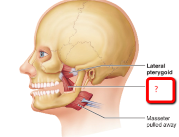
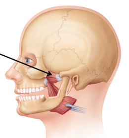
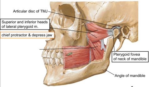
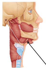
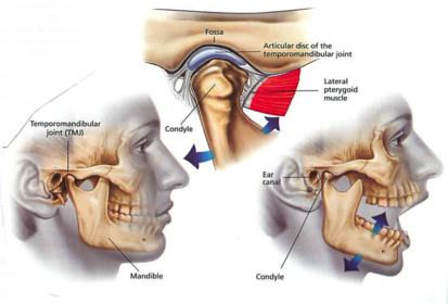
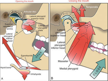
| Name Origin: Insertion: Function: Innervation: | Lateral pterygoid Origin: Has two heads; the superior head originates from the infratemporal crest of the infratemporal surface of the greater wing of the sphenoid, the inferior head originates from the lateral surface of the lateral pterygoid plate. Insertion: Neck of the mandible, capsule and disc of the temporomandibular joint (TMJ). Function: Protrudes and depresses the mandible. Innervation: Lateral pterygoid nerve of mandibular (V3). |
| Name Origin: Insertion: Function: Innervation: | Digastric Origin: Two bellies; the anterior belly originates from the anteromedial surface of the mandible, the posterior belly originates from the medial aspect of the mastoid process. Insertion: Both bellies attach to an intermediate tendon, linking to the hyoid bone. Function: Raise the hyoid bone, or, if the hyoid bone is fixed, depress the mandible as well as retract. Innervation: Anterior belly; mylohyoid nerve, originating from the inferior alveolar nerve, a branch of the mandibular (V3). Posterior belly; digastric branch of the facial nerve (cranial nerve 7) |
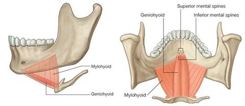
| Name Origin: Insertion: Function: Innervation: | Mylohyoid Origin: Mylohyoid line of the mandible. Insertion: Raphe and body of the hyoid bone. Function: Flat triangular muscle that forms the floor of the mouth and can raise the hyoid bone, floor of the mouth and tongue. If the hyoid bone is fixed, can depress the mandible. Innervation: Mylohyoid nerve, a branch of the inferior alveolar nerve of the mandibular (V3). |
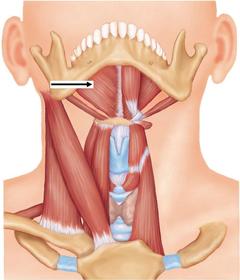
| Name Origin: Insertion: Function: Innervation: | Geniohyoid Origin: Inferior mental spine of the mandible. Insertion: Body of the hyoid bone. Function: Raise the hyoid bone anterosuperiorly or if the hyoid bone is fixed can depress and retract the mandible. Innervation: C1 via the hypoglossal nerve (cranial nerve 12). |
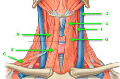
| Name A, B, G, E Origin: Insertion: Function: Innervation: | A: Omohyoid Origin/insertion: Consists of two bellies; the inferior belly originates from the upper surface of the scapula and then passes anterior deep to the sternocleidomastoid attaching to an intermediate tendon, held in place by a fibrous loop to the clavicle. The superior belly extends from the intermediate tendon to the body of the hyoid bone. B: Sternohyoid: Superficial Origin: Manubrium of the sternum and medial end of the clavicle. Insertion: Body of the hyoid bone. G: Sternothyroid: Deep to sternohyoid Origin: Manubrium of the sternum. Insertion: Oblique line of the thyroid cartilage. E: Thyrohyoid: Origin: Oblique line of the thyroid cartilage. Insertion: lower border of the body and lateral horn of the hyoid bone. Function: Anchor the hyoid bone (they fix and steady it) so that the suprahyoid muscles can act. They also depress the hyoid and larynx during swallowing and speaking. Innervation: They are supplied by branches arising from the deep branches of the cervical plexus. |
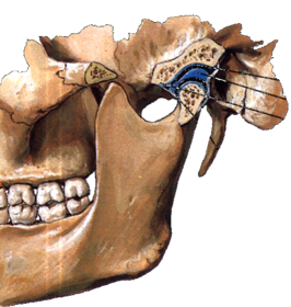
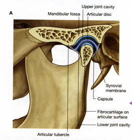
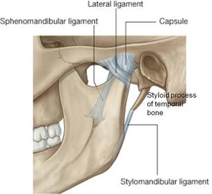
| Temporomandibular joint Type of joint: Articular surfaces involved (3): Describe joint cavity and capsule: | Type of joint: This is a modified hinge type of synovial joint. The articular surfaces involved: The condyle of the mandible, the articular tubercle and the mandibular fossa of the temporal bone. An articular disc divides the joint cavity into superior and inferior compartments. The joint capsule is loose and attaches to the margins of the articular area on the temporal bone and around the neck of the mandible. Laterally, the capsule is thickened as the lateral ligament. Synovial membrane lines the capsule, superior and inferior to the disc. |
| Temporomandibular joint Ligaments which attach mandible to cranium (3): | Laterally, the capsule is thickened as the lateral ligament. Stylomandibular ligament: runs from the styloid process to the angle of the mandible. Sphenomandibular ligament:: runs from spine of the sphenoid to the lingula of the mandible. |
| Temporomandibular joint Movements at the TMJ: Describe movements to cause jaw opening: | Movements of the mandible at the TMJ: Depression, elevation, protrusion, retraction, and lateral movements. When the mandible is depressed during opening of the mouth, the head of the mandible rotates on the inferior surface of the disc (hinge movement), and then both structures move anteriorly on articular surface (gliding movement) until the mandibular condyle lies inferior to the tubercle. |
| Temporomandibular joint: Muscles of elevation: Muscles of depression: Muscles of protraction: Muscles of retraction: | |
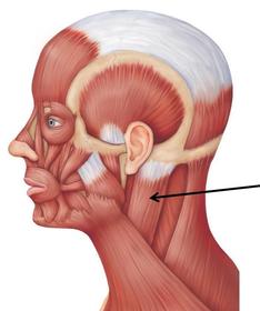
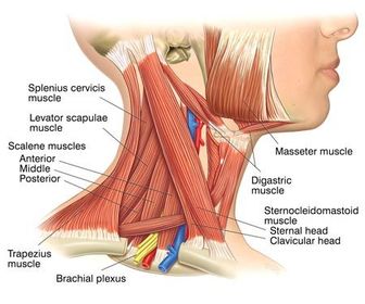
| Name Origin: Insertion: Function: Innervation: | Sternocleidomastoid Origin: From the lateral aspect of the mastoid process and the lateral half of the superior nuchal line. Insertion: Two heads; the sternal head inserts into the upper anterior surface of the manubrium of the sternum. The Clavicular head inserts into the superior surface of the medial third of the clavicle. Function: Unilateral: flex the neck and rotate the neck so that the head faces superiorly to the opposite side. Bilateral: bring the head forward. Innervation: Spinal root of accessory nerve (cranial nerve 11) providing motor fibres and C2 and C3 providing sensation. |
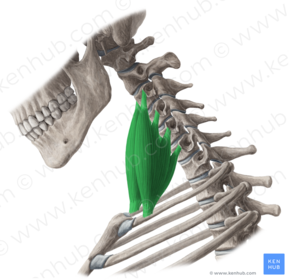
| Name Origin: Insertion: Function: Innervation: | Scalenus anterior Origin: From the transverse processes of the cervical vertebrae. Insertion: Into the scalene tubercle on the anterior aspect of the 1st rib. Function: Can flex the neck forwards as well as laterally. Raises the 1st rib during forced inspiration. Innervation: Lower branches of the cervical spinal nerves. |
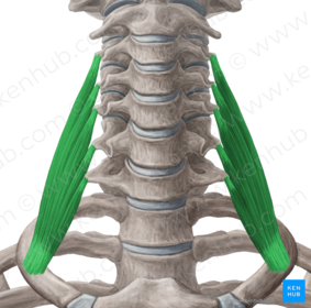
| Name Origin: Insertion: Function: Innervation: | Scalenus medius Origin: From the transverse processes of the cervical vertebrae. Insertion: Into the posterior surface of the 1st rib. Function: Can flex the neck laterally. Raises the 1st rib during forced inspiration. Innervation: Cervical spinal nerves. |
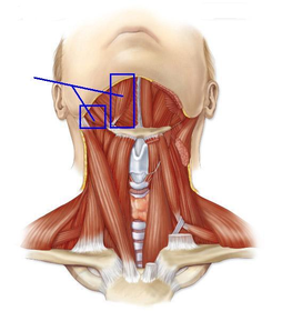
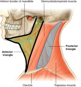
| Triangles of the neck | |
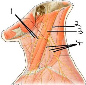
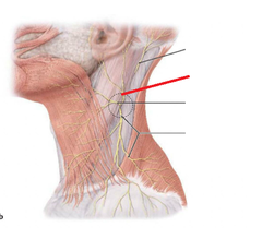
| Name Origin: Location: Groups of branches (2): Supplies: Cutaneous branches (4): | Cervical plexus Location: Is found in the posterior triangle. Origin: Formed by the union of the first four cervical spinal nerves joined together by simple loops. It lies deep the sternocleidomastoid muscle. Groups of branches: The branches of the plexus can be divided into deep and superficial groups. Deep branches supply muscles. Superficial branches are distributed to the skin of the anterior and lateral sides of the neck and the lateral aspect of the head. Cutaneous branches from the plexus emerge around the middle of the posterior border of the sternocleidomastoid muscle. There are four cutaneous branches: Transverse cervical nerve (1). Great auricular nerve (3). Lesser occipital nerve (2). Superior clavicular nerves (4). |
| Name Origin: Supplies: | Great auricular nerve (C2 and C3) Origin: One of the four cutaneous branches of the plexus. Supplies: Ascends across the sternocleidomastoid muscle and supplies the skin over the auricle, and angle of the mandible. |
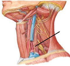
| Name Origin: Course: Supplies: | Phrenic nerve Origin: Predominantly from C4, but also has fibres from spinal nerve roots C3 and C5. Course: Forms at the upper lateral border of the scalenus anterior muscle and then travels over the muscle descending into the thorax anterior to the subclavian artery and posterior to the subclavian vein. Supplies: Sole motor supply to the diaphragm as well as sensory to the middle of the diaphragm. |
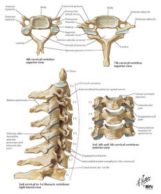
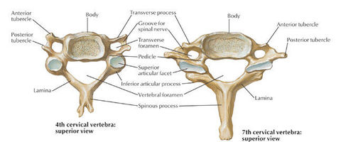
| Cervical vertebrae Number typical/atypical: Characteristics of typical (body, vertebral foramen, transverse process and spinous process): | Cervical vertebrae: C1-C7. They are typical vertebrae except for atypical C1 and C2. Characteristics: Body: Small and longer from side to side than anteroposteriorly; superior surface is concave and inferior surface is convex. Vertebral foramen (canal): Large and triangular. Transverse processes: Transverse foramina (small or absent in C7); vertebral arteries pass through, except C7. Spinous process: Short and bifid; process of C7 is long (vertebra prominens). |
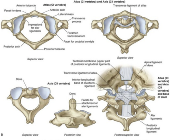
| The atlas and axis (C1 and 2) Descriptions: Joints and movements(2): Ligaments (2): | The atlas (C1): Has no spinous process or body and consists of two lateral masses connected by anterior and posterior arches. Its concave superior articular facets receive the occipital condyles. Atlanto-occipital joints: Permits nodding of the head on the vertebral column. The axis (C2): The distinguishing feature is the dens that projects superiorly from its body. Transverse ligament of the atlas: Strong band extending between the lateral masses of C1 holding the dens of C2 against the inner surface of the anterior arch of C1. Atlantoaxial joints: Permits the head to be moved from side to side. Dens acts as a pivot that allows the skull and C1 as a unit to rotate on C2. Alar ligaments: Extend from the sides of the dens to the lateral margins of the foramen magnum. They check excessive rotation of the head and atlas relative to the axis. |
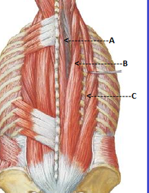
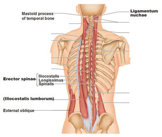
| Collective and individual names: | Erector spinae: Consists of 3 muscles: Iliocostalis (C). Longissimus (B). Spinalis (A). |
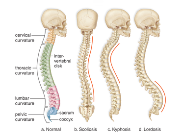
| Curvatures of the spine Normal types (2): Descriptions: Development: | Primary curvature (kyphosis; concave anteriorly, convex posteriorly): Develops during the foetal period. Secondary curvature (lordosis; convex anteriorly, concave posteriorly): Begins to appear before birth and are accentuated when an infant begins to hold its head erect. The secondary curvatures are caused mainly by differences in thickness between the anterior and posterior parts of the intervertebral discs. |
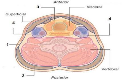
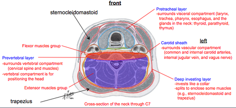
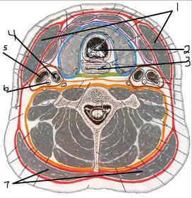
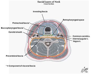
| Deep fasciae of the neck Name of and structures enclosed by layers (3): | Deep fasciae of the neck: 3 layers: 1. Investing layer of cervical fascia: Surrounds all structures in the neck and lies between the superficial fascia and muscles. Splits to enclose the SCM and trapezius muscles. 2. Pretracheal fascia: Limited to the anterior part of the neck. Lies deep to the infrahyoid muscles and splits to enclose the thyroid, trachea, pharynx and oesophagus. 3. Prevertebral fascia: Forms a tubular sheath for the vertebral column and the muscles associated with it. |
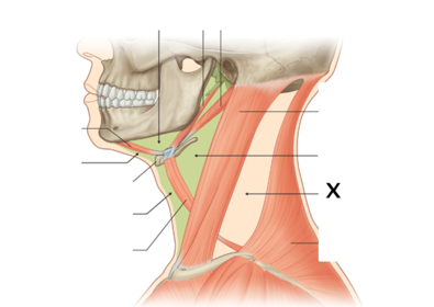
| Carotid sheath Course: Contents (6): | Carotid sheath(es) Tube of fascia that extends from the base of the skull to the root of the neck. It blends anteriorly with the investing and pretracheal fascia and posteriorly with the prevertebral fascia. It contains: Common and internal carotid arteries (surrounded by sympathetic fibres forming plexuses). Internal jugular vein and deep cervical lymph nodes. Vagus nerve (X). |
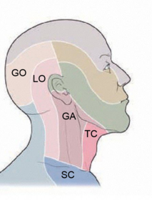
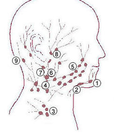
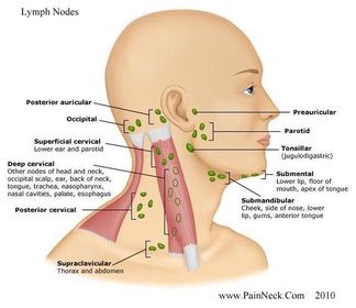
| Superficial lymph nodes: 1, 2, 5, 6, 8, 9 (9 is 2 nodes together). | The superficial lymph nodes are: Occipital (9), retroauricular (9), parotid (8), buccal (5), submental (1), submandibular (2) and superficial cervical (6); drain into the deep cervical nodes. |
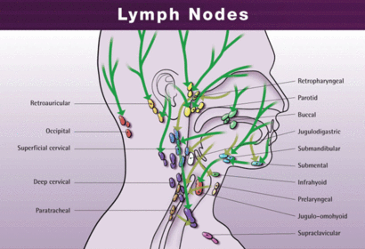
| Lymphatic drainage of the face Routes (3): Final route: Other deep lymph nodes (4): | Lymphatic vessels in the forehead and anterior part of the face accompany other facial vessels and drain into the submandibular lymph nodes, located along the inferior border of the mandible. From the lateral part of the face lymph drains into parotid nodes. Lymphatic from the central part of the lower lip and chin drain into submental nodes. All lymph from the head and neck eventually drains into the deep cervical lymph nodes. The main group is situated beneath the sternocleidomastoid muscle and forms a chain along the internal jugular vein. Other deep nodes include prelaryngeal, pretracheal, paratracheal and retropharyngeal nodes. |
Want to create your own Flashcards for free with GoConqr? Learn more.