6489629
Description
Flashcards by Ashutosh Kumar, updated more than 1 year ago
|
|
Created by Ashutosh Kumar
over 7 years ago
|
|
| Question | Answer |
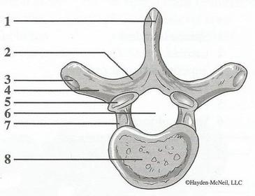
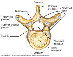
| Name 2, 7 and 4: Name 3 and what structure it articulates with: | 2: Lamina 7: Pedicle 4: Transverse process 3: Articular facet on the transverse process which articulates with the tubercle of the rib of the same level. |
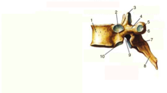
| Name 2, 10 and 5: Name the structures they articulate with: | 2: Superior costal facet, articulates with the head of the rib of the same level. 10: Inferior costal facet, articulates with the head of the rib of the level below. 5: The costal facet of the transverse process, articulates with the tubercle of the rib of the same level. |
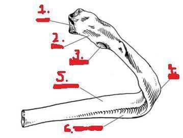
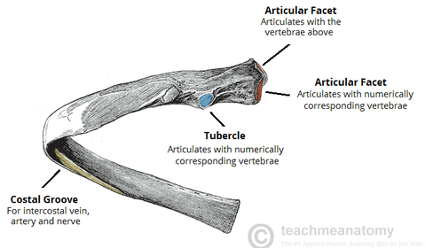
| Name 1, 2, 3, 4, 5 and 6: Name the structures 1 and 3 articulate with: Name what runs in 6: | 1: Head of the rib, articulates with the superior costal facet of the vertebral body of the same level and the inferior costal facet of the vertebral body of the level above. 2: Neck of the rib. 3: Tubercle of the rib, articulates with the transverse process costal facet of the vertebra of the same level. 4: Angle of the rib; region of the rib where the shaft bends forward. 5. Body of the rib. 6. Costal groove: a groove found on the inferior margin of the rib on the internal aspect. Running in it from superior to inferior is intercostal vein, artery and nerve. |
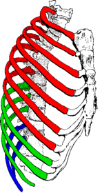
| Describe the type of ribs shown in red: Describe the type of ribs shown in green: Describe the type of ribs shown in blue: | Red ribs: These are true ribs, ribs 1-7, because they articulate with the sternum directly through independent costal cartilages. Green ribs: These are false ribs, ribs 8-10 because they articulate anteriorly with the costal cartilages of the ribs above and not directly with the sternum. Blue ribs: These are floating ribs, ribs 11 and 12, since they do not articulate with the sternum at all. |
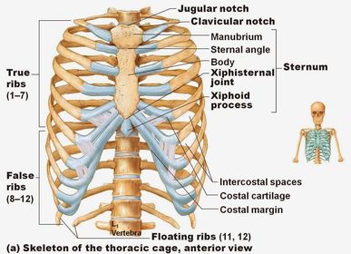
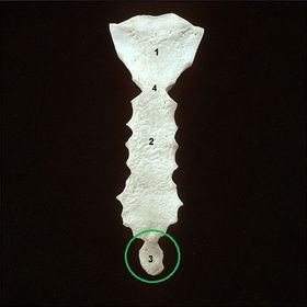
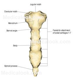
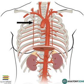
| Name 1, 2, 3 and 4: Name the surface landmarks: Name the rib level and thoracic vertebra level 4 corresponds to: | 1: Manubrium of the sternum. 2: Body of the sternum. 3: Xiphoid process of the sternum. 4: Sternal angle (joint between the manubrium and body of the sternum). Surface landmarks: The sternal angle and the xiphoid process. The sternal angle corresponds to the 2nd rib anteriorly and the inferior aspect of the body of the 4th thoracic vertebra posteriorly. |
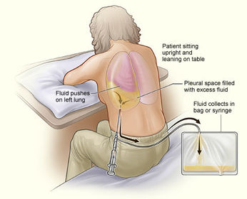
| Name the procedure shown here: Name the 3 indications: Describe the procedure: | Name: Thoracocentesis/thoracentesis/pleural aspiration/drainage Indications: 1. Pneumothorax (air in the pleural cavity). 2. Haemothorax (blood in the pleural cavity). 3. Pleural effusion (excess fluid in the pleural cavity). Procedure: Needle inserted into pleural space via intercostal space. |
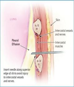
| When inserting an intercostal needle or chest drain, why is it important to stay just above a rib border? | The needle should be inserted just above the upper margin of the rib to avoid injury to the intercostal neurovascular bundle of the rib above, the main part of which runs in the costal groove along the inferior margin of the rib. |
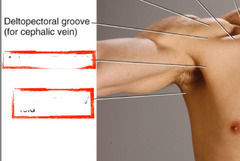
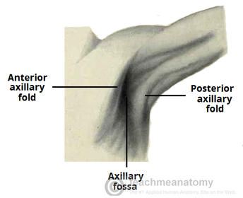
| Name the two folds demarcated by the two red boxes: Name what these folds are made by: | Anterior axillary fold, formed by the pectoralis major. Posterior axillary fold, formed by the teres major and latissimus dorsi. |
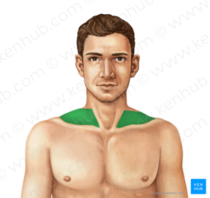
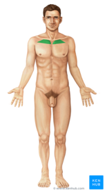
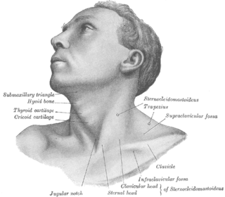
| Name: | Supraclavicular fossa: Indentation immediately above the clavicle. |
| Name: | Infraclavicular fossa: Indentation immediately below the clavicle. |
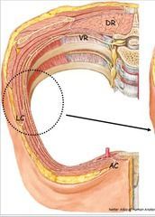
| Name the innervation and blood supply to the region labelled LC: Name the innervation and blood supply to the region labelled AC: | LC: Lateral subcutaneous tissue of the thorax, innervated and supplied by the lateral cutaneous nerves and arteries. AC: Anterior subcutaneous tissue of the thorax, innervated and supplied by the anterior cutaneous nerves and arteries. Both the lateral and anterior cutaneous nerves are branches of the thoracic spinal nerves. |
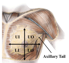
| Name the divisions of the breast: State the percentages of breast cancer involvement for each division: | 1. Upper outer quadrant (50%) 2. Upper inner quadrant (15%) 3. Lower outer quadrant (10%) 4. Lower inner quadrant (5%) 5. Centre (20%) 6. Axillary tail, which runs along the inferolateral border of the pectoralis major towards the axilla. |
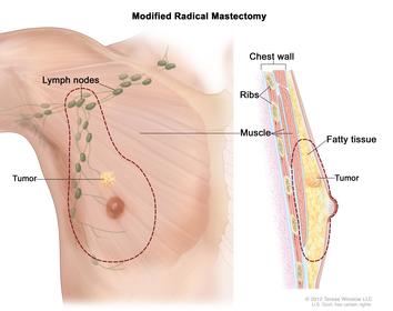
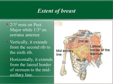
| Describe the average extent of breast vertically: Describe the average extent of breast horizontally: | Vertically: 2nd and 6th rib. Horizontally: From the lateral edge of the sternum medially to the midaxillary line laterally. |
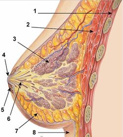
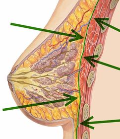
| Name 1-8: | 1: Chest wall. 2: Pectoralis major. 3: Lobule consisting of multiple acini/ductules. 4: Nipple. 5: Areola. 6: Lactiferous sinus. 7: Retromammary fat. 8: Skin. |
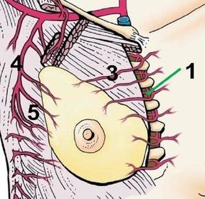
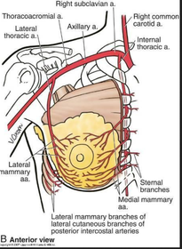
| Name the sources of blood supply to the breast: | Blood supply to the breast tissue is via perforating arteries which perforate the deep fascia overlying the pectoralis major and subsequent subcutaneous tissue since the breast tissue lies in the superficial fascia. These perforating arteries arise from: 1. Internal thoracic artery 2. Intercostal arteries 3. Lateral thoracic artery 4. Thoracoacromial artery |
| Name the region highlighted in green: | Retromammary space: Between the breast and deep fascia. |
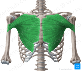
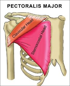
| Name the structure highlighted in green: Describe the structure: | Pectoralis major, consists of two heads; clavicular and sternocostal. |
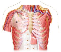
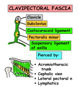
| Name the structure asterisked: Name what this structure lies deep to: Name the origin and insertion: Name the structures which pass through: Name the structures enveloped: | Clavipectoral fascia, lies deep to the pectoralis major muscle and extends from the clavicle superiorly to the axilla inferiorly forming the suspensory ligament of the axilla. The fascia envelopes the subclavius and pectoralis minor muscles. There are four things which pierce the fascia: 1. Cephalic vein. 2. Thoracoacromial artery. 3. Lateral pectoral nerve. 4. Lymphatics. (CALL) |
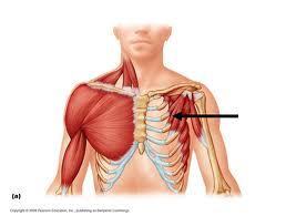
| Name: Structures which lie deep that are important in breast cancer: | Pectoralis minor Deep to this are lymph nodes in the axilla, important in the spread of breast cancer. |
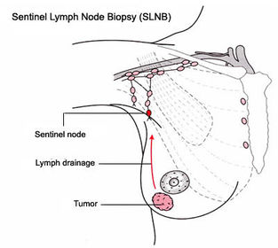
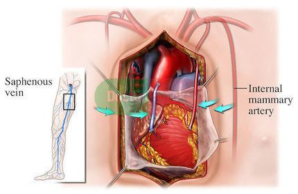
| Name the structure: Name the procedure in which the structure is commonly utilized: | |
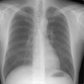
| Describe the outline of how to interpret a chest radiograph: | AP vs PA A, B, C, D, E, F, G, H Airways (consolidation) Bones (ribs; 6 anterior and 10-11 posterior; fractures) Cardiac size (the cardiac silhouette should be less than 50% of the thoracic diameter) Diaphragm (Right is higher than the left) Equal volumes (expansion of lungs into apices) Fine details (abnormalaties) Gastric bubble Hilum (left higher than right) |
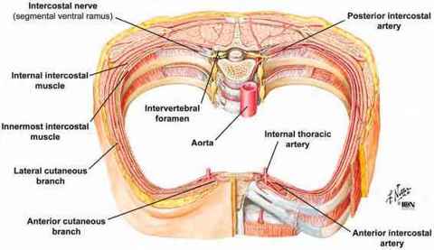
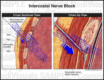
| Name the procedure shown: Describe the procedure: Describe the indications of the procedure: | Intercostal nerve block Procedure: Local anaesthesia can be injected around intercostal nerves posteriorly in an intercostal space. Because of overlapping sensory nerve territories, an effective intercostal nerve block usually requires the anaesthetic agent to be injected around both the intercostal nerve and its collateral branch in two or three adjacent spaces. Indications: Intercostal nerve blocks can be used to alleviate pain after thoracotomy (a surgical incision between the rib spaces) or rib fractures. |
| Why is the posterior region of an intercostal space selected? | The posterior region of an intercostal space is selected because the intercostal nerve is mainly anaesthetised anteriorly from the point of injection. By injecting local anaesthetic nearer the origin of the nerve, this maximises the area of anaesthesia produced. |
Want to create your own Flashcards for free with GoConqr? Learn more.