6490202
Description
Flashcards by Ashutosh Kumar, updated more than 1 year ago
|
|
Created by Ashutosh Kumar
over 7 years ago
|
|
| Question | Answer |
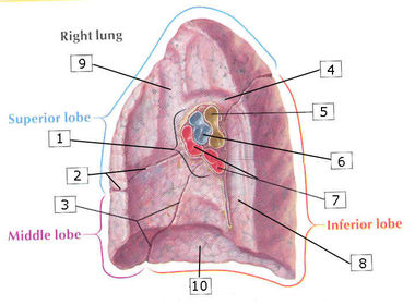
| 1. Root of the lung (a term which encompasses all the structures which enter and exit the lung at the hilum). 2. Horizontal fissure (between the upper and middle lobes, often incomplete). 3. Oblique fissure (between the lower and both middle and upper lobes). 4. Groove for the azygos vein (which drains into SVC). 5. RIght main bronchus (already dividing into upper and lower). 6. Pulmonary artery. 7. Pulmonary veins. 8. Groove for the esophagus. 9. Groove for right brachiocephalic vein draining into SVC. 10. Diaphragmatic surface. | |
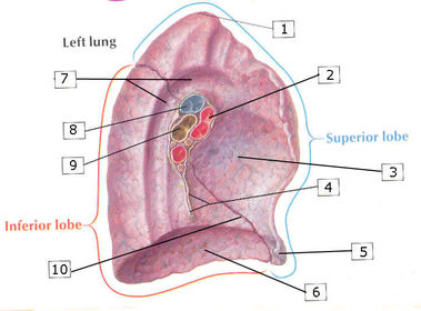
| 1. Apex 2. pulmonary veins 3. Cardiac impression 4. Pulmonary ligament 5. Lingula (anatomical parallel to the middle lobe of the right lung) 6. Diaphragmatic surface 7. Groove for arch and descending aorta 8. Pulmonary artery 9. Left main bronchus 10. Oblique fissure (between upper and lower lobes) | |
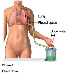
| Describe the procedure indicated: | Chest drain, inserted into the 5th intercostal space in the midaxillary line, to drain blood or fluid which has collected in the pleural cavity. |
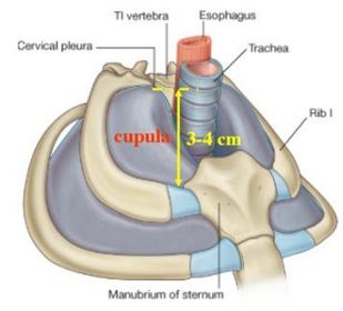
| Describe the location of the cupola and the implications for central venous catheter insertion: | The cupola/cervical parietal pleura covers the apex of the lung, which extends 3-4 cm above the first costal cartilage anteriorly, but is still below the superior border of the 1st thoracic vertebra posteriorly due to the anteriorly downward sloping superior thoracic aperture. Since the cupola extends above the medial end of the clavicle, during insertion of a central venous catheter into the internal jugular vein or subclavian vein, the apex of the lung can be accidentally punctured producing pneumothorax. |
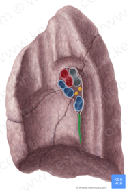
| Name the structure highlighted in green: Function: | Pulmonary ligament (extends inferiorly from the root of the lung where the visceral and parietal pleura are continuous with each other forming the ligament inferiorly) Function: The pulmonary ligament helps to accommodate the up and down movement of structures in the root of the lung during respiration and changes in the diameter of the pulmonary veins. It may also help stabilise the inferior lobe of the lung. |
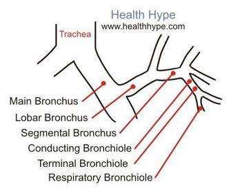
| Describe the structures which comprise the root of the lung: | The root is a term which encompass the structures that enter or leave the lung through the hilum, a depression in the medial surface of the lung. As a general rule: Superior: Pulmonary vein. Posterior: Bronchus. Anteroinferior: Pulmonary arteries. Also present are the tracheobronchial lymph nodes, bronchial arteries, nerves and lymphatics. |
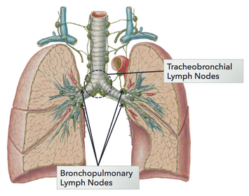
| Typically, the bronchopulmonary lymph nodes are a dark colour. Why is this? | Because they contain macrophages which have ingested inhaled soot particles and/or cigarette smoke. Nodes at the hilum of the lung are sometimes called bronchopulmonary nodes - these drain to tracheobronchial nodes around the primary bronchi and bifurcation of the trachea. |
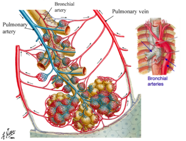
| What is the function of bronchial arteries? | The bronchial arteries are the nutrient vessels of the airways and lung whereas the pulmonary arteries carry deoxygenated blood to the alveoli. |
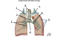
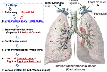
| Name 1-8: Describe the general pathway of lymphatic drainage of the lungs: | |
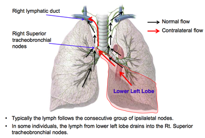
| Describe how the lymphatic drainage pathways of the upper and lower lobes of the left lung differ in some individuals: | Upper lobe: Intrapulmonary nodes. Bronchopulmonary (hilar) nodes. Inferior tracheobronchial (carinal) lymph nodes. Superior tracheobronchial lymph nodes. Tracheal nodes and/or aortic arch node. Bronchomediastinal trunk. Thoracic duct. Left jugular angle. Lower lobe: Intrapulmonary nodes. Bronchopulmonary (hilar) lymph nodes. Inferior tracheobronchial (carinal) lymph nodes. Right superior tracheobronchial lymph nodes. Right tracheal nodes. Bronchomediastinal trunk. Right lymphatic trunk. Right jugular angle. |
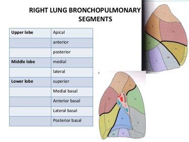
| Describe what a bronchopulmonary segment is: | A segment of the lung supplied by a segmental bronchus and its accompanying pulmonary artery branch. |
Want to create your own Flashcards for free with GoConqr? Learn more.