6645569
| Question | Answer |
| What are the four parts of the lower limb? Define each part. | Pelvic Girdle - attaches to the axial skeleton Thigh - between the pelvis and the knee Leg - between the knee and the ankle Foot - distal to the ankle |
| What are three main differences between the lower and upper limbs? | Function - lower is for weight bearing and locomotion Range of Movement - lower limb is less mobile, but much more stable Rotation during development - lower limb is rotated internally so that flexors are located posteriorly |
| Describe where the line of gravity is positioned relative to the lower limb. | Slightly posterior to the hip joint Slightly anterior to the knee joint Anterior to the ankle joint |
| What bones make up the pelvic girdle? | Two hip bones (os coxae) Sacrum Coccyx |
| What are the three main functions of the pelvic girdle? | 1. Connects the lower limb to the axial skeleton 2. Supports the weight of the upper limb 3. Protects the urogenital organs |
| What are three main differences between the shoulder girdle and the pelvic girdle? | 1. The pelvic girdle articulates directly with the vertebral column 2. The socket is deeper and more congruent in the pelvic girdle 3. The pelvic girdle is more stabile |
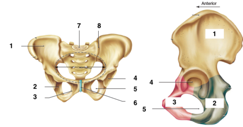
| What bones make up the hip bone? | Ilium Pubis Ishchium |
| Name | 1. Ilium 2. Ischium 3. Pubis 4. Acetabulum 5. Obturator Foramen 6. Pubic Symphysis 7. Sacrum 8. Sacroiliac Joint |
| What is the acetabulum? | A large depression on the lateral aspect of the hip bone which articulates with the femoral head - it is the point where the three bones of hip fuse |
| How do male and female hip bones differ? | Males have narrower and taller hip bones Females have shorter and wider hip bones |
| What is the obturator foramen? | A large opening between the ischium and pubis at each hip bone - it is covered by a membrane |
| What is the function of the ischium? | To support the weight of the body when sitting |
| 1. What type of joint is the pubic symphysis? 2. What bones are articulating at this joint? 3. Describe the stability of this joint. | 1. Cartilaginous joint 2. The left and right pubis of the hip bones (at the midline) 2. Very stable - can be loosened during pregnancy |
| 1. What type of joint is the sacroiliac joint? 2. What bones are articulating at this joint? 3. Describe the stability of this joint. | 1. Synovial plane joint 2. The sacrum and the ilium of the hip bone 3. Very strong and stable - can be loosed during pregnancy |
| What joints are loosened allowing more mobility during pregnancy? | Sacroiliac joint and pubic symphysis |
| 1. What type of joint is the hip joint? 2. What bones are articulating at this joint? 3. Describe the stability of this joint | 1. Synovial ball and socket 2. The acetabulum of the hip bone and the head of femur 3. Very stable |
| Identify and describe three main stabilising factors that allow the increased stability of the hip joint. | Deep and rounded surface of the acetabulum Acetabulur labrum (increases surface area of articulation) Strong ligaments (which are continuous with the joint capsule) |
| Identify the three ligaments that support the hip joint. | Iliofemoral Ligament Ischiofemoral Ligament Pubofemoral Ligament |
| 1. Where is the iliofemoral ligament located at the hip joint? 2. What attachments does it have? 3. What function does it serve? | 1. Anteriorly 2. Ilium and femur 3. Compresses the femoral head during extension |
| 1. Where is the ischiofemoral ligament and the pubofemoral ligament located at the hip joint? 2. What attachments does each ligament have? | 1. Posteriorly 2. Ischiofemoral: Ischium and femur Pubofemoral: Pubis and femur |
| What are two common injuries of the hip joint? | Neck of femur fracture (risk of avascular necrosis) Hip Dislocation (can be posterior or anterior) |
| 1. What type of bone is the femur? 2. How is the femur oriented? 3. How does this differ for males and females? | 1. Long bone (the largest in the body) 2. Since the hips are extended lateral, the femur is angulated medially 3. Angulation is greater in females due to the broader pelvis |
| 1. Head 2. Neck 3. Greater Trochanter 4. Lesser Trochanter 5. Lateral Epicondyle 6. Medial Epicondyle 7. Intercondylar Fossa 8. Medial Condyle 9. Lateral Condyle | |
| What is the trabecular system of the femur? | Trabecular pattern of growth which develops in the direction of force giving the bone extra strength - this occurs in the head of femur |
| 1. What type of bone is the patella? 2. Where is it located? 3. What function does it serve? | 1. Sesamoid bone 2. Anterior surface of the knee joint within the tendon of the quadriceps femoris 3. Protects the knee joint and provides leverage for the thigh muscles |
| 1. What is special about the knee joint? 2. What bones articulate at this joint? | 1. It consists of two articulations within the one joint capsule 2. Femoral condyles and tibial condyles at the tibiofemoral joint Distal femur and the patella at the patellofemoral joint |
| 1. What type of joint is the tibiofemoral joint? 2. Describe the stability of this joint. | 1. Synovial hinge joint 2. Fairly stable - supported by menisci, bursae, collateral ligaments and cruciate ligaments (most stable in extension - ligaments are pulled taut) |
| 1. What are the two menisci that support the knee joint? 2. Where are they located and where do they attach? | 1. Medial and lateral menisci 2. Medial meniscus: between the medial condyles of the femur and tibia Lateral meniscus: between the lateral condyles of the femur and tibia Both menisci attach at the intercondylar region (midline) of the tibial plateau |
| 1. Describe the structure of the menisci of the knee joint. 2. What function do they serve? | 1. C-shaped, thicker in the periphery and taper down towards the intercondylar region 2. Increases surface area of articulation at the tibiofemoral joint Absorbs shock |
| 1. What type of ligaments are the cruciate ligaments of the knee joint? 2. Identify the two cruciate ligaments? 3. In what plane do they provide support? | 1. Intracapsular ligaments 2. Anterior cruciate ligament and posterior cruciate ligament which cross over 3. Sagittal plane |
| Describe the location and function of the anterior cruciate ligament (ACL) | Location: extends posteriorly from the anterior part of the intercondylar region of the tibia and attaches to the femoral lateral condyle Function: prevents anterior displacement of the tibia relative to the femur |
| Describe the location and function of the posterior cruciate ligament (PCL) | Location: extends anteriorly from the posterior part of the intercondylar region of the tibia and attaches to the femoral medial condyle Function: prevents posterior displacement of the tibia relative to the femur |
| . 1. What type of ligaments are the collateral ligaments of the knee joint? 2. Identify the two collateral ligaments? 3. In what plane do they provide support? | 1. Extracapsular ligaments 2. Medial (tibial) collateral ligament and lateral (fibular) collateral ligament 3. Coronal |
| Describe the structure and location of the medial collateral ligament (MCL). | Structure: flat and broad Location: attaches to the medial femoral epicondyle and the medial surface of fibular head - also adheres to the joint capsule and the medial meniscus |
| Describe the structure and location of the lateral collateral ligament (LCL). | Structure: cord like Location: attaches to the lateral femoral epicondyle and the lateral surface of the fibular head - is seperated from the joint capsule by a tendon |
| What is the function of the two collateral ligaments of the knee joint? | MCL: Resists valgus forces (forces applied laterally - moving the knee medially) LCL: Resists varus stress (forces applied to medially - moving the knee laterally) |
| What are bursae? | Synovial fluid filled sacs which reduces friction between moving structures |
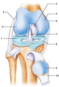
| 1. Lateral meniscus 2. Lateral femoral condyle 3. Lateral (fibular) collateral ligament 4. Posterior cruciate ligament 5. Medial femoral condyle 6. Medial (tibital) collateral ligament 7. Anterior cruciate ligament 8. Medial meniscus 9. Patella 10. Quadriceps tendon | |
| Describe the 'unhappy triad' injury | Damage to the medial collateral ligament which consequently results in the injury of the medial meniscus (due to attachment) and the ACL - common in contact sports |
| Identify and describe the location and function of the bones of the leg. | Tibia: located medially - the larger weight bearing bone Fibula: located laterally - non-weight bearing, mainly for muscle attachment |
Want to create your own Flashcards for free with GoConqr? Learn more.