6653179
Description
Flashcards by Ashutosh Kumar, updated more than 1 year ago
|
|
Created by Ashutosh Kumar
over 7 years ago
|
|
| Question | Answer |
| Describe the function of endocrine cells: Presence of ducts: How is specificity achieved: The function of the endocrine system: | Endocrine cells synthesize and secrete chemical messengers (hormones) into the bloodstream which then regulate the activity of specific target cells located elsewhere in the body. Primary function: cell to cell communication. No ducts (ductless glands) in contrast with exocrine glands which have ducts. Specificity is provided by receptors located on the target cells (in contrast with the nervous system). Therefore, the message (hormone) is broadcast globally (via the bloodstream), but only those cells that have the specific receptors can receive the message. The function of the endocrine system is to enable homeostasis. |
| Describe the anatomical distribution of endocrine tissue: | Anatomical distribution of endocrine tissue: Discrete glands: Pituitary gland, thyroid gland, parathyroid gland and adrenal gland. These are all ductless as they secrete the chemical messenger, the hormone into the bloodstream. As a result, they require extensive blood supply. Clusters of endocrine cells associated with non-endocrine tissue: Pancreas, heart, gonads, placenta, brain and adipose tissue. Isolated endocrine cells: In the gut and the respiratory tract. |
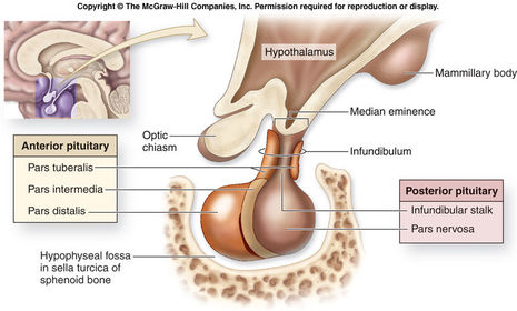
| Describe the constituents of the pituitary gland and list the hormones secreted: Describe whether the hormones act on exocrine or endocrine glands: Describe whether the hormones are trophic: | The pituitary gland consists of three different parts: Anterior pituitary gland (adenohypophysis, pars distalis): Growth hormone (GH) (somatotroph). Prolactin (mammotroph). Thyroid stimulating hormone (TSH, thyrotrophin). Adrenocorticotropic hormone (ACTH, corticotrophin). Follicle-stimulating hormone (FSH, gonadotrophin). Luteinizing hormone (LH, gonadotrophin). Prolactin is the only hormone of the list which does not act on an endocrine gland. Instead, prolactin acts on an exocrine gland. The latter four hormones (TSH, ACTH, FSH and LH) are all trophic hormones, which means they stimulate growth of the gland (hyperplasia). Intermediate gland (pars intermedia): Melanocyte stimulating hormone (MSH). This gland is significantly reduced in humans (somewhat vestigial). Posterior pituitary gland (neurohypophysis, pars nervosa): Oxytocin. Vasopressin (antidiuretic hormone). |
| Describe the location of the pituitary gland: | |
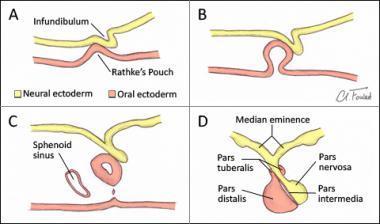
| Describe the embryological development of the pituitary gland: | The anterior and intermediate lobes are formed as up growth of oral ectoderm known as ‘Rathke’s pouch’. When this ball of cells comes into contact with the developing posterior pituitary (which is growing down from the brain) it begins to differentiate and the most dorsal/posterior part becomes the intermediate lobe, while the ventral/anterior part proliferates extensively to form the anterior lobe. The lumen of Rathke’s pouch forms a narrow cleft between the two lobes. In addition, the epithelial tissue from Rathke’s pouch wraps around the stalk or infundibulum to form the pars tuberalis. The posterior pituitary gland is a down growth of neural cells from the floor of the third ventricle (neural ectoderm). |
| Describe the arrangement of cells and blood vessels in the anterior pituitary gland: | Anterior pituitary gland: Typical endocrine tissue. Loosely arranged cords of cells. Fenestrated capillaries and sinusoids. The discrete bands of cells are separated by capillaries. These capillaries have a larger lumen compared to capillaries elsewhere in the body. In addition, these capillaries are fenestrated. This is to enable secretions from the anterior pituitary gland cells to enter the bloodstream. |
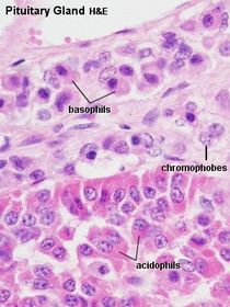
| Describe the histological arrangement of cells in the anterior pituitary gland: | Traditional histology: Staining is dependent on the presence of glycosylated proteins in different cells taking up specific dyes. Acidophils: These are either somatotrophs (GH) or lactotrophs (prolactin) and typically make up approximately 40% of the cell population. Basophils: These are generally larger cells, making up approximately 10% of the cell population, and are either gonadotrophs (LH or FSH), thyrotrophs (TSH) or corticotrophs (ACTH). Chromophobes: Identified by lack of staining by either acidic or basic dyes, these cells are the smallest of the anterior pituitary gland cell types, and typically make up 50% of the total cells. These cells are thought to represent degranulated or resting forms of the chromophilic cells described above, thus the absence or low level of staining is due to the reduced amount of secretory product present in the cell. |
| Describe the appearances of the cells of the anterior pituitary gland and reasons why on an H&E stained section: | In a traditional hematoxylin and eosin (H&E) staining, the cytoplasm of these cells would appear pink/red (acidophils taking up the acid eosin stain) and blue (basophils taking up the basic stain hematoxylin). Note, as in all cells, the acidic structures in the nucleus (nucleic acids) in all cell types, including chromophobes will also stain with the basic stain, hematoxylin (blue/purple). |
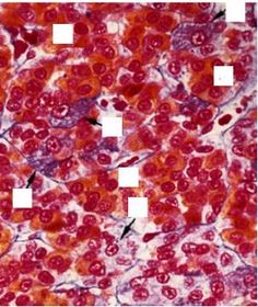
| Describe the strategy for delineating the cell types when an unfamiliar dye is used: | When faced with an image of an unfamiliar stain, it can be helpful to remember the percentages of cell types. Many of the cells in this adjacent image are stained orange. Recalling that 40% of the cells in the anterior pituitary gland are acidophils, it is likely that these cells are acidophils. There are also a few purple stained cells. Recalling that 10% of the cells in the anterior pituitary gland are basophils, it is likely that these purple cells are basophils. The remaining unstained cells are chromophobes, recalling that in the anterior pituitary gland 50% of the cells will be not take up the stain at any one point in time. |
| List the percentages of each cell type (somatotrophs etc): | Somatotrophs (50%). Mammotrophs (20%). Corticotrophs (20%). Gonadotrophs (5%). Thyrotrophs (5%). It should be noted that all cells contain secretory material and chromophobes are degranulated cells. |
| Describe the usefulness of histological staining in pituitary adenomas: | Histological staining is important for identifying whether a pituitary tumour is functional or nonfunctional. Chromophobic staining could indicate the the tumour is a non-functional tumour or that the tumour is functional and hyperactive, secreting material at the same rate at which it is formed resulting in very little intracellular material to stain causing a chromophobic appearance. |
| Describe two other techniques that could be used to identify the cell types in the anterior pituitary gland: | Specific anterior pituitary cell types are now more commonly identified by immunohistochemical techniques using antibodies to the specific hormones. It is also possible to distinguish the different cells by electron microscopy, based on differences in the shape, size and texture of their secretory granules, and by the characteristics of their intracellular organelles. |
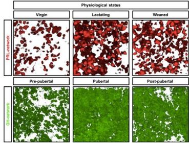
| Describe the idea of cell networks: | The pituitary cell types form integrative networks between the same cell types. For example, GH secreting cells form an extensive network with each other. These integrative networks between cells of the same type are plastic. For instance, it can be seen that during pregnancy the prolactin secreting cells proliferate and the number of cells persist post-pregnancy. Similarly, the GH secreting cells proliferate during puberty and persist post puberty. |
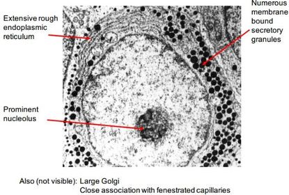
| Describe the ultrastructural features of a peptide secreting cell: | Features: Prominent nucleolus, extensive rough endoplasmic reticulum (to synthesize protein), prominent golgi (to modify, package and traffic protein) and numerous membrane bound secretory granules containing the peptide hormone. |
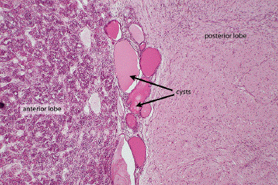
| Describe the histological appearance of the intermediate lobe: | The intermediate lobe is represented by large “colloid follicles” present in cysts in the cleft region between the anterior and posterior pituitary lobes, and by smaller follicles on the posterior side of the cleft. Under high power, cuboidal epithelial cells can be observed lining these follicles and extending a small distance into the posterior lobe. These cells appear to either be chromophobic or basophilic. |
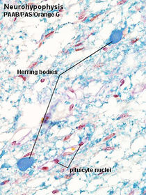
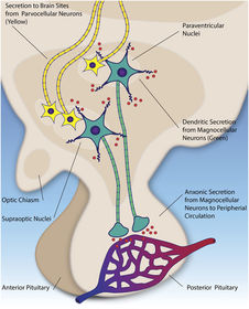
| Discuss the anatomy of the posterior pituitary gland: | In contrast to the anterior lobe, the posterior lobe does not exhibit the usual characteristics of endocrine tissue. Rather, the posterior lobe contains the unmyelinated axons and nerve terminals of neurosecretory neurons, the cell bodies of which are located in the supraoptic and paraventricular nuclei in the hypothalamus. Although the nerve fibres of the hypothalamo-hypophyseal tract are not stained as such, the neurosecretory material which has travelled down the axons gives a granular blue staining to the area. These aggregated neurosecretory granules (called Herring bodies) contain the hormones oxytocin and antidiuretic hormone (ADH). The neurosecretory neurons release these hormones in close proximity to fenestrated capillary networks (neurosecretion). In addition, the posterior lobe contains fibroblasts and pituicytes (neuroglial-like cells, unique to the posterior pituitary gland which provide support), and an extensive network of capillaries. |
| Discuss the reasons why an adenoma of the lactotrophs is more likely than any other cell type: | Many of the hormones released from the target endocrine gland negatively feedback on both the pituitary gland and hypothalamus, thus achieving control of their secretion. An exception is prolactin. Prolactin secretion from the anterior pituitary gland is tonically inhibited by the brain via dopamine secretion. Therefore, if there is damage to the infundibulum, the mammotrophs in the anterior pituitary gland will no longer be inhibited. In addition, prolactin is the only hormone that acts on an exocrine gland as opposed to an endocrine gland. Prolactin regulates its own secretion via short loop negative feedback. Moreover, it is these differences between the regulation of prolactin secretion compared to the other hormones that makes prolactinomas the most common pituitary adenoma. |
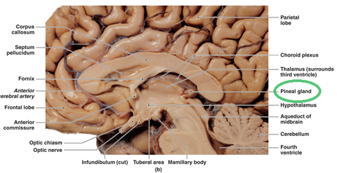
| Describe the anatomy of the hypothalamus: Define the bordering structures rostrally, caudally and dorsally: | Anatomy of the hypothalamus: Located at the base of the diencephalon. Divided into two symmetrical sides by the third ventricle. The neurons are arranged in numerous anatomically defined nuclei. Widespread connections. The rostral edge of the hypothalamus (lamina terminalis) is limited by the optic chiasm and anterior commissure. Dorsally, the hypothalamus is separated from the thalamus by the hypothalamic sulcus. The caudal edge of the hypothalamus is marked by the pineal gland and mammillary body. |
| Compare and contrast the magnocellular and parvicellular neurons. | The magnocellular neurons (large neurons) are located in the hypothalamus and project all the way to the posterior pituitary gland. The magnocellular neurons are large because they need to produce enough hormone to reach the extremes of the body via the systemic circulation. Parvicellular neurons: The parvicellular neurons are located in the hypothalamus and project only as far as the median eminence. The parvicellular neurons are smaller because they only have to produce enough hormone to reach the anterior pituitary gland via the median eminence. |
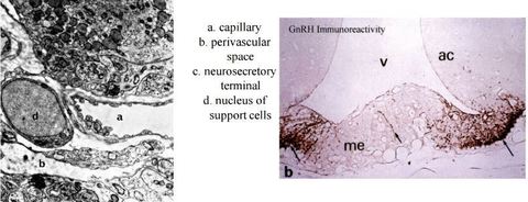
| Describe the median eminence location and layers: | The median eminence forms the floor of the third ventricle and looks like a swelling on the infundibulum when viewed externally. There are 3 major layers: Ependymal cells: tanycytes line ventricle and extend down into the perivascular space. Internal layer: axons of the hypothalamo-hypophyseal tract. External zone: nerve terminals of the neurosecretory axons. |
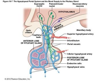
| Describe the blood supply of the pituitary gland: What significance does the blood supply of the anterior lobe have: | Superior hypophyseal arteries (originating from the internal carotid artery and posterior communicating artery) supplies the median eminence and infundibular stalk. Inferior hypophyseal arteries (originating from the internal carotid artery) supply the posterior lobe. The anterior lobe has no direct supply. Instead, the blood flows down from the median eminence to supply the anterior pituitary gland. This is important for the function of the anterior pituitary gland. This is because the hypothalamic parvicellular neurons can secrete hormones into the median eminence. Since this blood will directly reach the anterior pituitary gland, it will therefore be undiluted. As a result, the hypothalamic parvicellular neurons only need to secrete small amounts of hormone to achieve a high concentration in the median eminence blood. Thereafter, this blood flows down to the anterior pituitary gland causing a large response to the hormone in the blood. Therefore, it could be said there is an amplification effect. |
Want to create your own Flashcards for free with GoConqr? Learn more.