6657299
Description
Flashcards by Ashutosh Kumar, updated more than 1 year ago
|
|
Created by Ashutosh Kumar
over 7 years ago
|
|
| Question | Answer |
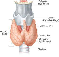
| Describe the thyroid gland and its location: | The thyroid gland is a bi-lobed gland lying anterior to the upper part of the trachea. |
| Describe the types of hormones produced by the thyroid gland: | It produces two distinct types of hormone: the amino acid derived thyroid hormones, thyroxine and triiodothyronine, and the polypeptide hormone calcitonin. These hormones are secreted from different cell types located within the same gland. |
| Compare and contrast the amino acid derived thyroid hormones: | There are 4 iodine molecules in T4 and 3 in T3. T4 is the inactive form whereas T3 is the active form. Therefore, there must be deiodination in the thyroid gland. |
| Describe the chemical classification of hormones: Describe the significance of the chemical structure: | Peptides: from three amino acids to large proteins. Amines: derivatives of tyrosine (an amino acid). Steroids: synthesized from cholesterol. Different chemical properties result in a different tissue structure, different mode of transport in the plasma and different cellular mode of action. |
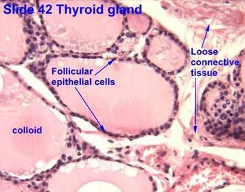
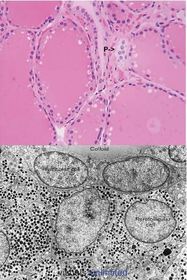
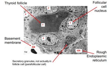
| Describe the anatomy of the thyroid gland as observed through lower power microscopy: | Anatomy of the thyroid gland: Under low power the thyroid hormone-producing thyroid follicles can be seen. They consist of a layer of cuboidal epithelial cells surrounding a glycoprotein complex called thyroglobulin or thyroid colloid. The thyroid follicles are surrounded by a fine network of capillaries associated with thin, fibrous connective tissues. |
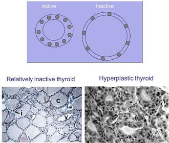
| Describe how the appearance of the follicles reflects the secretory state of the thyroid gland: | There is characteristic variability in the size of the follicles. This is because the size of each follicle reflects its state of secretory activity. Follicles of the active thyroid are small, with reduced colloid material and the cells lining the follicles are relatively ‘tall’. By contrast, the follicles of the resting thyroid tissue are large and distended by stored thyroglobulin and the lining cells are flattened. |
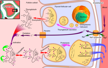
| Describe the process of synthesis of T4 and T3: | 1. Thyroglobulin is synthesized by RER, packaged by the golgi apparatus and then exocytosed into the follicle lumen. 2. Iodide is concentrated by the follicular epithelial cells and released into the follicle lumen, where it undergoes oxidation to iodine. 3. Iodine then combines with thyroglobulin at tyrosine residues (extracellular synthesis since a low pH is needed for mass oxidation). 4. TSH stimulation causes uptake of droplets of iodinated thyroglobulin by the follicular epithelial cells. 5. Lysosomes break down iodinated thyroglobulin into T4 and T3, passing into the bloodstream. |
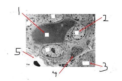
| Describe the function of the parafollicular cells: How can they be distinguished from adjacent follicular cells: What is the stimulus for and function of the secreted hormone: | The parafollicular cells of importance are the calcitonin producing C cells. These cells are generally larger than other epithelial cells and are very pale staining. The C cells contain dense-core secretory granules characteristic of peptide secreting endocrine cells and are readily distinguishable from follicular cells by electron microscopy. Calcitonin release is stimulated by high serum calcium (independent of the pituitary gland). |
Want to create your own Flashcards for free with GoConqr? Learn more.