6665414
Description
Flashcards by Ashutosh Kumar, updated more than 1 year ago
|
|
Created by Ashutosh Kumar
over 7 years ago
|
|
| Question | Answer |
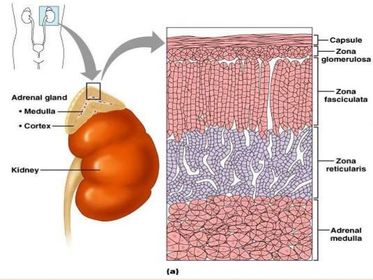
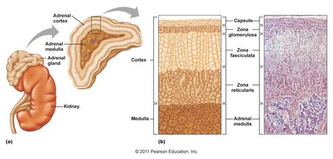
| Describe the location of the adrenal glands: Describe broadly the two divisions of the adrenal gland: Name the layers of the trilaminar embryo the two divisions arise from: Describe the coverings of the adrenal gland: | The adrenal gland is also known as the suprarenal gland, as it is located on the superior pole of each kidney. Each gland is a small, flattened structure, embedded within the perirenal fat. Functionally, each gland is made up of two distinct parts: the outer adrenal cortex, which secretes steroid hormones (e.g glucocorticoids such as cortisol or a mineralocorticoid such as aldosterone) and the inner adrenal medulla, which secretes catecholamines (e.g Noradrenaline/adrenaline). The two parts have a distinct embryological origin, with the cortex derived from the mesoderm, and the medulla derived from the neural ectoderm, closely related to the sympathetic nervous system. The whole gland is surrounded by thick layers of dense fibrous connective tissue (adrenal capsule), containing many small arteries. A prominent vein is characteristically observed in the centre of the adrenal medulla. |
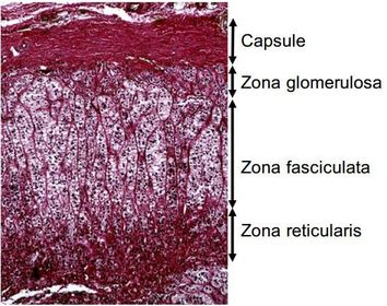
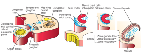
| Describe the appearance of the adrenal cortex under medium magnification: | Zona glomerulosa: The narrow outer layer, made up of oval or circular clumps of cells just inside the capsule (aldosterone secretion). Zona fasciculata: The wider intermediate zone consisting of parallel columns or cords of secretory cells (glucocorticoid secretion, principally cortisol in the human), separated by sinusoidal capillaries. Zona reticularis: The narrow innermost layer containing branching irregularly arranged cells which are deeply stained (androgen precursors, dehydroepiandrosterone (DHEA) and androstenedione) and glucocorticoids. |
| Describe the appearance of the adrenal cortex under high power magnification: | Under high power, all zones can be seen to contain numerous capillary vessels. The steroid secreting cells of the zona glomerulosa and zona fasciculata appear foamy due to the presence of numerous lipid droplets in the cytoplasm. In contrast, cells of the zona reticularis contain more densely stained cytoplasm due to a relative absence of lipid droplets. |
| Describe the control of the layers of the adrenal cortex: | The zona glomerulosa is controlled by plasma sodium whereas the zona fasciculata and zona reticularis is controlled by adrenocorticotropic hormone (ACTH) from the anterior pituitary gland. |
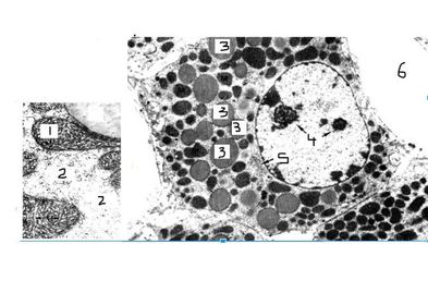
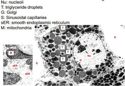
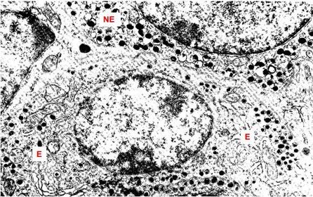
| List the characteristic features of a steroid secreting cell: | Often more than one nucleolus. Large extensive smooth endoplasmic reticulum. Large golgi apparatus. Mitochondria with tubular cristae as opposed to membranous. Cytoplasm filled with lipid droplets. Close association with capillaries. |
| Describe the function of the golgi apparatus: | The golgi apparatus is involved in generating membrane to pack the lipids into. This is because as opposed to a peptide hormones, steroid hormones cannot be stored. The reason for this is because steroid hormones are lipid soluble and can therefore diffuse through the cell membrane. Instead, cholesteryl esters are stored and used to make the hormone when required. All steroid hormones are derived from the steroid nucleus; a 19 carbon chain from cholesterol. Moreover, the steroid hormone once in the plasma is then bound to a steroid binding globulin e.g cortisol binding globulin. |
| Describe the anatomical arrangement of the adrenal medulla: | The adrenal medulla contains closely packed clumps of secretory cells that have a strongly basophilic cytoplasm (pale stained in contrast to the strongly eosinophilic adrenal cortex cells) and are larger than cortical cells. A fine network of capillaries branch throughout the medulla and also numerous larger venules draining blood from the sinusoids of the cortex into the central medullary vein. |
| Describe the function of the adrenal medulla secretory cells: | The adrenal medulla cells secrete the catecholamines adrenaline and noradrenaline under control of the sympathetic nervous system. Stored hormone is released in response to nervous stimulation. |
| Describe the origin of the term 'chromaffin cells' of the adrenal medulla: | When nervous tissue is fixed in chrome salts, stored hormone granules containing noradrenaline are oxidised to form a brown colour. This is the origin of the name “chromaffin cells” of the adrenal medulla and is now used to describe both the adrenaline and noradrenaline secreting cells in this tissue. |
| Compare the secretion from the chromaffin cells to that of the sympathetic nervous system: | In contrast to the SNS, the adrenal medulla predominantly produces adrenaline whereas the SNS produces noradrenaline. The reason for this is because the adrenal medulla is bathed in blood from the adrenal cortex, therefore containing glucocorticoids secreted by the cortical cells. The glucocorticoids cause the conversion of noradrenaline to adrenaline. |
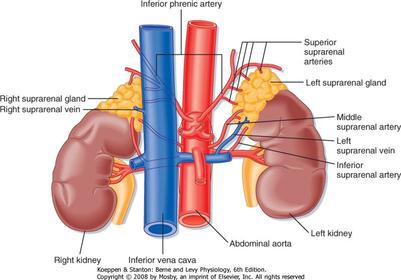
| Describe the arterial supply to the adrenal gland: Describe the venous drainage of the adrenal gland: | Superior (inferior phrenic), middle (abdominal aorta) and inferior (renal) suprarenal arteries. Right suprarenal vein drains into the inferior vena cava whereas the left suprarenal vein drains into either the left renal vein or inferior phrenic vein. |
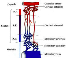
| Describe how the vascular structure influences the type of catecholamine secreted: | From the capsular artery, there are two vascular networks: 1. There is a dense vascular plexus formed in the zona glomerulosa, which then drains into the zona fasciculata. This is rich in glucocorticoids, and via sinusoids, drains to the adrenal medulla. The areas of the adrenal medulla supplied by this blood will secrete adrenaline. This is because glucocorticoids convert noradrenaline into adrenaline. 2. There are metarterioles which directly supply regions of the adrenal medulla. This blood is low in glucocorticoids, therefore, regions supplied will secrete noradrenaline. |
| Describe the embryological development of the adrenal gland: | Cortex: Mesothelial origin, from cells located at the cranial end of the mesonephros. Very large in the foetus playing an important role in development. Medulla: Neural crest cells migrate to from the sympathetic ganglia. Some invade the developing adrenal gland. The cells are effectively postganglionic sympathetic neurons but do not develop neuronal processes. Innervated by preganglionic fibres to the thoracic spinal cord T5-T11. |
| List the glands derived from Ectoderm: Mesoderm: Endoderm: | Ectoderm: Adrenal medulla, Posterior pituitary gland and parafollicular C cells. Mesoderm: Anterior pituitary gland, Adrenal cortex and gonads. Endoderm: Thyroid and parathyroid gland. |
Want to create your own Flashcards for free with GoConqr? Learn more.