7417137
Description
Flashcards by Megan Popp, updated more than 1 year ago
|
|
Created by Megan Popp
almost 9 years ago
|
|
| Question | Answer |
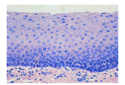
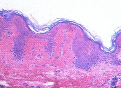
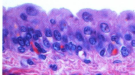
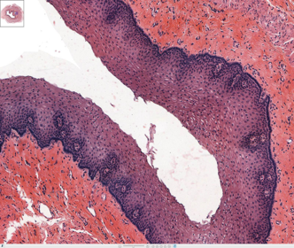
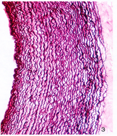
| describe this type of epithelium | stratified squamous nonkeratinized epi. LM |
| Describe this type of epithelium | simple squamous |
| describe this type of epi | simple cuboidal |
| describe the type of epi | simple columnar |
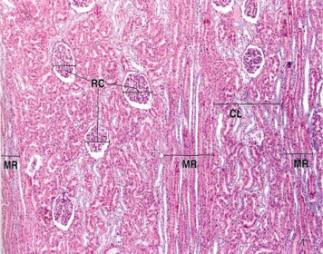
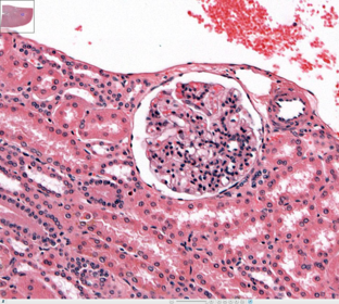
| where is this located? Now describe it based on location. | Simple squamous epi lining the Bowman's Capsule of Kidney "Spaghetti and meatballs" |
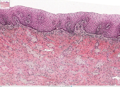
| Thin Keratinized tissue | |
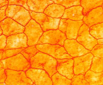
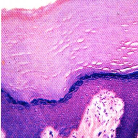
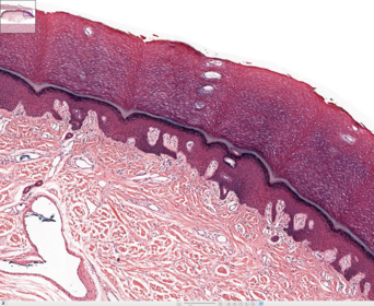
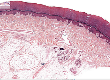
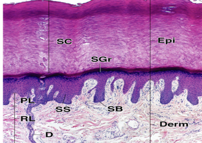
| THICK keratinized tissue, like on palm of hand | |
| Describe this type of epi | Stratified cuboidal Location: seat glands and ducts |
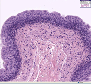
| Describe this type of epi | Transitional epithelium Dome shaped cells on top layer, some can be binucleated |
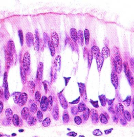
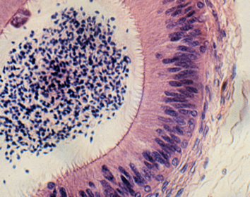
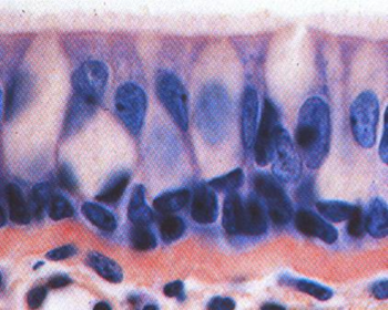
| name this type of epi | PCCE Pseudostratified ciliated columnar epi |
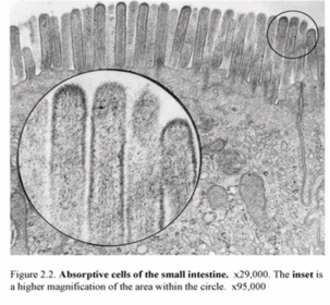
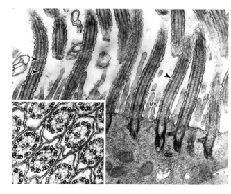
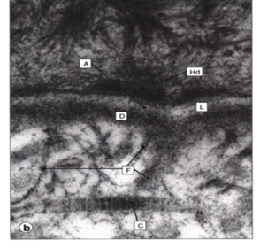
| What is this? what is the fuzzy gray stuff between the two? | microvilli Glycocalyx |
| What is the line connecting the bottoms of the microvilli? | Terminal Web |
| When you look at this think "sperm" so then you know its in the epididymis, so it has to have stereocilia | |
| What is the cone shaped parts? | sterocillia, found in epididymis |
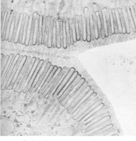
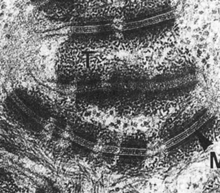
| What is this? what are the dark lines within it? B is what? | Cillia Darker lines within them are microtubules B- basal body that it grows from Cilia have a 9+2 arrangement |
| What are the round, hollow structures? | Centrioles, the base of cilia, arranged in 9 triplets of microtubules |
| Describe this epi | PCCE |
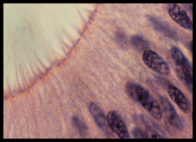
| Describe the fuzzy dark stuff on each cell | Desmosomes (Macula adherens) |
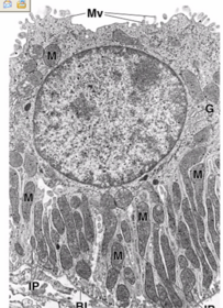
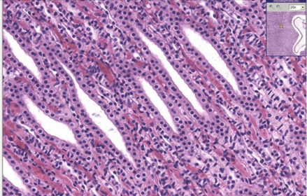
| What is the main cell specialization and what is its function? | Basal infolding/invagination with longitudinal mitochondria Function: active ion transport/exchange |
| PCCE | |
| What types of collagen can be seen here? | Darker area: BL, type 4 lighter, anchoring: reticular, type 7 |
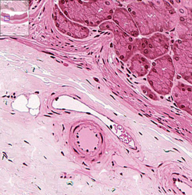
| ID the tissue lining the tube | simple squamous epithelium (endothelium in blood vessels) |
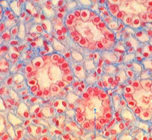
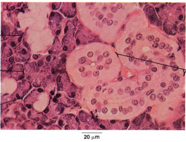
| What type of tissue surrounds the glomerulus of the kidney? What about away from it? | (spaghetti and meat balls) Simple squamous epithelium simple cuboidal epithelium |
| ID the tissue type | simple cuboidal epi |
| ID the tissue type | simple columnar epi |
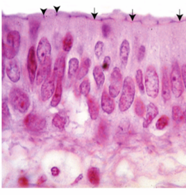
| ID the cell surface specialization. Id the tissue on the cell surface | Specialization - cilia Type: PCCE, with goblet cells |
| ID the tissue on the surface | (many layers of flat cells) stratified squamous nonkeratinized |
| ID Tissue on the surface | stratified squamous nonkeratinized |
| ID tissue on the surface | stratified squamous keratinized, this is because they lack cell organelles |
| ID tissue on the surface | Transitional Dome shaped, binucleated |
| Name the cell specialization | flagella |
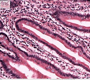
| what type of epi? and what is the cell specialization between them? | Simple columnar epi, with terminal bars between the columnar cells |
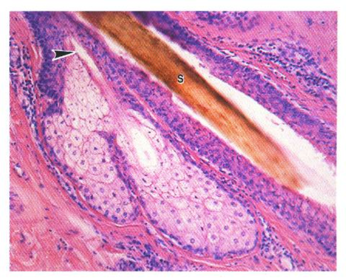
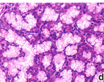
| Name the branching of the duct | Simple branching acinar Sebaceous gland |
| Classify this by number of cells | unicellular - goblet found in digestive system (colon) |
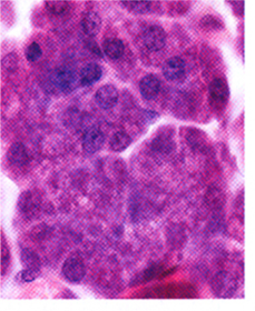
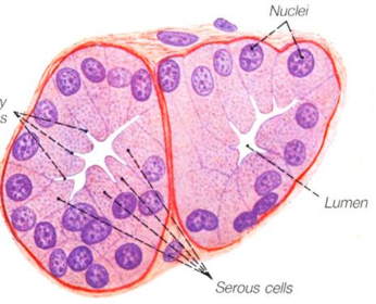
| What type of secretion does this cell have? | Serous - stained mostly dark purple/pink |
| Serous cells | |
| what type of secretion does this cell have? | mucous |
| mucous cell | |
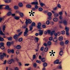
| How can you tell what this secretes? | It secretes both mucus and serous. You can tell by the serous demilunes that are surrounding the mucus cells |
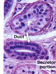
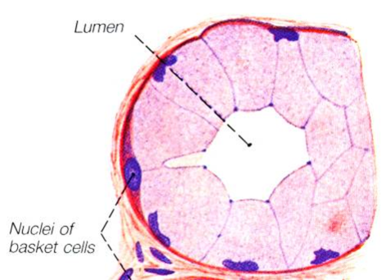
| what type of duct is shown by the "*"? | intercalated duct, look at the simple cuboidal epi |
| what kind of duct can you see here? | striated duct, look at the simple columnar epithelim |
| what type of duct is this? | interlobular duct - on the outside, has striated columnar epi |
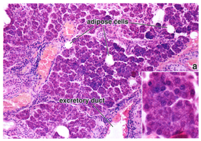
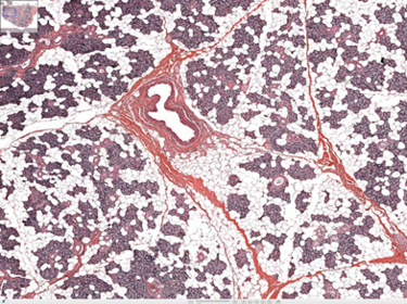
| parotid gland lots of fat cells | |
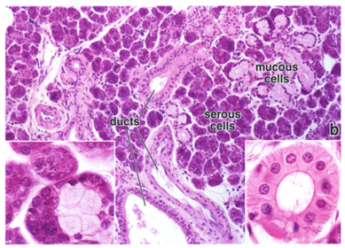
| submandibular gland (S,m) | |
| sublingual gland (s, M) | |
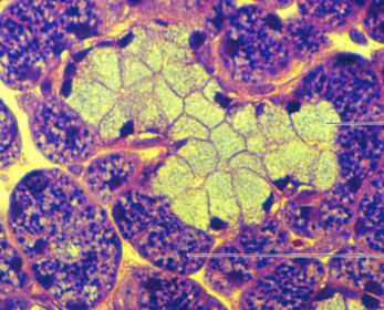
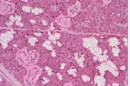
| how can you tell this is the parotid gland? | it only does serous secretion and you can see fat |
| What is the mode of secreation?What type of secretion does this do? | Salivary glands do MEROCRINE It has less m then S so its submandibular gland that does mixed because you can also see the serous demilune surrounding the mucus cell |
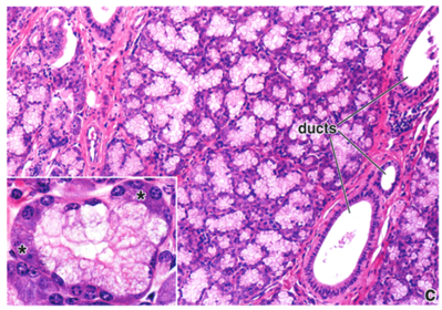
| All salivary glands have ________ branching | compound tubular alveolar |
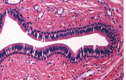
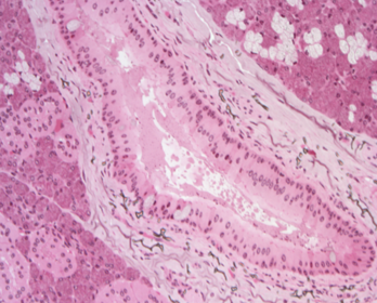
| Classify tissue lining tube. What kind of duct? | stratified columnar epi interlobular duct in salivary gland |
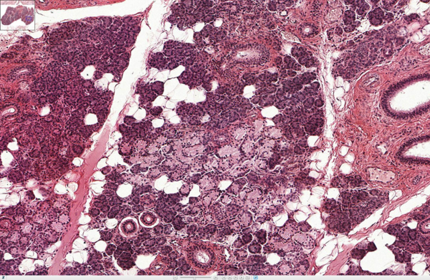
| Submandibular - S,m white: fat purple/pink: serous pale in between: mucus | |
| ID type of cell | unicellular Goblet cells |
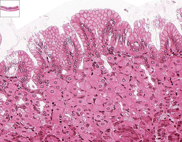
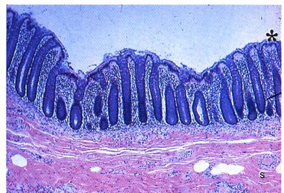
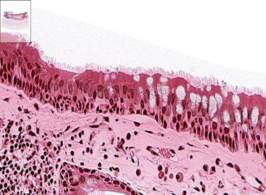
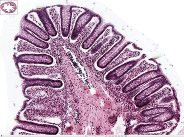
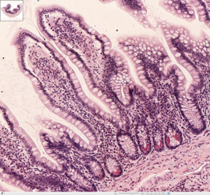
| ID surface epi type | simple columnar epi on the surface of villi with goblet cells |
| What type of duct is shown here? | simple duct, like a test tube |
| What type of branching is seen here? | simple branch acinar (looks like grapes) - you can also see simple coil tubular in lower right |
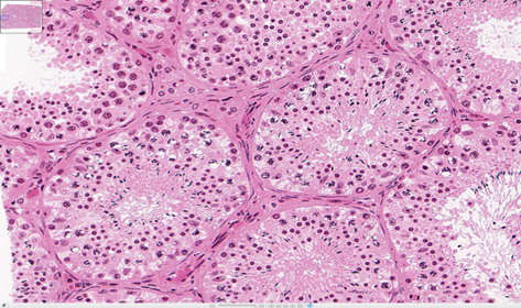
| what mode of secretion does this do? | sperm in seminiferous tubule does cytocrine secretion whole LIVE cell |
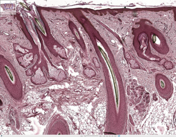
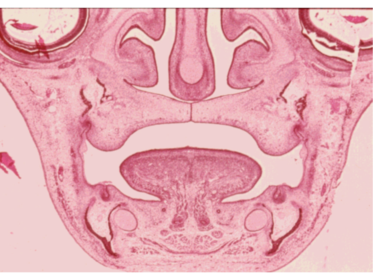
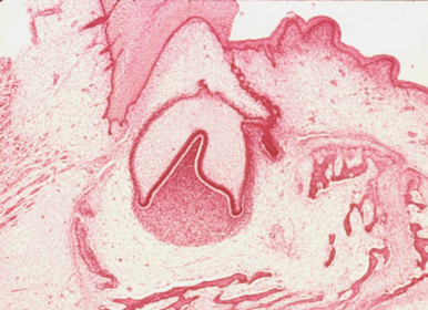
| Tooth germ, mesenchyme | |
| tooth germ | |
| embryonic mesenchyme | |
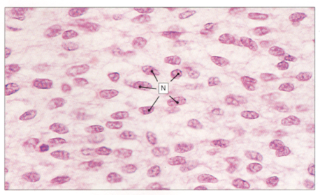
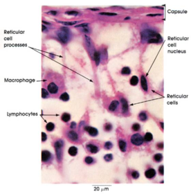
| reticular cells with reticular fibers | |
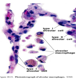
| alveolar microphages | |
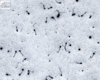
| dust cells within macrophages in lungs (black dots) | |
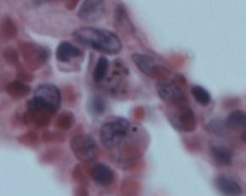
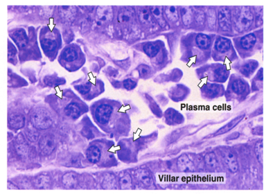
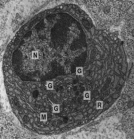
| plasma cells | |
| plasma cells have a lot of..... | RER |
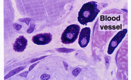
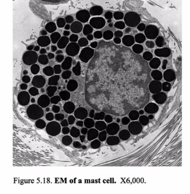
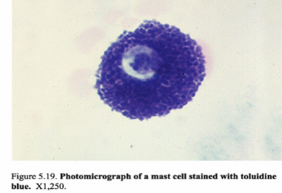
| mast cells | |
| mast cells | |
| mast cells | |
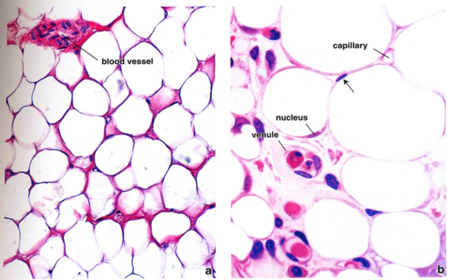
| white/yellow fat unilocular adipose tissue | |
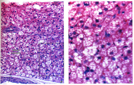
| Brown fat with multilocular adipose tissue | |
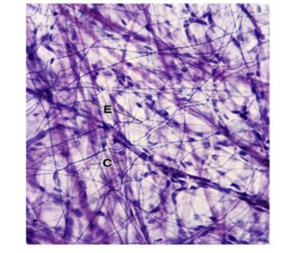
| collagen and elastin fibers | |
| Name the types of fiber and the collagen type | collagen, big bundles so its type 1 |
| silver staining of reticular fibers | |
| reticular fibers | |
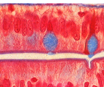
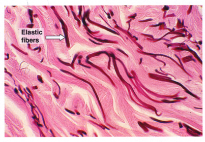
| elastic fibers, formed by smooth muscle cells | |
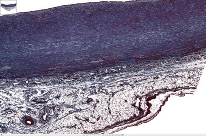
| dark wavy elastic fibers in the walls of the aorta. you can also see fat tissue | |
| Umbilical cord, mucoid tissue all around 2 umbilical arteries and one umbilical vein | |
| Describe the type of CT | Loose connective tissue, within the intestines |
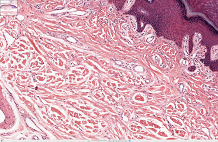
| Describe the type of CT | dense, irregular collagenous CT |
| what are the red fibers? blue dots? | Collagen - type 1 blue elongate nuclei are fibroblasts/fibrocysts |
| Be able to ID the layers | |
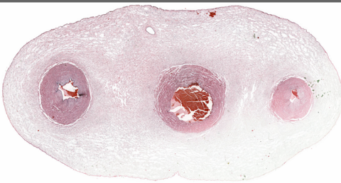
| what type of CT? what are the blue dots? | Tendon dense regular collagenous CT, collagen type 1 (red) blue dots are fibroblast/fibrocysts |
Want to create your own Flashcards for free with GoConqr? Learn more.