
Flashcards
Cardiac Cycle
Cards in this deck (35)
- 1Why do we need a CVS? (______________ ________)Cardiovascular system,
Because diffusion alone to supply our tissues with nutrients is too slow. - 2What method of _____ transport does the CVS rely on, and what does it involve?passive,
CONVECTION - movement of fluid down a PRESSURE gradient - 3What are the component of the CVS and what do they do? (4)1. Heart - provides force
2. Arteries - distribute blood
3. Capillaries - site of exchange
4. Veins - reservoir of blood - 4What is CO?Cardiac Output, the volume of blood ejected from the heart per minute.
CO = HR x SV - 5What is caused when there's an imbalance between R and L outputs?Congestive Heart Failure
- 6Why do both HR and SV need to be raised if CO is to be raised?Raising HR alone can only elevate CO to a certain point, and then can decrease it as heart muscle is pumping so fast it has inadequate time to fill ventricles.
- 7What is the equation for flow? (DARCY'S LAW)flow = change in pressure / resistance
- 8How can this be applied to blood flow?Change in pressure = Arterial pressure - CVP. CVP is negligible, so P is approx. BP.
Resistance = TPR - 9How can this be rearranged to help us?Rearranging Darcy's Law gives:
Blood flow = BP/TPR.
Blood flow = CO
THEREFORE, rearranging gives:
BP = CO x TPR - 10What is the first stage of the cardiac cycle?Diastole - atria contract due to high pressure opening the tricuspid and mitral valves, and ventricles fill.
- 11When the ______ in the ventricles exceeds that in the atria, ________ close and ________ begins in a closed chamber. This is called _____________ ___________Pressure,
tricuspid and mitral valves,
contraction,
isovolumetric contraction - 12The volume in the LEFT ventricle during isovolumetric contraction is called the ___ _______ _______. It is approx ____mlEnd Diastolic Volume,
120 - 13Once the ______ in the ventricles is higher than that in the _____ and _________ _____, the _______ valves open and blood is ejected (rapidly at first, then slower).pressure,
aorta,
pulmonary trunk,
semilunar - 14The _____ in the _____ and ___________ _______ becomes higher than in the _______ so the ________ valves close. There is little blood in the ventricles at this point.pressure,
aorta,
pulmonary trunk,
ventricles,
semilunar - 15This is called __________ ________ and the volume remaining in the LEFT ventricle is called the ____ _______ _______ and is approx ___mlisovolumetric relaxation,
end systolic volume,
40 - 16What is the ejection fraction (EF), and what is it normally?The proportion of the full ventricular volume which is being ejected with each beat. Normally 55-65%.
- 17What would cause a low EF?Anything which has damaged the myocardium would cause inefficient pumping and therefore lower EF.
- 18At which stages do rapid increases and decreases in LV pressure occur?Isovolumetric contraction and isovolumetric relaxation respectively.
- 19What determines the opening and closing of valves?The pressure difference between the chambers/vessels.
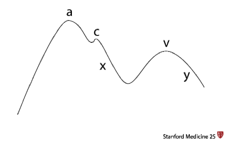
- 20—This shows Atrial and JVP through diastole(A+C) and systole (X,V+Y)
- 21What does the A wave represent?Increased pressure due to atria contracting - peaks then falls as blood is being emptied into ventricles so pressure in atria drops
- 22Why does the C wave show a slight increase in pressure?During ventricular contraction, atria are pulled open slightly to allow more blood to fill, so atrial pressure increases briefly.
- 23What is the X wave showing?Systole - atrial pressure is lowest here as atria are virtually empty. As atria fill, pressure begins to increase again.
- 24What is the V wave?The V wave peaks and shows the atria are full and pressure is high. At this point the cuspid valves are closed.
- 25What is the Y wave?This shows the start of diastole again, as cuspid valves open and atria begin to empty.
- 26—This is a volume/pressure loops in the LV.
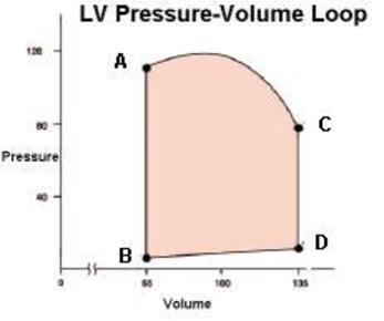
- 27What does the width of the shape represent?The SV
- 28What does the area of the shape represent?The 'stroke work' - that is the energy of consumed during the cardiac cycle
- 29What do the vertical lines represent?Isovolumetric contraction and relaxation.
- 30What is a simple way of working out the stroke work?Work = change in pressure x change in volume
- 31What is happening when we hear the traditional heart sounds?Valves are closing
- 32What is S1?'lubb' - AV valves close
- 33What is S2?'dupp' - semilunar valves close
- 34When can a third heart sound sometimes be heard and why?In neonates, pregnant women, anaemia. Due to turbulent blood flow as it is flowing faster or around tighter corners (in the case of neonates as heart is smaller).
- 35What is a 4th heart sound a sign of?Some sort of adult pathology. Due to forced atrial contraction and reduced ventricular compliance.
Information
- Description
- Intro to the physiology of the cardiac cycle
- Created by
- Hannah Tribe
- Views
- 301
- Tags
- Rating