9520460
Description
Flashcards by Marissa Alvarez, updated more than 1 year ago
|
|
Created by Marissa Alvarez
almost 7 years ago
|
|
| Question | Answer |
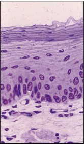
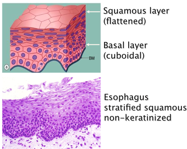
| Where is this epithelium found, what are its important features and how does it function within its organ? | Esophagus & oral cavity stratified squamous **lubricated protective lining |
| Extent of epithelial tissue (epithelial tissue is EVERYWHERE) | Epidermal outer surface of skin Entire GI track Entire airway Urinary track Reproductive tracks Dispersed small glands Major internal organ glands, including liver and pancreas Lines entire blood vessel and lymphatic systems Lines internal body cavity and many organs |
| Major Functions of Epithelium | **Protection** Secretion Sensory Reception Absorption Excretion Reproduction |
| Diseases directly related to epithelia | Cancer: Pancreatic ductal adenocarcinoma Breast cancer from the lactiferous duct Basal cell carcinoma of the epidermis Metastatic melanoma of epidermal melanocytes Influenza: infection of respiratory epithelium Reflux disease: acid erosion of esophageal epithelium Renal failure: damage to epithelial filtration barrier Cystic fibrosis: genetic defect in chloride channel |
| General Features of Epithelial Tissues | Covers surfaces or lines cavities Rests on a basement membrane Lacks direct vascular supply Contains little or no extracellular matrix Cells linked by junctional complexes Polarized Forms exocrine and endocrine glands |
| Classification of Epithelia | 1. Cell Layers 2. Cell Shape |
| 1. Number of cell layers | One layer: simple Two or more layers: stratified All cells rest on basement membrane but not all cells reach the surface: pseudostratified Specialized multilayer: transitional |
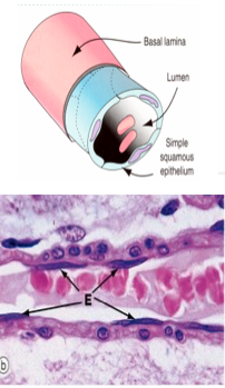
| 2. Cell Shape Shapes of cells in SIMPLE epithelium | Squamous Cuboidal Columnar |
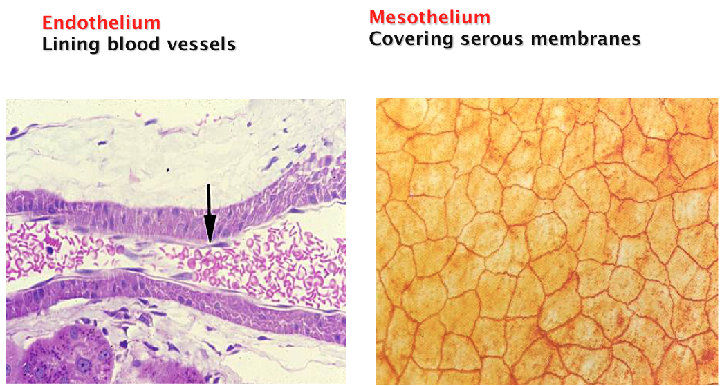
| Shapes of cells in SIMPLE epithelium SQUAMOUS | -lining of blood vessels -FLAT, long-hollow cell -Larger, flat nuclei ex: endothelium of arteriole |
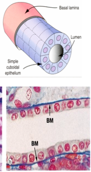
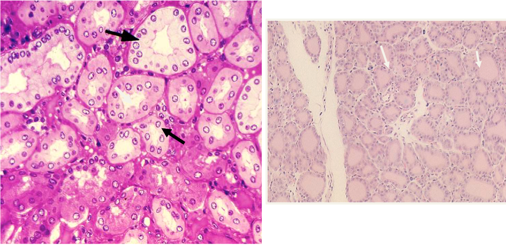
| Shapes of cells in SIMPLE epithelium CUBOIDAL | -cube shaped ex: renal collecting tuble |
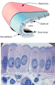
| Shapes of cells in SIMPLE epithelium COLUMNAR | Tall cells ex: Gall bladder |
| REMEMBER! Epithelia are named based on the cell shape of the | superficial / OUTSIDE layer |
| Shape of cells in STRATIFIED epithelia Stratified squamous | |
| Shape of cells in STRATIFIED epithelia Stratified cuboidal | |
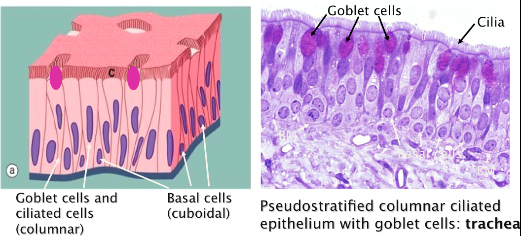
| Shape of cells in PSEUDOstratified epithelia | |
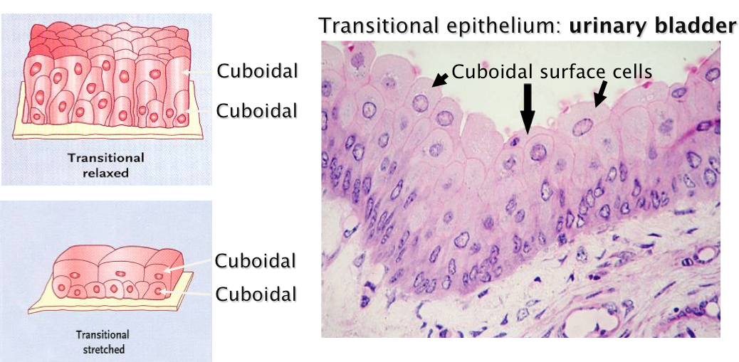
| Shape of cells in TRANSITIONAL epithelia | ***Transitional epithelium is an exception: the name is derived from a functional change in height, not the shape of the surface cells. "pillow top" |
| Major types of epithelia SIMPLE Squamous Endothelium VS Mesothelium | |
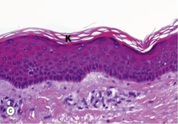
| Major types of epithelia Stratified squamous, keratinized: SKIN | |
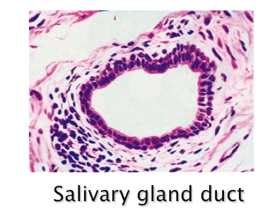
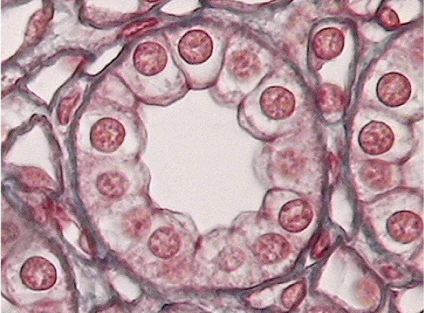
| Major types of epithelia SIMPLE Cuboidal Kidney tubules, thyroid gland (ducts) | |
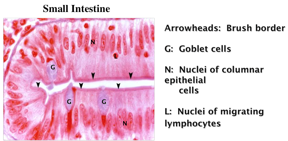
| Major types of epithelia Simple columnar with BRUSH border and GOBLET cells: (small intestine) | |
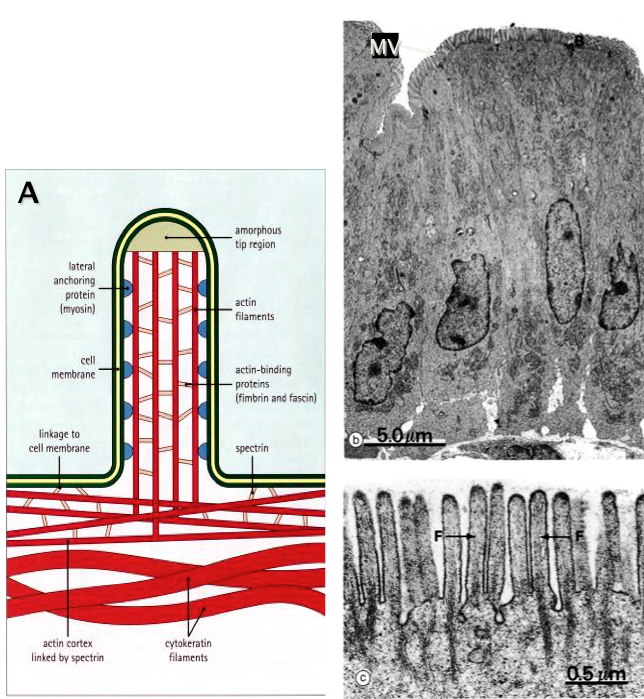
| Apical Cell surface specializations: Microvilli (short, straight, and stiff, filled w/actin) | Microvilli are NON-MOTILE and only 0.5-1.0 µm in length Internal actin microfilaments are linked to each other, the membrane and the terminal web Cells of the small intestine and renal proximal tubule have thousands of microvilli per cell creating a BRUSH OR STRIATED BORDER **The main role of microvilli is to INCREASE SURFACE AREA |
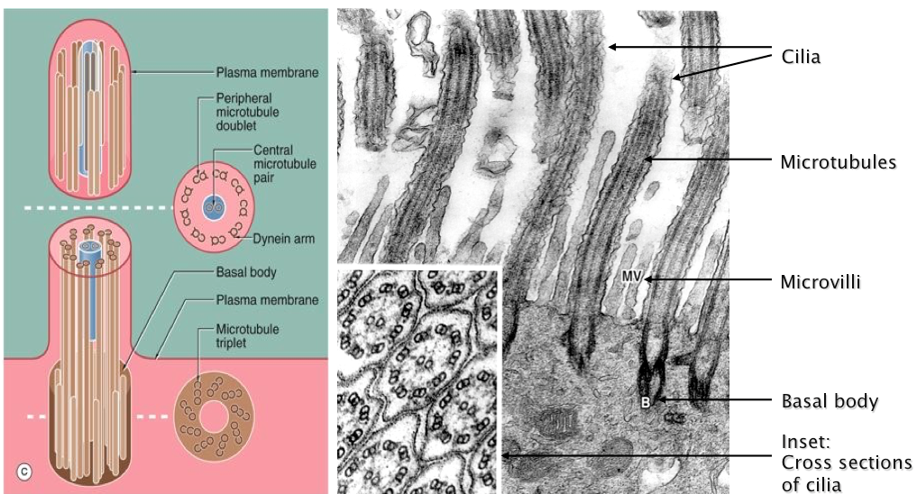
| Apical Cell surface specializations: Cilia (much longer, can MOVE, strong, & diff. composition) | Cilia are found on many cell types especially in the UPPER AIRWAY and oviduct Cilia are up to 10 µm long and usually beat in one direction MOTILITY |
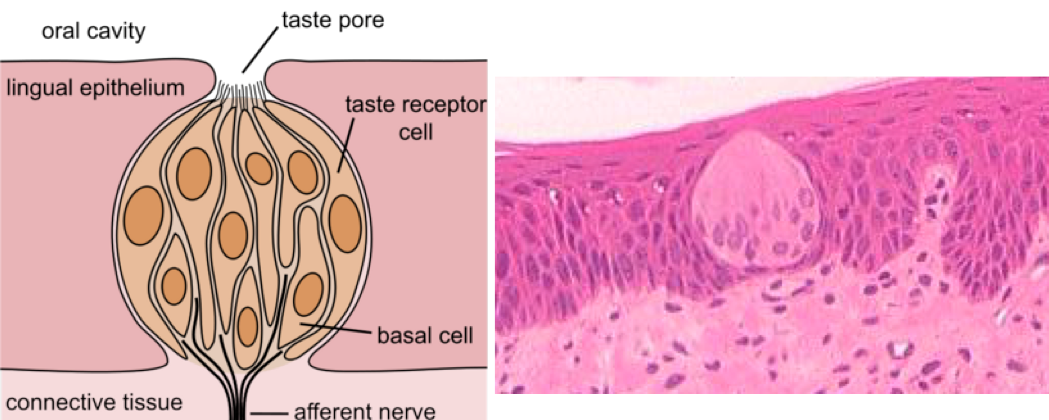
| Sensory Specialization: Taste Buds | |
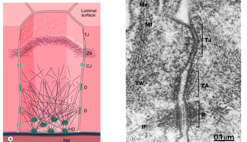
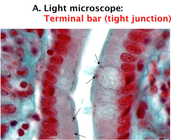
| How do epithelial cells attach and communicate? | Epithelial cells are joined at the lateral surfaces by the JUNCTIONAL COMPLEX Tight junctions (TJ): more narrow, barrier b/w inside & outside of cell Zonula adherens (ZA): belt of actin Desmosomes (D): a well, prevents from sloughing off are often found near the apex of the cell Hemidesmosomes (HD) anchor the epithelia to the basement membrane (BM) |
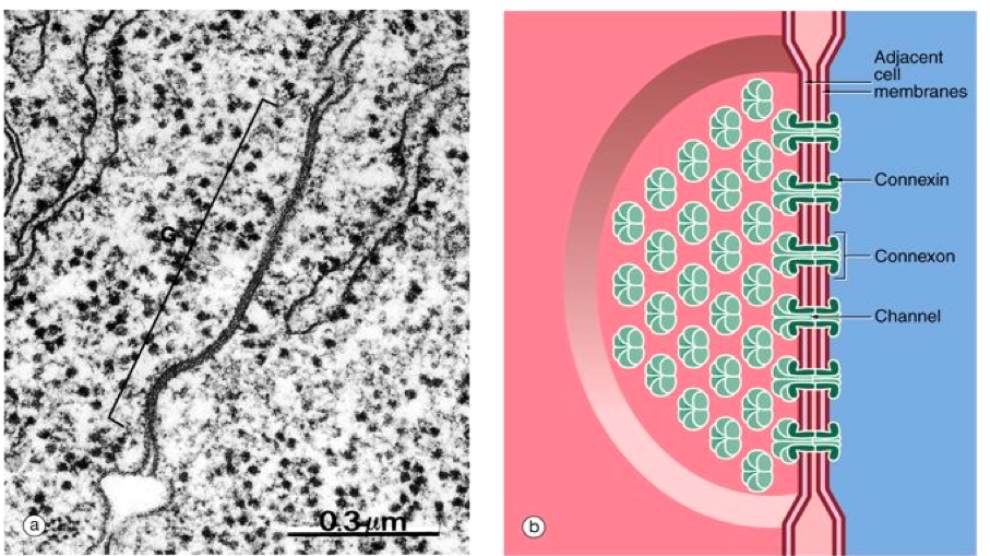
| Gap junction (communicating junction) | Gap junctions provide 1nm gated channels between adjacent cells Small molecules and ions <1.5 kDa can pass through open channels **Connexins! |
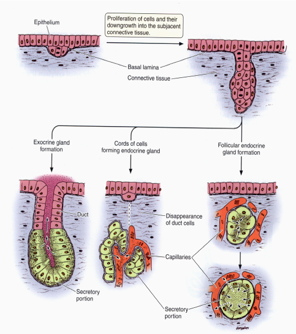
| GLANDS Specialized epithelial structures Exocrine VS Endocrine | Exocrine: secrete products via DUCTS onto the epithelial surface from which they originated EX: Pancreas Endocrine: ductLESS; secrete products into blood vessels, lymphatic vessels, or surrounding space for distribution |
| Formation and classification of glands | Epithelial structures specialized for secretion Exocrine defined by BOTH duct and secretory unit Endocrine are ductLESS glands |
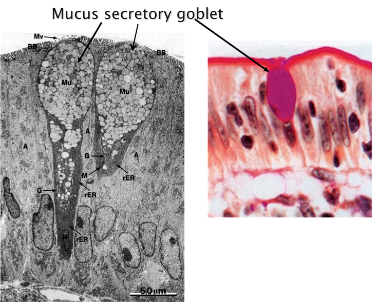
| Single-cell EXOcrine gland: the goblet cell | -secretes mucous -uses ducts |
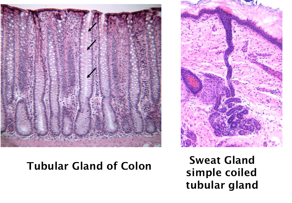
| MORE EXOcrine Gland examples Tubular gland of colon & Sweat Gland | |
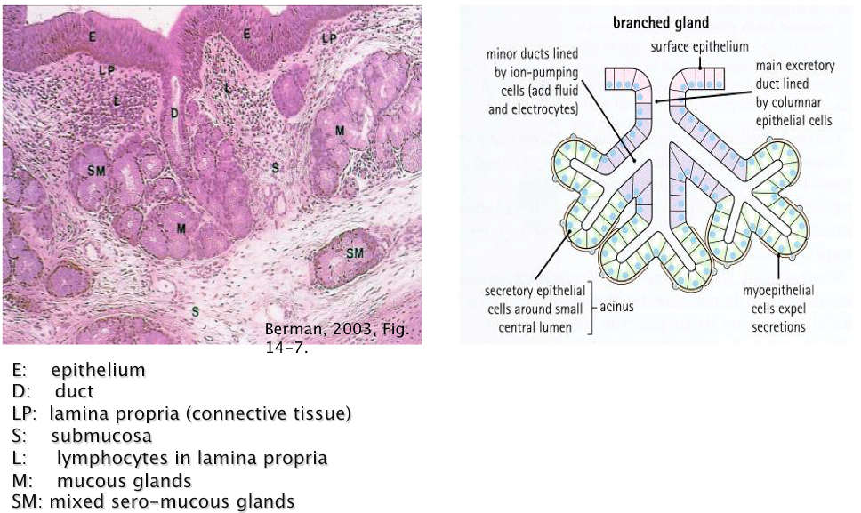
| Example of Branched Exocrine Gland: Tracheal glands | -more complex, specialized -dual exocrine gland (mucous and protein) |
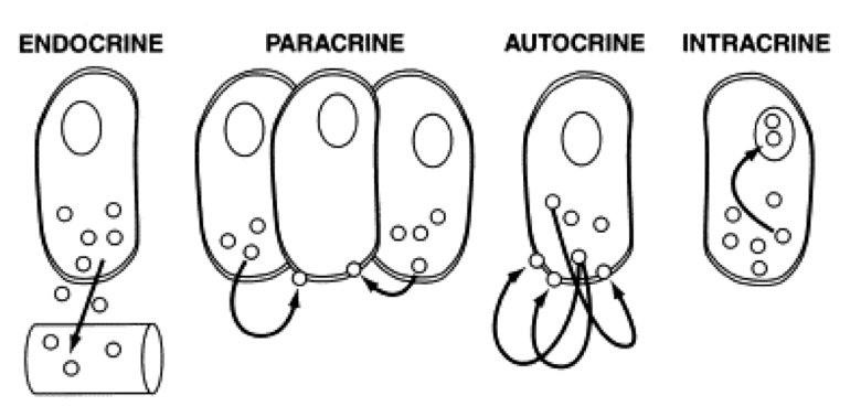
| Endocrine Single Cell endocrine, paracrine, autocrine, intracrine | |
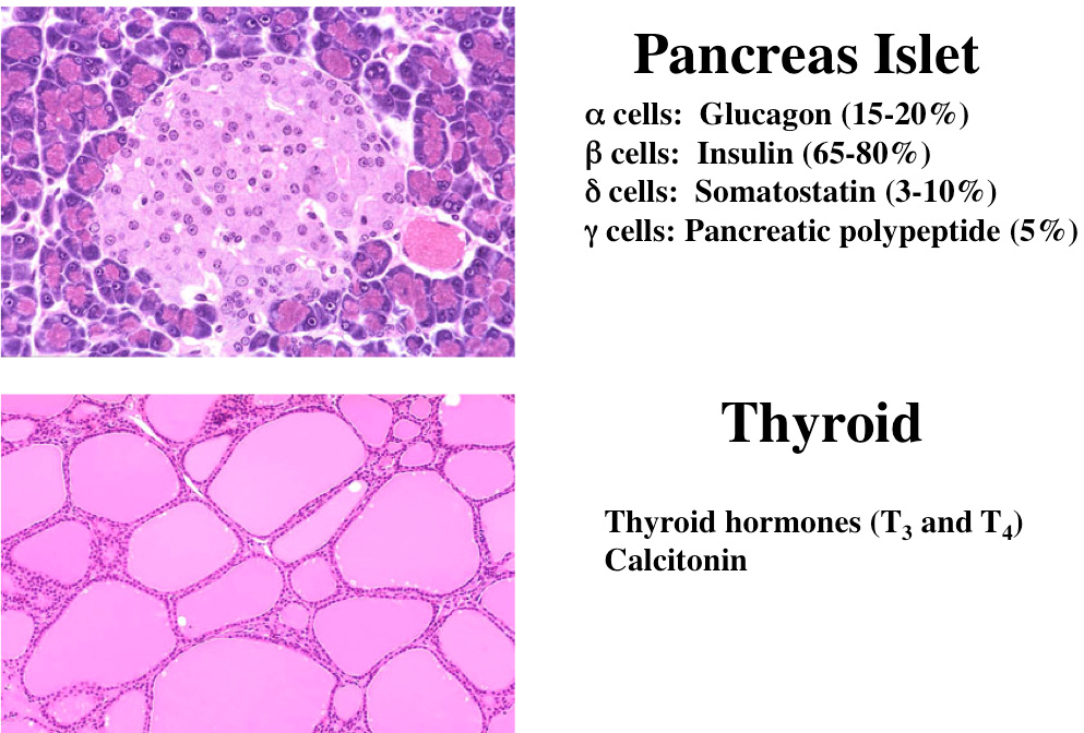
| ENDOcrine Multicellular Pancreas Islet (no secretory ducts, blood vessels present) & Thyroid (simple cuboidal epithelium) | |
| Exam Question What Type of Epithelium is this? A. Stratified squamous B. Simple cuboidal C. Simple squamous D. Transitional | B. Simple cuboidal (kidney tubules & thyroid gland) |
Want to create your own Flashcards for free with GoConqr? Learn more.