9521222
Description
Flashcards by Marissa Alvarez, updated more than 1 year ago
|
|
Created by Marissa Alvarez
almost 7 years ago
|
|
| Question | Answer |
| Connective Tissues | |
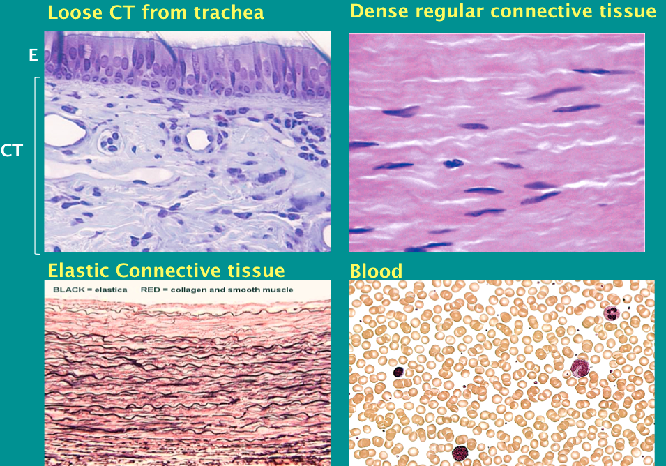
| Forms of Connective Tissue Connective tissue proper | 1. Loose – irregular 2. Dense – regular or irregular |
| Forms of Connective Tissue Special connective tissues | Adipose tissue Elastic tissue Blood Mucous tissue |
| Forms of Connective Tissue Supporting (skeletal) connective tissues | Cartilage Bone |
| Functions of Connective Tissue | exchange of metabolites and waste products electrolyte balance storage of energy reserves mechanical support shock absorption insulation source of heat defense against pathogens repair after injury |
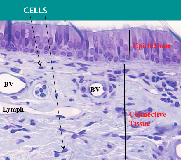
| Distinctive features of connective tissues | Consist of cells and extracellular matrix (ECM) **The ECM contains fibers and GROUND substance Cells density is LOW Blood vessels, lymphatics, and nerves are present |
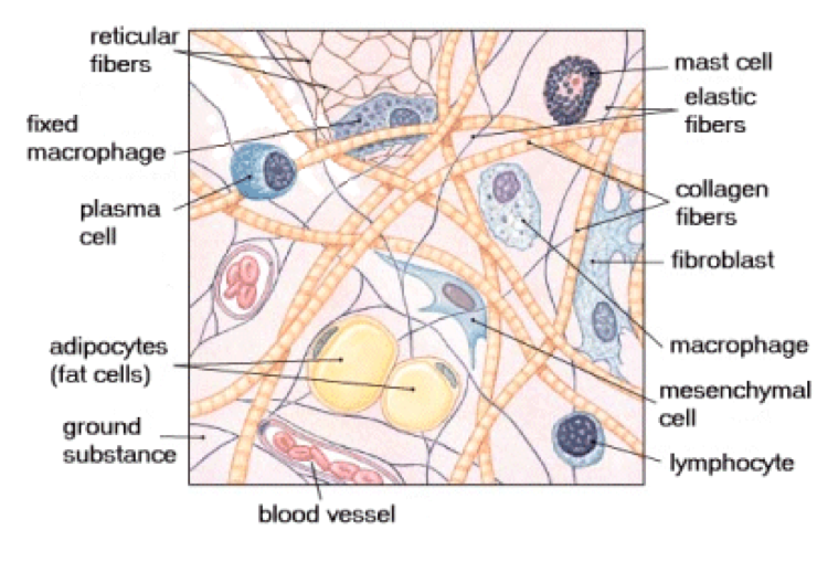
| Connective Tissue: Loose irregular (areolar) CT (Resident cells & Fibers) | Resident cells: Fibroblasts Fat cells Macrophages Mast cells Plasma cells Fibers: Collagen Reticular Elastic Ground substance |
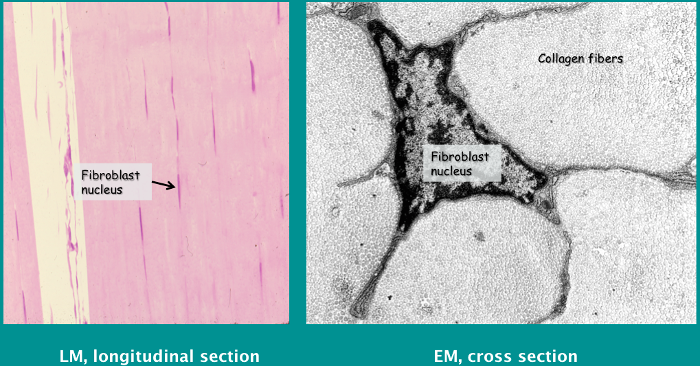
| Loose irregular CT Resident Cell: Fibroblasts | - synthesize extracellular matrix materials: precursors for collagen and elastic fibers components of ground substance - important in development and wound healing (from the CT itself) |
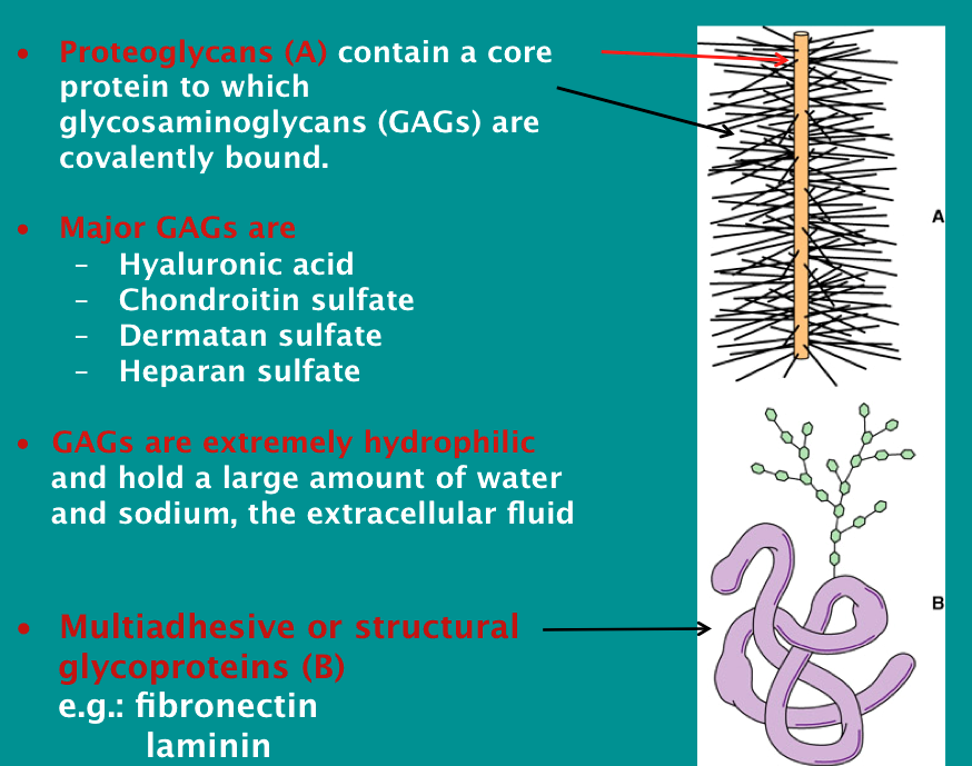
| Ground Substance | |
| Loose irregular (areolar) CT Fibers 1. COLLAGEN | Main fiber type and MOST abundant protein in human body Synthesized and secreted as a precursor (tropocollagen) by fibroblasts Assembled into mature fibers spontaneously in the extracellular space. These fibers have greater tensile STRENGTH per unit weight than steel At least 27 types exist, of these the most important are: Type I Type II Type III Type IV Mutations of the genes coding for collagen lead to serious inherited diseases – e.g., Ehlers-Danlos syndromes (hyper-elastic skin) |
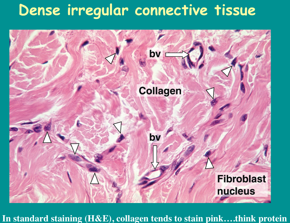
| DENSE irregular connective tissue | |
| Type I Collagen: | most common (in internal organs, skin, fascias, tendons, ligaments, bone) |
| Type II Collagen | cartilage (main component of hyaline and elastic cartilage) in hyalin cartilage (see images later on in slide set!) |
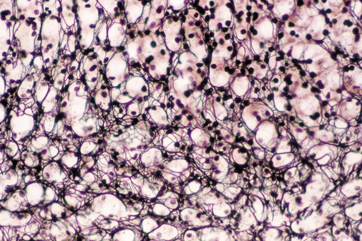
| Type III Collagen | reticular fibers They can only be demonstrated by special stains such as silver impregnation Reticular fiber network from adrenal cortex, silver stain. Nuclei are also black, cytoplasm is unstained. |
| Type IV Collagen | in basement membranes (does not form fibrils) |
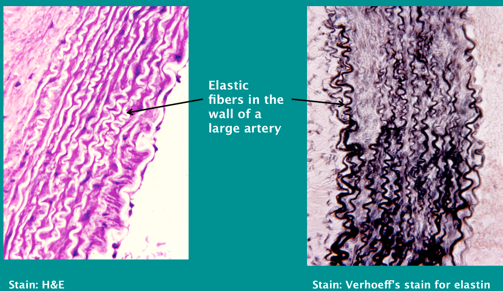
| Fibers: Elastin | can form fibers or fenestrated membranes. "loose rubber band" Different protein altogether (NOT collagen) |
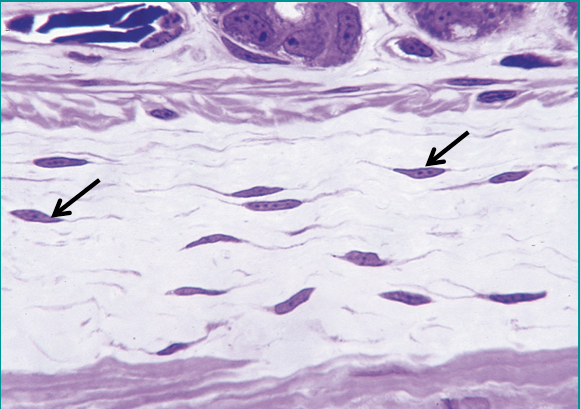
| Dense REGULAR connective tissue: TENDON (made by fibroblasts + collagen + ground substance) *very straight, matrix of proteins | |
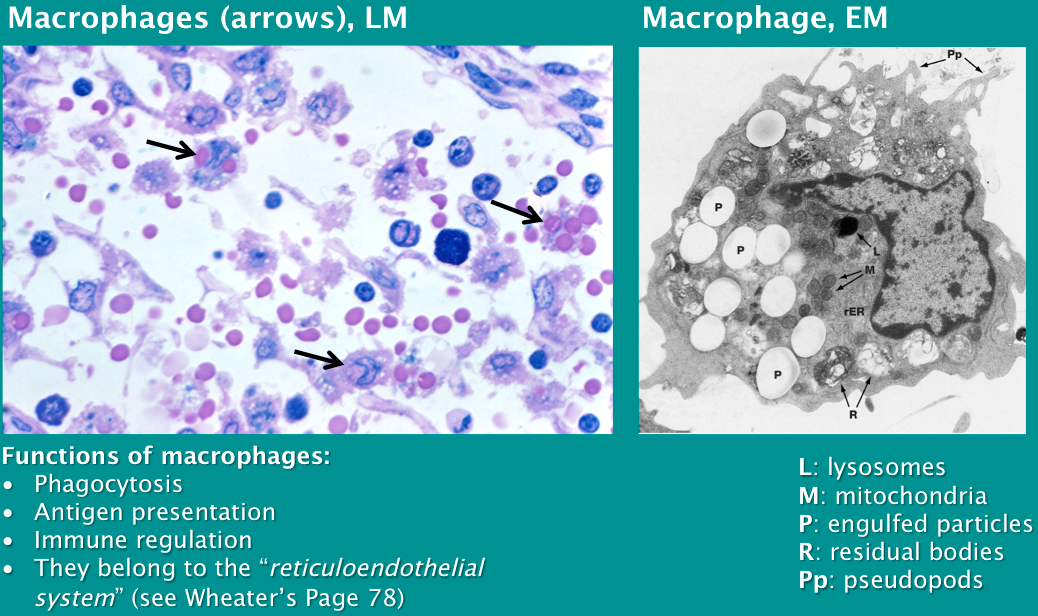
| Defense cells in connective tissue Resident, intrinsic, or fixed cells: | Macrophages (from blood monocytes) Mast cells (functionally analogous but not identical to basophils in blood) Plasma cells (from B lymphocytes) |
| Defense cells in connective tissue Migrating or wandering cells | All leukocytes circulating in blood enter connective tissues: Lymphocytes Monocytes Granulocytes Neutrophils Basophils Eosinophils |
| After tissue injury, macrophages in regional lymph node engulf erythrocytes (RBC's) MACROPHAGES *main function = Phagocytose (resident cell) | |
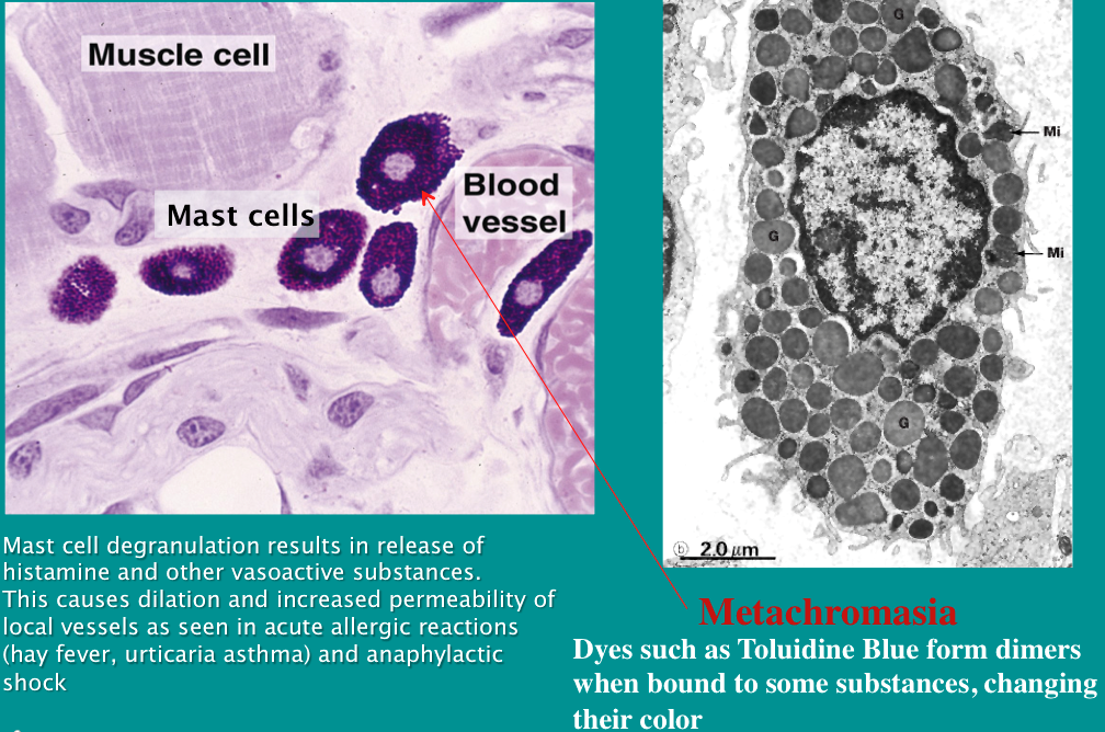
| MAST Cells Main function: release histamine via granules (resident cell) | -mediates immune response "trigger" -Metachromasia: acidity of granules change Toluidine blue dye to a reddish color |
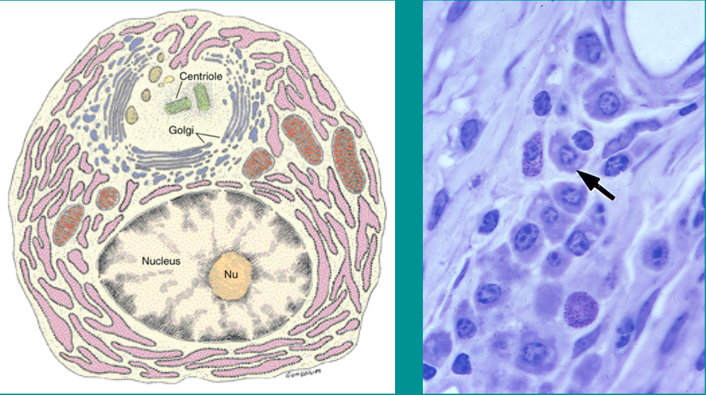
| Plasma Cells Main function: antibody production (differentiated B cells) Resident cells (clock-face nucleus and clearing indicating Golgi) | |
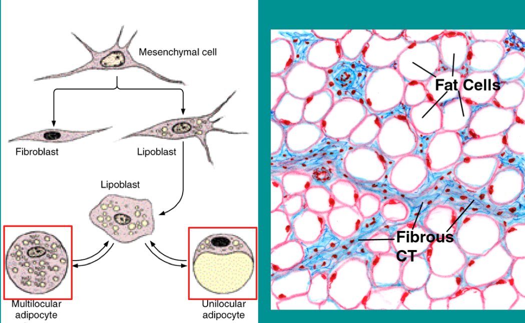
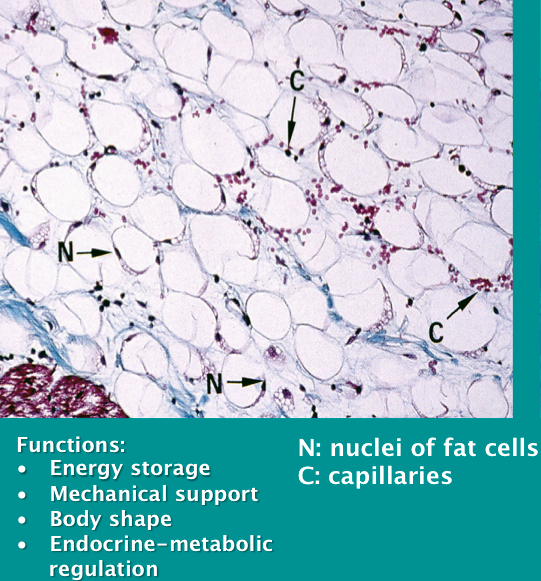
| Fat cells (Adipose Tissue) 2 Types: 1. Unilocular (yellow/white fat) 2. Multilocular (brown fat) | |
| Fat cells (Adipose Tissue) 1. Unilocular (yellow/white fat) | |
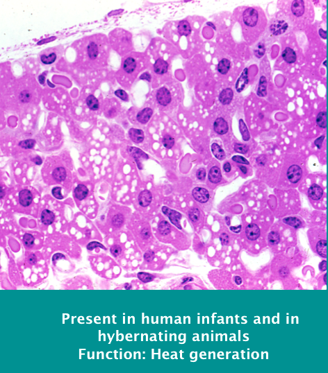
| Fat cells (Adipose Tissue) 2. Multilocular (brown fat) HEAT generation | |
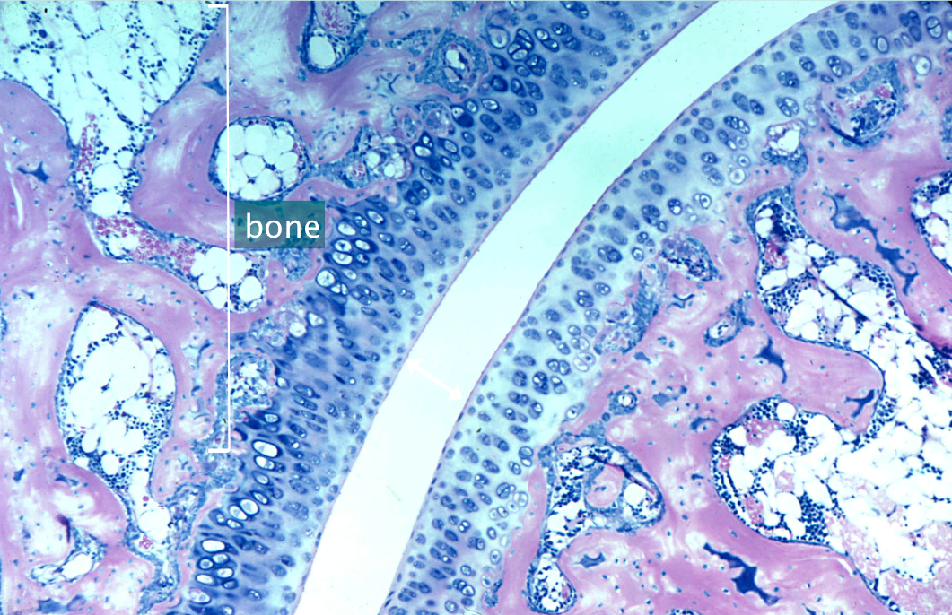
| CARTILAGE Type II Collagen -Two types: Elastic & Hyalin Cartilage | structural support shock absorption reduce friction in joints template for bone formation (endochondral ossification) |
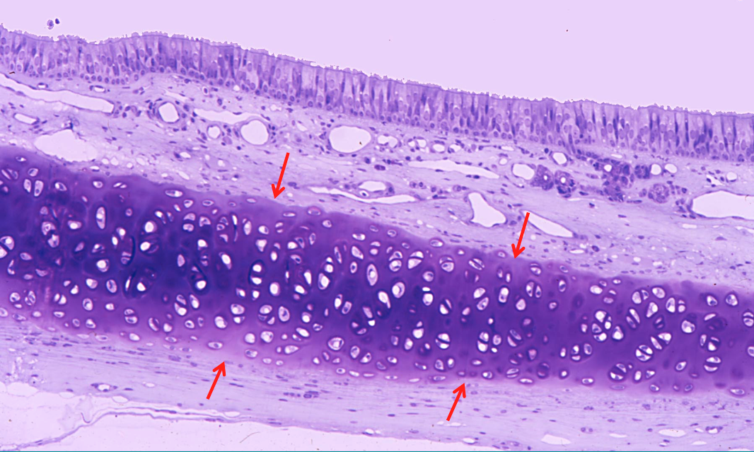
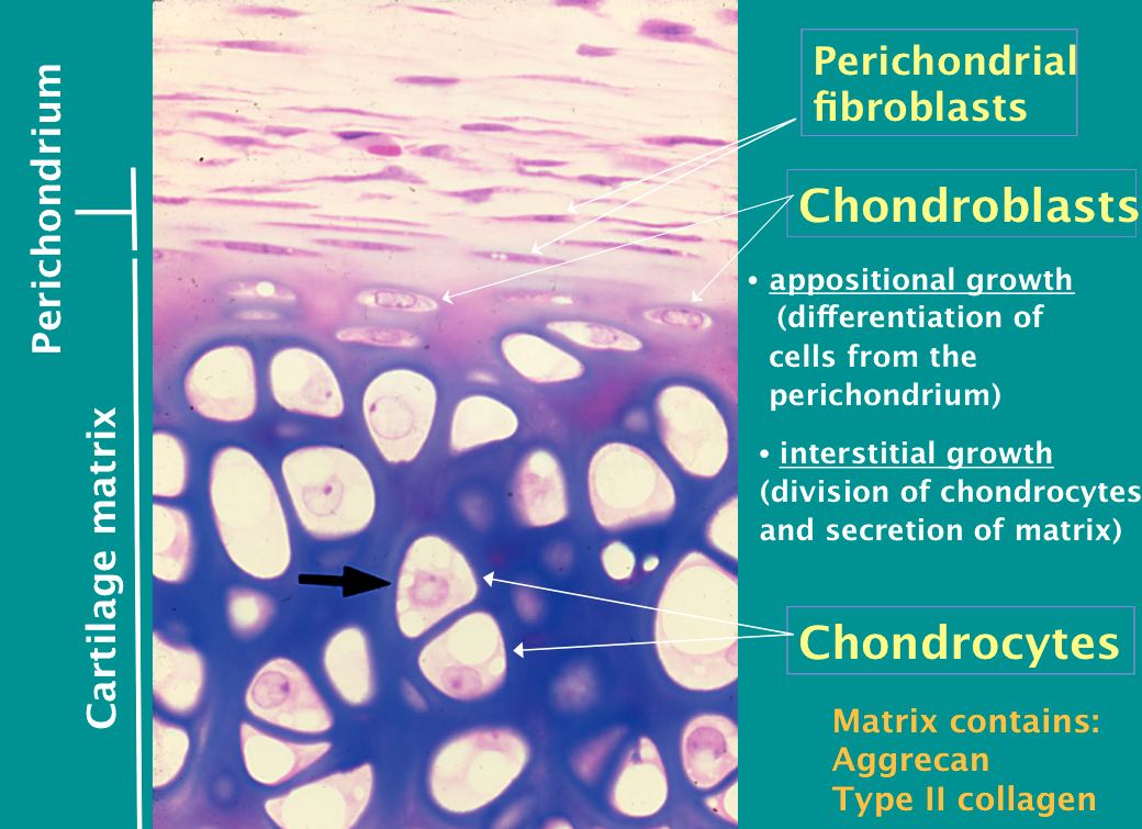
| Hyaline Cartilage (in the NOSE) Type II Cartilage -surrounded by chondroblasts, then there are perichondrial fibroblasts around the chondroblasts | |
| Perichondrium and Cartilage Matrix | |
| Joint Space / Articular Cartilage (empty space = synovial fluid) **LACKS perichondria fibroblasts & chondroblasts (Cannot regenerate - painful: because underlying bone!) | |
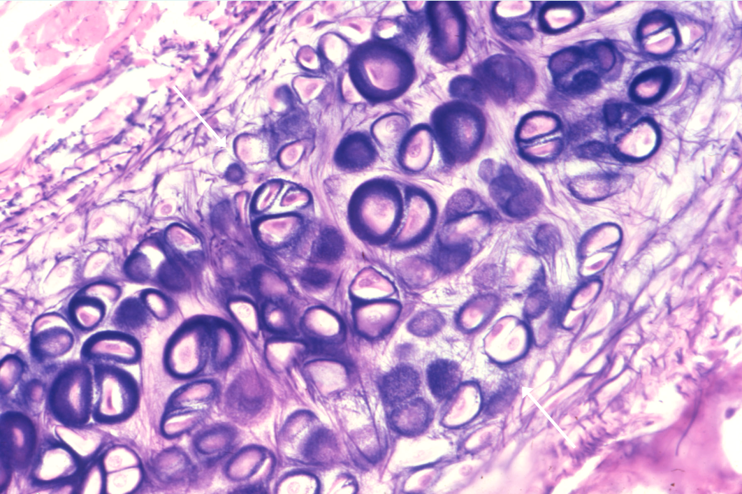
| Elastic Cartilage (Epiglottis, part of the ear) -basically hyalin cartilage BUT has a fibrous look, a form of elastic fibers | |
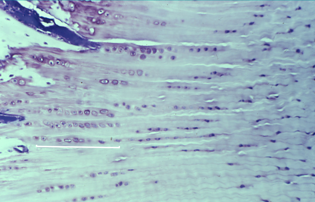
| Fibrocartilage **Contains both Types Collagen I and II (cells lined up in rows - pattern present) | Pubic symphysis Meniscus of joints Part of intervertebral joint TMJ *Will replace severely torn Hyalin Cartilage* (forms in regions of injury/degeneration within hyalin cartilage) |
| EXAM Question: Hyaline Cartilage is composed of: A. Elastic Fibers B. Collagen I C. Collagen II D. Adipose Cells | C. Collagen II |
Want to create your own Flashcards for free with GoConqr? Learn more.