9551357
Description
Flashcards by Kristina Redd, updated more than 1 year ago
|
|
Created by Kristina Redd
over 8 years ago
|
|
| Question | Answer |
| What is the main function of muscle tissue? | Contraction to generate motion (muscle must span a joint to achieve this) |
| What does sarco- refer to? | "muscle component" |
| What does myo- refer to? | "muscle component" |
| Describe the activity of skeletal muscle. | Strong, quick discontinuous voluntary contractions |
| Describe the activity of cardiac muscle. | strong, quick continuous involuntary contraction |
| Describe the activity of smooth muscle. | Weak, slow involuntary contraction |
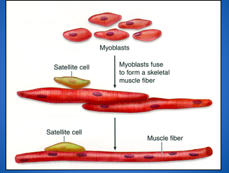
| How do skeletal muscle fibers develop? | myoblasts --> fusion of myoblasts --> formation of muscle fiber |
| _______ cells are the source of muscle regeneration after injury. | Satellite |
| _______ fuse to form a skeletal muscle fiber. | Myoblasts |
| Myofibrils contain microfilaments that contain ____ and ____ . | actin, myosin |
| What type of filament is actin: thin or thick? | thin |
| What type of filament is myosin: thin or thick? | thick |
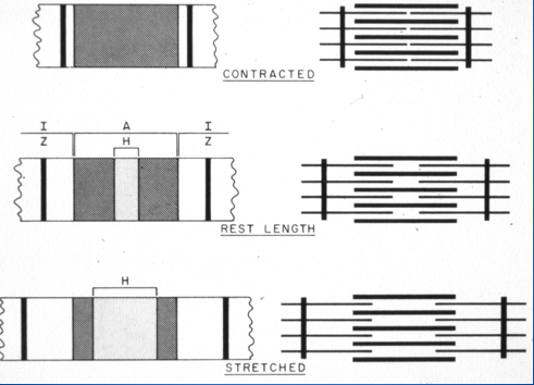
| A Z line to Z line makes up a _____ . | sarcomere |
| ____ band is made up of all myosin and a small amount of actin. | A band |
| Why does the A band have a dark band and a density that causes a darkness to the stain? | High concentration of myosin |
| This band is made up of all actin, which makes its stain light. | I band |
| This line is a density of protein that actin binds to on the A band. It's a scaffolding of the entire muscle fiber. | Z line |
| True/False: The Z line changes during contraction. | False. During contraction, the Z line pretty much doesn't change. |
| Coined as the leverage point of the entire muscle. | Z line |
| During contraction, a repetition binding-movement-release cycles between the ____ heads and ____ filaments occurs. | myosin head actin filaments |
| What ion plays a significant role in activation? | Ca2+ |
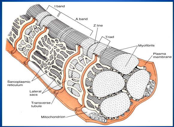
| Where are calcium sacs stored in the muscle? | sarcoplasmic reticulum |
| What is a triad and what does it control? | Special internal membrane system that controls muscle contraction by regulating calcium release. |
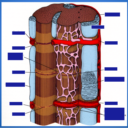
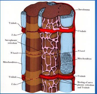
| What are invaginations of the sarcolemma (plasma membrane) or a membrane-enclosed tube that goes down into the cell hundreds of times within a muscle cell? | T-tubules |
| Where do T-tubules occur in mammalian skeletal muscle? | A/I junction |
| What do t-tubules form with two terminal cisternae of the sarcoplasmic reticulum? | triads |
| How do triads at each A/I junction coordinate an impulse? | Sarcomeres (Z line to Z line) must contract or relax at the same time |
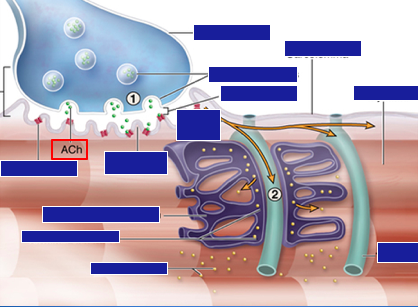
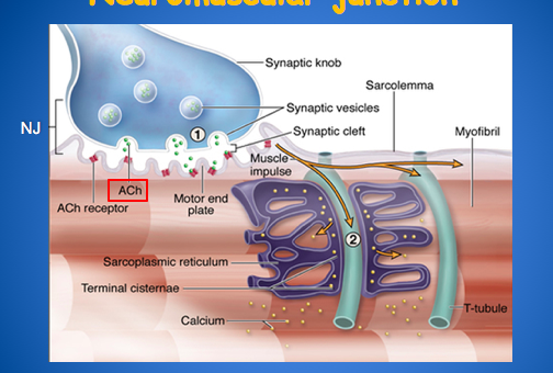
| How does an impulse run down the neuromuscular junction? | 1. Nerve impulse --> Ach released into synaptic cleft --> Ach binds to Ach receptor on sarcolemma of motor end plate --> triggering of muscle impulse 2. impulse spreads from sarcolemma to T-tubules --> Ca released from terminal cisternae of sarcoplasmic reticulum |
| Describe skeletal muscle: fibers? nuclei? fiber innervation? | long, syncytial fibers peripheral nuclei fibers don't branch each fiber is innervated |
| Describe cardiac muscle: fibers? nuclei? fiber innervation? | cells attached end-to-end to form fibers central nuclei fibers branch no innervation (stimulus spreads from cell-to-cell via Purkinje fiber coordination) |
| Where do T-tubules run in cardiac muscles? | T-tubules run at Z-lines |
| What is the difference in cardiac muscle t-tubules and muscle cells' t-tubules? | Cardiac muscle's t-tubule is a diad instead of a triad like the muscle cell. |
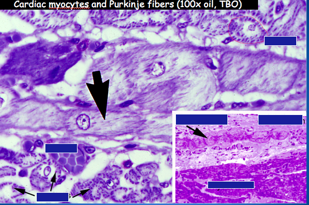
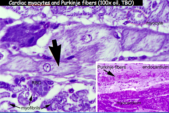
| ____ _____ are modified cardiac myocytes interconnected by gap junctions (electrical synapses). | Purkinje fibers |
| Where can smooth muscle be found? | Found in larger blood vessels, the GI system, ureter, bladder, reproductive systems and many other locations |
| What type of muscle Controls peristaltic motions of tubular structures? | smooth muscle |
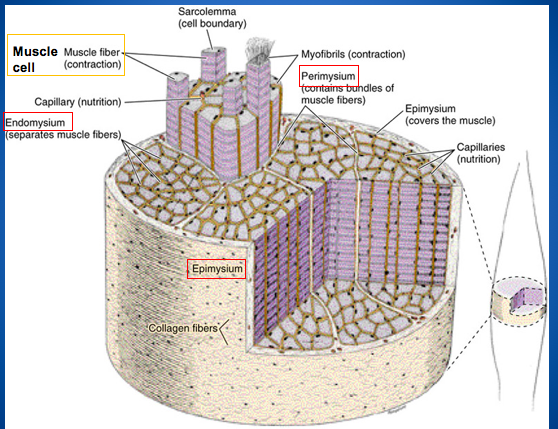
| Which part of a muscle cell is responsible for contraction? | myofibrils |
| The portion of the muscle cell that surrounds each muscle fiber by a small amount of connective tissue created by fibroblasts. | Endomysium |
| What provides nutrition to a muscle cell? | capillaries |
| This layer of the muscle cell surrounds the whole muscle. | epimysium |
| This layer of the muscle cell surrounds fascicles and bundles of muscle fibers. | perimysium |
| Where is the nucleus in a muscle cell? | The nucleus is located at the cell's periphery (pushed off to the side). |
| This layer of the muscle cell separates muscle fibers. | Endomysium |
| What functions as the cell boundary in the muscle cell? | cell boundary |
| In what layer do collagen fibers exist in a muscle cell? | epimysium |
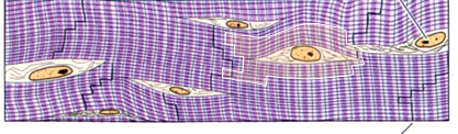
| Identify the type of muscle tissue and indicate the stair-steps. | Cardiac muscle tissue & intercalated disks |
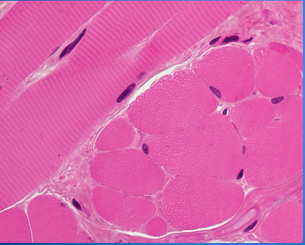
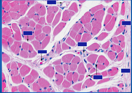
| Identify this muscle type and types of sections. | Skeletal muscle tissue w/ striations at longitudinal and cross sections |
| Identify the missing parts of this muscle cell. | |
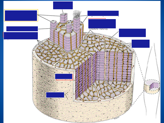
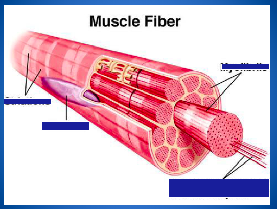
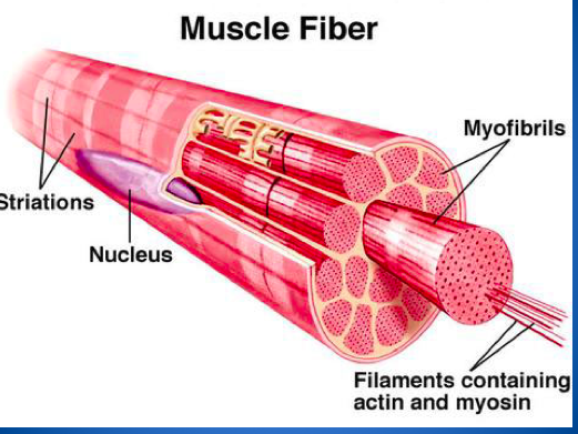
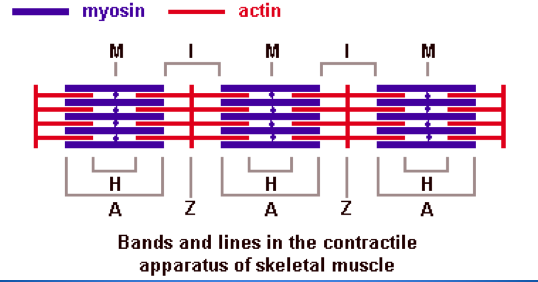
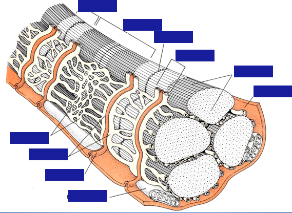
| Identify the parts of the muscle fiber. | |
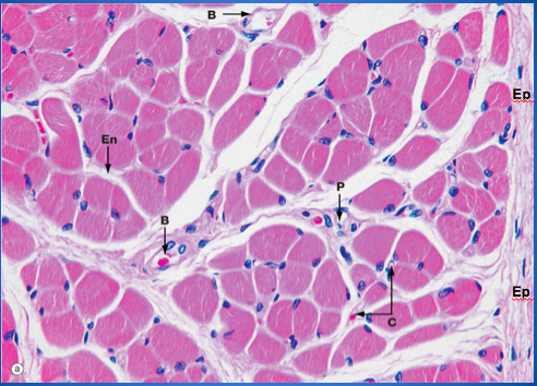
| Identify the organization of a skeletal muscle: epimysium, perimysium, endomysium, blood vessels, and capillaries. | |
| Identify the organization of a skeletal muscle. | |
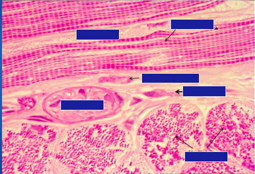
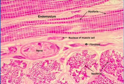
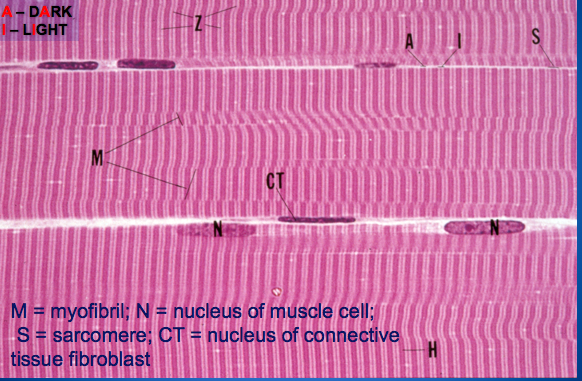
| Identify myofibril, nucleus, sarcomere, connective tissue, and fibroblast. | |
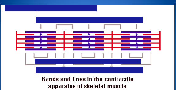
| Identify. | |
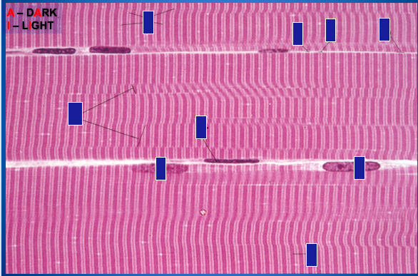
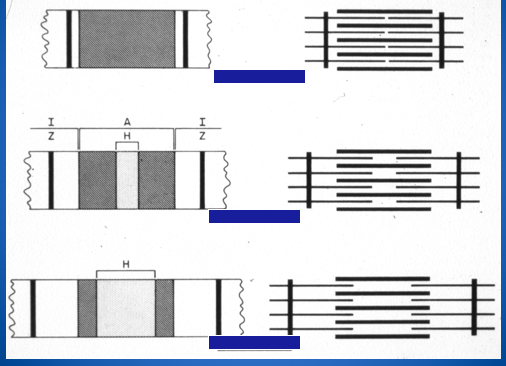
| These indicate changes in band widths during contraction. Find contracted, rest length, and stretched. | |
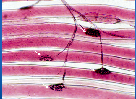
| What does this picture indicate and in what type of muscle tissue? | Muscle cell with motor end plates (arrows) demonstrated by silver impregnation. |
| Identify these parts of a skeletal muscle. | |
| Identify parts of the neuromuscular junction. | |
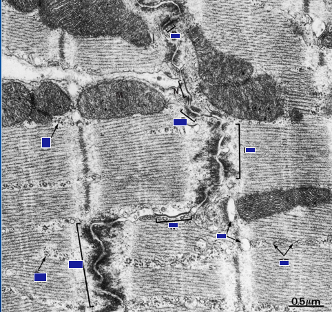
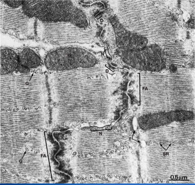
| Identify the parts of a cardiac muscle t-tubule system. | |
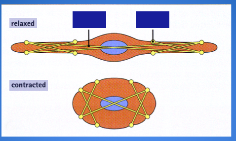
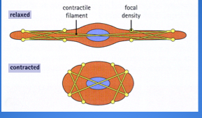
| Name the parts of a smooth muscle contraction. | |
| Name the parts of the cardiac muscle associated with intercalated disks. | |
| Identify this tissue. | Cardiac muscle |
Want to create your own Flashcards for free with GoConqr? Learn more.